Novel Substituted Indazoles towards Potential Antimicrobial Agents
Jaganmohana Rao Saketi1 , S N Murthy Boddapati1,2
 , Raghuram M3
 , Geetha Bhavani Koduru4
and Haribabu Bollikolla1*
1Department of Chemistry, Acharya Nagarjuna University, Guntur-522510, A. P.,India.
2Department of Chemistry, Sir C R Reddy College, P G Courses, Eluru, A.P-534007, India.
3Department of Botany and Microbiology, Acharya Nagarjuna University,Guntur-522510, A.P., India.
4Department of Chemistry, J. M. J. College for Women, Tenali, Guntur Dist, A.P., India.
Corresponding Author E-mail: dr.b.haribabu@gmail.com
DOI : http://dx.doi.org/10.13005/ojc/370234
Article Received on : 07-Jan-2021
Article Accepted on :
Article Published : 11 Mar 2021
The in vitroantimicrobial properties of a series of N-methyl-3-aryl indazoles (5a-5j) were screened. In this present work, we describe our efforts towards the development of potent antimicrobial activity of synthesized indazole derivatives. The antimicrobial activities of the prepared compounds were investigated against four bacterial strains: Xanthomonascampestris, Escherichia coli, Bacillus cereus, Bacillus megaterium, and a fungal strain Candida albicans. The biological evaluation studies of these indazole derivatives revealed that some of these tested compounds have shown moderate to goodin vitroantimicrobial activities.
KEYWORDS:Antimicrobial Activity; Indazole; Well Diffusion
Download this article as:| Copy the following to cite this article: Saketi J. R, Boddapati S. N. M, Raghuram M, Koduru G. B, Bollikolla H. Novel Substituted Indazoles towards potential Antimicrobial Agents. Orient J Chem 2021;37(2). |
| Copy the following to cite this URL: Saketi J. R, Boddapati S. N. M, Raghuram M, Koduru G. B, Bollikolla H. Novel Substituted Indazoles towards potential Antimicrobial Agents. Orient J Chem 2021;37(2). Available from: https://bit.ly/3bBK42V |
Introduction
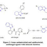
Due to the omnipresent nature of nitrogen containing heterocycles they are the key scaffolds of many biologically important molecules and pharmaceutical products. These nitrogen atoms containing heterocycles are part of the world’s largest selling drugs.1 Nowadays, it is well realized that indazole based motifs are an imperative family of nitrogen containing heterocycles with a broad array of agricultural, biological and industrial applications.2 The structurally varied indazole analogs have been achieved enormous consideration previously, just as in ongoing period, as a result of their wide scope of biological properties, such as anti-hypertensive,3 anti-inflammatory,4 anti-HIV,5 antimicrobial,6 anti- angiogenesis,7 anticancer anticancer,8 anti-tubercular,9 neuroprotective,10 and anti-protozoal,11 activities (Figure 1). Some indazole derivatives were also reported as estrogen12 and 5-HT1A receptors.13Additionally, in the area of modern drug design and discovery, indazoles are useful bioisosteres for benzimidazoles and indoles.14 Especially, a number of indazole derivatives were reported as potent anti-bacterial and antimicrobial agents (Figure. 2).15-20
 |
Figure 1: Pharmacological properties of derivatives with indazole scaffold. |
 |
Figure 2: Some antimicrobial and antibacterial, antifungal agents with indazole skeleton. |
Moreover, the usage of many antimicrobial agents is limited not only by the quickly rising drug resistance but also by the substandard status of current treatments of fungal and bacterial infections. Thus, the development of new compounds for resistance to bacteria and fungi has become one of the most significant areas of antimicrobial research. The increasing resistance towards the existing antimicrobial agents resulted in an essential and imperative call for the discovery of new motifs for the infectious treatment with diverse modes of action that possibly will target the resistant and sensitive microbial stains together.21 When considering the real picture of solving this resistance problem, the screening for potential antimicrobial agents among the novel courses of chemical entities is one of the promising approaches.22 Considering all the above mentioned issues into accountchemists around the world reportedvarious methods for the construction of the indazole heterocycles. Very recently our group reported the synthesis and anticancer activity ofN-methyl-3-aryl-indazole derivatives using Pd catalyst.23In continuation to our efforts in developing structurally diverse heterocyclic compounds as antimicrobial agents24-27 herein, we report the antimicrobial activities of someN-methyl-3-arylindazoles (5a-5j).
Experimental
Recently our group has reported23 the construction of various N-methyl-3aryl-indazole derivatives 5a-5j using the below Scheme 1 and the structures of the obtained indazoles (Figure 3), were confirmed by physical parameters like melting point and spectral data.
 |
Scheme 1: Reported pathway for the synthesis of titled indazoles. |
Reagents and conditions:
(i) KOH, I2, DMF, 25 °C, 2 h, 79 % (ii) MeI, KOH, acetone, 0 °C, Overnight, 60-75% (iii) Pd(PPh3)4, NaHCO3, DMF, 80 °C, Overnight, 60‒75 %.
 |
Figure 3: Prepared indazoles derivatives 5a-5j |
Material and methods
The as synthesized compounds23 were tested for their antibacterial activity against two types of bacterial strains Xanthomonas campestris, Escherichia coli (Gram-negative) and Bacillus megaterium (Gram-positive), and also against one fungal strain Candida albicans by well
diffusion method. The Microbial Type Culture Collection (MTCC) was provided by the Institute of Microbial Technology, based at Chandigarh. All compounds were dissolved in solvent dimethylsulfoxide (DMSO) to get desired final concentration. The antimicrobial activity of the compounds under examination was compared with the activity of the standard antimicrobial drug streptomycin. A control test was also performed with DMSO and found that the solvent did not affect the tested bacteria.
Procedure for antimicrobial activity
The in vitro microbial activity of prepared compounds 5a-5j was determined by the well diffusion method28 . The antibacterial analysis was done with twenty four hours old bacterial cultures. The pour plate method was used to prepare the required bacterial culture plates for the study. The culture plates were obtained by the addition of about 0.3 mL of each microbial suspension into sterile Petri plates prior to the addition of the molten state nutrient agar. Wells with eight millimeter diameter were prepared by sterile cork borer after solidification of the medium. The samples for investigation were prepared by dissolving 2 mg in 500 µL DMSO. Wells were filled with 100 µL of the sample. At 37 oC, each plate was incubated for 24 hours. Following the completion of incubation, the diameter of the zone of inhibition was calculated. Triplicate measurements were done for every sample and microbial species. A similar concentration of standard antibiotic, Streptomycin, was utilized as a positive control. The average zone of inhibition was determined and contrasted with that of the standard. A similar process was followed for examining the antimicrobial activity against the other organisms.Three replicates were maintained for each treatment. Values were given as means ± standard deviation.
To carryout Minimum Inhibitory Concentration analysis, a series of 25, 50, 75, and 100 μL sample dilutions were taken separately in four different test tubes having 10 mL of nutrient broth. A loop full of bacterial inoculum was included in every test tube and then incubated at room temperature for 24 h. After incubation, the MIC was measured by evaluating the optical density (OD) at 600 nm utilizing a spectrophotometer. The concentration at which minimal growth (OD) of the organism occurred was recorded for each of the tested microorganisms.
Results
Antimicrobial activity
The in vitroantimicrobial potency of the synthesized N-methyl-3-aryl indazoles 5a-5j (Figure 3) was determined by the well diffusion method.28 The compounds were tested against bacterial strains Xanthomonascampestris, Bacillusmegaterium (gram positive), Escherichia coli (gram negative), and the fungal strain Candida albicans. Table 1 represents the diameter of zone of inhibition exhibited by the tested compounds for the bacterial strains at a concentration of 100 µL as per the procedure.
Table 1: Antimicrobial evaluation of the test compounds (5a-5j).
|
|
|
Diameter of the Zone of inhibition ( in cm) |
|||
|
S.No. |
Compound |
XCa |
ECb |
BMc |
CAd |
|
1 |
5a |
2.1± 0.02 |
1.5± 0.10 |
1.5± 0.10 |
1.4± 0.10 |
|
2 |
5b |
1.3± 0.01 |
1.2± 0.10 |
— |
1.6± 0.10 |
|
3 |
5c |
1.3± 0.10 |
1.4± 0.11 |
1.1± 0.10 |
1.5± 0.10 |
|
4 |
5d |
1.6± 0.11 |
1.4± 0.11 |
1.1± 0.11 |
1.5± 0.11 |
|
5 |
5e |
1.7± 0.10 |
1.1± 0.13 |
— |
1.3± 0.10 |
|
6 |
5f |
2.2± 0.11 |
1.3± 0.14 |
1.2± 0.10 |
1.4± 0.10 |
|
7 |
5g |
1.5± 0.14 |
1.4± 0.02 |
1.1± 0.05 |
1.3± 0.10 |
|
8 |
5h |
1.6± 0.13 |
1.3± 0.12 |
1.2± 0.06 |
1.5± 0.11 |
|
9 |
5i |
2.3± 0.14 |
1.4± 0.20 |
1.3± 0.09 |
1.2± 0.11 |
|
10 |
5j |
1.4± 0.14 |
1.6± 0.02 |
1.6± 0.09 |
1.5± 0.20 |
|
Streptomycin (Standard as positive control) |
2.8± 0.15 |
3.9± 0.02 |
3.7± 0.12 |
3.8± 0.12 |
|
XCa-Xanthomonas campestris; ECb-Escherichia coli; BMc-Bacillus megaterium; CAd-Candida albicans
– – : no zone of inhibition.* Results are expressed as Mean± Standard deviation of three replicates.
The outcome of biological studies of the tested compounds 5a-5j reveals that they possess antimicrobial activities. From the results of this screening, it was found that the compounds 5i, 5f, and 5a showed superior activity against Xanthomonascampestris with zone of inhibition as 2.3, 2.2, 2.1 cm respectively, when compared with the zone of standard streptomycin as 2.8 cm. The compounds 5j, 5a, and 5h showed excellent activity against Bacillus megaterium with zone of inhibitions 1.6, 1.5, and 1.2 cm respectively compared with the zone of standard streptomycin as 3.7 cm. The compounds 5j and 5a showed good activity against Escherichia coli with zone of inhibitions 1.6 cm and 1.5 cm compared with the zone of standard streptomycin as 3.9 cm. While the compounds 5b, 5c,and 5d are moderately active against Candida albicans with a zone of inhibition as 1.6 cm, 1.5 cm, and 1.5 cm respectively compared with the zone of standard of streptomycin as 3.8 cm. Based on the zone of inhibition studies it was assumed that (i) the presence of electron donating methoxy and hydroxyl groups at fourth position of the phenyl ring (5f, 5i) is accountable for betterantimicrobial activity (ii) also an unsubstituted phenyl attached to indazoleC3-position (5a) showed good antimicrobial activity.
Minimum Inhibitory Concentration (MIC)
Based on the above results, the authors further tested the compounds 5a-5j for the minimum inhibitory concentration (MIC) of those compounds for which a high zone of inhibition was recorded in the above test, to control the tested microbes Xanthomonascampestris, Escherichia coli, Bacillusmegaterium,and Candida albicans.The MIC values for the tested compounds were presented below in Table 2.
Table 2: MIC test for test compounds 5a-5j along with Zone of inhibition (in cm).
|
Compound |
Concentration of the compound (µL) |
Zone of inhibition (diameter in cm) |
|||
|
Organism growth (OD) at different concentrations of compound |
|||||
|
25µL |
50µL |
75µL |
100µL |
||
|
Bacillus magaterium |
|
|
|||
|
5j |
0.213 |
0.152 |
0.090 |
— |
1.6 |
|
Candida albicans |
|||||
|
5b |
0.109 |
0.108 |
— |
— |
1.6 |
|
5c |
0.426 |
0.225 |
0.054 |
— |
1.5 |
|
5d |
0.105 |
0.032 |
— |
— |
1.5 |
|
Xanthomonascampestris |
|||||
|
5a |
0.100 |
— |
— |
— |
2.1 |
|
5e |
0.956 |
0.795 |
0.096 |
— |
1.7 |
|
5f |
0.332 |
— |
— |
— |
2.2 |
|
5h |
0.413 |
0.263 |
0.103 |
— |
1.6 |
|
5i |
0.514 |
— |
— |
— |
2.3 |
– – : no zone of inhibition
Discussion
The above antimicrobial screening data demonstrated that the MIC of compound 5j to prevent the growth of Bacillusmegaterium was 100 µL. Similarly, the growth of Candida albicans was prevented at a MIC of 100µL of 5c and at 75 µL of 5b and 5d compounds. However, to control the Xanthomonas campestris the MIC of 50µL of 5a, 5f, and 5i compounds was sufficient.From these studies it can be concluded that (a) samples 5a, 5f, 5i showed potential antimicrobial activity at low MIC levels to control the gram negative bacterium, Xanthomonas campestris;(b) Compounds 5b and 5d showed potential antimicrobial activity at low MIC levels to control the pathogenic fungi Candida albicans.
Conclusion
The synthesis of indazole derivatives inspires us to develop the heterocycles which are having therapeutic importance using simple reagents. Initially, diverse analogs of N-methyl-3-aryl indazoles were designed and synthesized. Next, the antimicrobial activities of the obtained analogs were tested. Most of the compounds are active against various bacterial strains.The compounds 5a, 5b, 5i,and 5j showed excellent inhibitory activity against different tested microbial strains. Finally, we believe that this call of indazole derivatives presents an interesting profile to promote experimental investigations predominantly in the area of antimicrobial research.
Acknowledgement
The authors like to express their thanks to Acharya Nagarjuna University, N Nagar, Guntur, for providing research facilities.
Conflict of Interest
The authors declare no conflict of interest.
Funding sourses
None
References
- Mills, A.D.; Nazer, M.Z.; Haddadin, M.J.; Kurth.; M.J. J. Org. Chem.,2006, 71, 2687-2689.
CrossRef - Zhang, T.; Bao, W. J. Org. Chem., 2013, 78, 1317-1322.
CrossRef - Saczewski, F.; Kornicka, A.; Rybczyn´ska, A.; Hudson, A.L.; Miao, S.S.; Gdaniec, M.; Boblewski, K.; Lehmann, A. J. Med. Chem., 2008, 51, 3599-3608.
CrossRef - Villanueva, J.P.; Mulia, L.Y.; Sanchez, I. G. Molecules, 2017, 22, 1864.
CrossRef - Kim, S.H.; Markovitz, B.; Trovato, R.; Murphy, B.R.; Austin, H.; Willardsen, A.J.; Baichwal, V.; Morham, S.; Bajji, A. Bioorg. Med. Chem. Lett., 2013, 23, 2888-2892.
CrossRef - Li, X.; Chu, S.; Feher, V.A.; Khalili, M.; Nie, Z.; Margosiak, S.; Nikulin, V.; Levin, J.; Sprankle, K.G.; Tedder, M.E.; Almassy, R.; Appelt, K.; Yager, K.M. J. Med. Chem., 2003, 46, 5663-5673.
CrossRef - Sun, Y.; Shan, Y.; Li, C.; Si, R.; Pan, X.; Wang, B.; Zhang,J. Eur. J. Med. Chem., 2017, 141, 373-385.
CrossRef - Chu, Y.Y.; Cheng, H.J.; Tian, Z.H.; Zhao, J.C.; Li, G.; Chu, Y.Y.; Sun, C.J.; Li, W.B. Chem. Biol. Drug Des.,2017, 90, 609-617.
CrossRef - Karalı, N.; Gürsoy, A.; Kandemirli, F.; Shvets, N.; Kaynak, F.B.; Ozbey, S.; Kovalishyn, V.; Dimoglo. A. Biorg. Med. Chem., 2007, 15, 5888-5904.
CrossRef - Lin, Y.C.; Chou, L.C.; Chen, S.C.; Kuo, S.C.; Huang, L.J.; Gean, P. W. Bioorg. Med. Chem. Lett., 2009, 19, 3225-3228.
CrossRef - Gerpe, A.; Aguirre, G.; Boiani, L.; Cerecetto, H.; González, M.; Azar, C.O.; Rigol, C.; Maya, J.D.; Morello, A.; Piro, O.E.; Aran, V.J.J.; Azqueta, A.; Cerain, A.L.; Monge, A.; Rojas, M. A.; Yaluff, G. Bioorg. Med. Chem., 2006, 14, 3467-3480.
CrossRef - Angelis, M.D.; Stossi, F.; Carlson, K.A.; Katzenellenbogen, B.S.; Katzenellenbogen, J.A. J. Med. Chem., 2005, 48, 1132-1144.
CrossRef - Andreonati, S.; Sava, V.; Makan, S.; Kolodeev, G. DiePharmazie, 1999, 54, 99-101.
- Clutterbuck, L.A.; Posada, C.G.; Visintin, C.; Riddal, D.R.; Lancaster, B.; Gane, P.J.; Garthwaite, J.; Selwood, D.L. J. Med. Chem., 2009, 52, 2694-2707.
CrossRef - Yakaiah, T.; Lingaiah, B.P.V.; Naraiah, B.; Kumar, K.P.; Murthy, U.S. Eur J Med Chem., 2008, 43, 341-347.
CrossRef - Minu, M.; Thangadurai, A.; Wakode, S.R.; Agrawal, S.S.; Narasimhan, B. Bioorg Med Chem Lett., 2009, 19, 2960-2964.
CrossRef - Parekh, C.; Modi, A.; Pillai, J.; Patel, B.A. Int. J. Drug Res. Tech., 2012, 02, 279-288.
- Ghaemi, M.; Pordel, M.Chem. Heterocycl. Comp., 2016, 52, 52-57.
CrossRef - Du, S.J.; Lu, H.Z.; Yang, D.Y.; Li, H.; Gu, X.L.; Wan, C.; Jia, C.Q.; Wang, M.; Li, X.Y.; Qin, Z.H. Molecules, 2015, 20, 4071-4087.
CrossRef - Actelion Pharmaceuticals Ltd., US 9, 2017, 624, 206.
- Holmes, A.; Moore, L.; Sundsfjord, A.; Steinbakk, M.; Regmi, S.; Karkey, A.; Guerin, P.; Piddock, L. Lancet., 2016, 387, 176-187.
CrossRef - Payne, D.J.; Gwynn, M.N.; Holmes, D.J.; Pompliano, D.L. Nat. Rev. Drug Discov., 2007, 6, 29-40.
CrossRef - Rao, S.J.M.; S N Murthy, B.; Raghuram, M.; Adil, S.F.; Rafi, S.M.; Alduhaish, O.; Siddiqui, M.R.H.; HariBabu, B. Applied Sciences, 2020, 10, 3792.
CrossRef - Bhavanam, L.R.; Kotra, V.; Reddy, M.S.; Krishna, K.B.M.; HariBabu B. Chemistry Select, 2019, 4, 441-450.
CrossRef - Ravi Kumar, G.; Kurmarayuni, C.M.; Manideepa, I.; Ramya Krishna P.; HariBabu B. Mediterranean Journal of Chemistry, 2019, 8, 261-269.
CrossRef - Mutchu, B.R., Kotra, V., Onteddu, S.R., Boddapati, S.N.M., HariBabu B.Chemistry Africa,2019, 2,15-20.
CrossRef - HariBabu, B., Narayana,K.J.P., Satyanarayana, P.V.V., Journal of Pure and Applied Microbiology, 2009, 3, 215-218.
- Valgas, C.; D’Souza, S.M.; Smania, E.F.; Smania,A.J. Brazilian Journal of Microbiology, 2007, 38, 369-380.
CrossRef

This work is licensed under a Creative Commons Attribution 4.0 International License.


