Colorimetric Detection of Pesticide Residues in Water Using Zinc oxide (ZnO) Nanoparticles
Danilet VI Muncal Mendoza*
Natural and Applied Sciences Department-College of Arts and Sciences, Nueva Ecija University of Science and Technology General tinio St., Cabanatuan City, Nueva Ecija, Philippines.
Corresponding Author E-mail: niletmendoza@yahoo.com
DOI : http://dx.doi.org/10.13005/ojc/380620
Article Received on : 01 Nov 2022
Article Accepted on : 19 Dec 2022
Article Published : 22 Dec 2022
Reviewed by: Dr. kalaimathi .M
Second Review by: Dr. S. Vidhya
Final Approval by: Dr. B.K Sharma
Zinc oxide nanoparticles were synthesized through green chemistry using lemon extract as reducing agent. The sensitivity of the synthesized nanoparticles against pesticide residues in water was determined. The nanoparticles were characterized based on the absorbance. Characteristic peaks were observed at 281 nm and 328 nm attributed to the exciton absorption peak and maximum absorbance that confirmed the size of the product. The nanoparticles were mixed with different concentration of pesticides in water at varying ratio. The color change was observed in relation to its sensitivity to the solutions. Evident change in color from pale yellow to colorless were observed for both 400 ppb of clothianidin and imidacloprid mixed with ZnO nanoparticles at 1:1 ratio. Absorbance spectra revealed that there is a difference in the interaction of the two pesticides with ZnO nanoparticles. However, same visual color change were observed for both pesticides from pale yellow to colorless. This suggests that the ZnO nanoparticles were not selective as colorimetric detector for clothianidin and imidacloprid.
KEYWORDS:Colorimetric Detection; Nanotechnology; Pesticide Residues; Water Pollutant; Zinc Oxide Nanoparticles
Download this article as:| Copy the following to cite this article: Mendoza D. V. M. Colorimetric Detection of Pesticide Residues in Water Using Zinc oxide (ZnO) Nanoparticles. Orient J Chem 2022;38(6). |
| Copy the following to cite this URL: Mendoza D. V. M. Colorimetric Detection of Pesticide Residues in Water Using Zinc oxide (ZnO) Nanoparticles. Orient J Chem 2022;38(6). Available from: https://bit.ly/3FNssiv |
Introduction
Pesticide residues occurrence in water systems is one of the persistent environmental problems worldwide which also pose great risk to health and safety. Occurrence of pesticides in water systems are mainly due to soil erosion and run off from fields. A review on pesticide residues detection worldwide based on published researches from 2000 to 2020, revealed that 31 countries including the Philippines have water systems contaminated with pesticide residues1. This not only suggests the need for the regulations regarding pesticide use and additional systems for water purification but also the need for continuous monitoring and evaluation of pesticide residues. The Philippines is considered as an agricultural country where almost 50% of its land area is being utilized by agriculture2. As such, rampant use of pesticides in the country that seeps to water systems are also evident3,4.
Detection of pesticide residues in food and water has been the focus of several researches due to the excessive use of pesticides parallel to the emergence of resistance of pests to pesticides. It is a known fact that pesticides have acute and chronic health risks associated to its exposure and intake. Pesticide residues in water usually is detected using sophisticated equipment such as LC-MS and GC-MS as reflected in the previous studies. Recent advances in the field of nanotechnology provided an alternative on the use of nanoparticles in colorimetric detection of pollutants. Several methods have been developed with the aim of simplifying the procedure of detection without the use of sophisticated equipment. Thus, colorimetric detector for the detection of pesticide residues attracted attention for research and innovation. Colorimetric detection aids in on-site detection of analyte real-time using the naked-eye. This offers the advantages of being economical, practical, with comparable sensitivity and selectivity, and it does not require a trained technician or specialized equipment for the detection5,6. There are several methods and strategies involved in the development of colorimetric sensors. Detection of pesticide residues based on nanoparticles is one of these.
Nanoparticles have strong extinction coefficient and localized surface plasmon resonance (LSPR) that enables it to change color in different environment. The size changes the surface plasmon resonance (SPR) of metal nanoparticles. It is the depiction of changes that occurs in the quantum scale of the resonance effect created due to the interaction of conduction electrons of metals7. It is dependent on the size, the shape or assembly and the composition of the dispersion medium. This phenomenon causes the nanoparticles to have a different optical, chemical and electronic properties as compared to the bulk material. The SPR serves as a measuring aid in determining changes in the refractive index on the surface of the nanoparticle8. It enables these changes to be visible to the naked-eye through exhibition of different colors in the visible region in the electromagnetic spectrum. Thus, nanoparticles have broad applications as colorimetric sensors for detection of analyte. Studies have been conducted in the development of silver nanoparticles modified with citrate in the detection of fungicide residues in water, potato and wheat samples9,10 and silver nanoparticles reduced using sucrose as sensor probe for hydrogen peroxide11. Gold nanoparticles have also been developed as colorimetric sensors for organophosphates12,13 and malathion14. The color change observed was from red to grey or purple attributed to the aggregation of gold nanoparticles in the presence of organophosphates.
Metal oxides nanoparticles have also been used both for sensing and remediation in the environment field due to its superconducting properties15. These nanoparticles tend to have photocatalytic activities that is being utilized for both sensing and remediation of pesticide residues. Zinc oxide nanoparticles have attracted attention worldwide due to its ease of synthesis and characteristics which can be utilized in various industries. It was determined to possess high chemical and thermal stability as well as optical transparency and luminescent properties16. Several studies have been conducted for its antimicrobial17 and therapeutic activity18. Recently, it was also determined to have the advantages as source of zinc for soil as nanofertilizers19,20,21 owing to its bioavailability and low toxicity in comparison to other metal oxide nanoparticles. Zinc oxide nanoparticles also showed significant photocatalytic activity and affinity to organic dyes22,23. ZnO nanoparticles have been modified with 1-butyl-3-methylimidazolium tetrafluoroborate for the removal of toxic dyes24. The same modification on ZnO nanoparticles was conducted and was determined to be effective adsorbent of naphthalene and efficient removal of pesticides25. ZnO nanoparticles have also been used and determined to be effective as nano-photocatalyst in the degradation of methyl parathion and parathion26 and permethrin27. These are considered as neurotoxic pesticides. These physical and chemical properties of zinc oxide nanoparticle provides it with advantages as a colorimetric detector application without having harmful threat to living organisms and posing as a pollutant in the future.
Green synthesis involves the use of organic materials or less toxic alternative to chemicals in the synthesis of a product. Plant materials such as stem, leaf, root, fruit and seeds extract have been utilized for green synthesis of nanoparticles due to its economical advantage in addition to its environmental viability in large scale production28. Several nanoparticles have been synthesized utilizing this advantages of green synthesis such as the synthesis of magnesium oxide nanoparticles using Tridax Procumbens and Myristica fragrance29 and cupper oxide nanoparticles using Illicium verum and Pollianthes tuberosa extracts as anticancer agent30.However, the efficiency of green synthesis is dependent on it optimization due to variations caused in changes in temperature, pH and incubation time for the synthesis31. Green synthesis of ZnO nanoparticles is usually confirmed by the appearance of a yellowish mixture. Several studies have been conducted in synthesizing ZnO nanoparticles using extracts from NPs synthesized from extracts of Illicium verum32, Agathosma betulina, Moringa oleifera, Pongamia pinnata, Plectranthus amboinicus, Nephelium lappaceum and Calatropis gigantea.
The study explores the characteristics of zinc oxide (ZnO) nanoparticles as a less toxic alternative for detecting pesticide residues in water. It includes the characterization of ZnO, sensitivity to different pesticide concentration and selectivity based on absorbance changes. The study is limited to determination of the sensitivity and selectivity of zinc oxide nanoparticles against the two pesticides commonly used in the production of palay. Particularly, the study focused only on the determination of two active ingredients of the pesticides, Clothianidin and imidacloprid.
Materials and Methods
Chemicals and equipment
Zinc oxide (ZnO) and other chemical reagents were used without any further purification. Distilled and deionized water was used as the solvent to prepare all the solutions.
A Perkin Elmer Lambda 365 UV-Vis Spectrophotometer was used for the characterization, sensitivity and selectivity studies.
Synthesis and Characterization of Zinc oxide Nanoparticles
Zinc oxide nanoparticles was synthesized using the optimized conditions determined by Hossain et al33 where lemon (Citrus limon) extract was used as reducing agent. The lemon fruits were cut into pieces and dried at 60 ◦C for 10 h. The dried lemon pieces were then osterized to obtain powder. The powder was mixed with distilled water at a ratio of 100ml per 1 g of lemon powder. The suspension was stirred continuously using a magnetic stirrer at 60-70◦C for 4 h. The suspension was allowed to cool at room temperature. It was then filtered using Whatman No. 1 filter paper. The extract was used as reducing agent for the synthesis of the nanoparticles.
About 4.07g of ZnO was dissolved in 10ml of ethylene glycol and diluting using distilled water to 100ml. The solution was mixed with the lemon extract with 1:1 ratio gradually at room temperature for 4 h until a colloid was observed. The colloid was diluted with distilled water with 1:10 ratio prior to its characterization.
The absorbance of the synthesized ZnO nanoparticles was determined using Perkin Elmer Lambda 365 UV-Vis Spectrophotometer. The absorbance at wavelength 250 to 500 nm at 0.5 nm resolution and scan rate of 120 nm/ minute was obtained.
Sensitivity and Selectivity of Zinc oxide Nanoparticles
The two most commonly used pesticides for palay in the region were used in the sensitivity studies. active ingredients are clothianidin and imidacloprid. Different concentrations of each pesticides were prepared using distilled water as solvent in accordance with the reported United States Environmental Protection Agency (US EPA) toxic dose as presented in table 1.
Table 1: Concentrations of pesticides used for the sensitivity studies.
|
Pesticide |
US EPA toxic dose |
Concentrations used |
|
Clothianidin |
320 ppb |
200, 250, 300, 350, and 400 ppb |
|
Imidacloprid |
283 ppb |
200, 250, 300, 350, and 400 ppb |
*ppb = parts per billion
The pesticide solutions were mixed with ZnO nanoparticles with 1:1 ratio. Changes in the absorbance of the ZnO nanoparticles and the initial spectra of the pesticide were determined by scanning the absorbance at wavelength 200 to 500 nm at 0.5 nm resolution.
Results and Discussion
Zinc oxide Nanoparticles Synthesis and Characterization
Zinc oxide nanoparticles were synthesized through green synthesis approach using lemon extract as reducing agent. The use of lemon extract offered the advantage of using a non-toxic and readily available reducing agent. Based on the review study of Basnet et al. 34 (2018), the polyphenols such as the tannins, glycosides, and flavonoids present in the plant extract forms complex with zinc ions precursors reducing zinc ions to be in zero valency. Consequently, the hydroxyl groups in the polyphenols causes the occurrence of hydrolysis reactions leading to the formation of zinc hydroxide. Zinc oxide nanoparticles result from the calcination and decomposition reactions that occurs afterwards. Lemon peels extract is determined to be rich in phytochemicals including carbohydrates, alkaloids, tannins, oils, proteins, cardiac glycosides, steroids, phenols, flavonoids and amino acids35,36.
The absorbance spectrum of the synthesized ZnO nanoparticles was obtained thru UV-Vis spectrophotometer from 300 to 500 nm as shown in Figure 1.
 |
Figure 1: Absorbance spectrum of ZnO nanoparticles. |
The characteristic peak of ZnO nanoparticles at 328 nm confirmed its size in the nanometer range. Another peak was observed at 281 nm. This peak can be considered as an excitonic absorption peak which occurred due to ZnO nanoparticles that lie below the band gap wavelength37. It can be gleaned that the several small absorption peaks are evident aside from the two normal absorption peaks of ZnO nanoparticles. This is due to the polydispersed nature of the sample which closely resembles a suspension. A side peak at around 362 nm was also evident in the spectrum. The side peak is due to the intrinsic band-gap absorption of the nanoparticles upon its electron transitions from the valence band to the conduction band38,39. These transitions due to band gaps are characteristics of ZnO nanoparticles that are absent in other nanoparticles.
Sensitivity of ZnO Nanoparticles to Pesticides
Sensitivity studies of the ZnO nanoparticles was conducted using different concentrations of pesticides commonly used in the rice fields. The pesticides used contained two different active chemical; clothianidin and imidacloprid.
Clothianidin solutions with concentration ranging from 200 to 400 ppb were prepared and were mixed with ZnO nanoparticles at 1:1 ratio. The appearance as obtained from a Huawei Leica smartphone with a white background was shown in Figure 2.
 |
Figure 2: Clothianidin and ZnO nanoparticles at 1:1 ratio. |
The color of the solutions ranges from pale yellow to almost colorless at increasing clothianidin concentration. Without a white background, the change in color cannot be seen clearly. Visible color change was observed at the solution with highest clothianidin concentration (figure 3).
 |
Figure 3: Difference in color of solutions at 1:1 ratio of (A) pure ZnO nanoparticles with distilled water and (B) ZnO nanoparticles with 400 ppb clothianidin. |
Thus, visible limit of detection was determined at around 400 ppb. This value is higher than the EPA determined toxicity limit of clothianidin for chronic exposure which is 320 ppb. This suggests that the limit of detection of ZnO nanoparticles still needs to be improved for application in colorimetric field testing.
Imidacloprid solutions with concentration ranging from 200 to 400 ppb were prepared and were mixed with ZnO nanoparticles at 1:1 ratio. The appearance as obtained from a Huawei Leica smartphone with a white background was shown in Figure 4.
 |
Figure 4: Imidacloprid and ZnO nanoparticles at 1:1 ratio. |
The color of the solutions ranges from pale yellow to almost colorless at increasing imidacloprid concentration. Similar to that of clothianidin, without a white background, the change in color cannot be seen clearly. However, the change in color is more gradual in comparison to clothianidin. Visible color change was observed at the solution with highest imidacloprid concentration (figure 5).
 |
Figure 5: Difference in color of solutions at 1:1 ratio of (A) pure ZnO nanoparticles with distilled water and (B) ZnO nanoparticles with 400 ppb imidacloprid. |
The visible limit of detection was determined at around 400 ppb. This value is higher than the EPA determined toxicity limit of imidacloprid for chronic exposure which is 283 ppb. This suggests that the limit of detection of ZnO nanoparticles still needs to be improved for application in colorimetric field testing.
Selectivity studies of ZnO nanoparticles
As based on the color change observed for both pesticides, it can be noted that the ZnO nanoparticles is not selective for it provided similar change from pale yellow to colorless for both pesticides. This non-selectivity observed was further confirmed by analyzing the absorbance spectra of the samples.
The change in color can be explained upon looking at the changes in the absorbance spectrum of ZnO nanoparticles upon addition of clothianidin (figure 6).
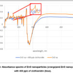 |
Figure 6: Absorbance spectra of ZnO nanoparticles (orange)and ZnO nanoparticles with 400 ppb of clothianidin (blue). |
As can be seen in figure 7, evident change in part of the absorbance spectra of ZnO nanoparticles occurred upon addition of 400 ppb of clothianidin. The characteristic excitonic absorption peak at 281 nm disappeared. This suggests that the positively charged electron hole created due to the absorption of energy that causes the electron to move from the valence band to the conduction band was placed with possibly an electron from clothianidin. This further suggests that the electronic properties of the nanoparticles will also change. While the disappearance of the pale yellow color may be attributed to the decrease in the absorbance of the solution at 420 to 450 nm.
The change in color can be explained upon looking at the changes in the absorbance spectrum of ZnO nanoparticles upon addition of imidacloprid (figure 8).
 |
Figure 7: Absorbance spectra of ZnO nanoparticles (blue) and ZnO nanoparticles with 400 ppb of imidacloprid (green). |
It can be gleaned that even if the change in color for both the pesticides was from pale yellow to colorless, the change in absorbance spectra were not exactly similar. The exciton absorption peak at 281 nm was still evident in the case of ZnO nanoparticles mixed with imidacloprid. The peak however, prominently decreased in absorbance. The results suggest that the positively charged electron holes were still present. This may be attributed to the larger structure of imidacloprid in comparison with clothianidin (figure 8).
 |
Figure 8: Chemical structure of (A) imidacloprid and (B) clothianidin. |
Moreover, it can be gleaned from the structures that imidacloprid has only 2 rotatable bond count which is lower than the 4 rotatable bond count for clothianidin. This implies that the structure of clothianidin may twist more readily and squeeze into the holes in the nanoparticles better than imidacloprid.
However, despite the difference in the interaction of the two pesticides with ZnO nanoparticles, the absorbance spectra of the samples at 420 to 450 nm were the same. Hence, the change in color observed was the same for both. In terms of colorimetric detection using the naked-eye, the ZnO nanoparticles were not selective against the two pesticides used in the study. Thus, modification of the nanoparticle may be explored to develop a more selective colorimetric detector. Other confirmatory analyses such as the use of IR, XRD, SEM techniques may also be conducted for future studies.
Acknowledgement
The author would like to express gratitude to the Natural and Applied Sciences Department and to the administration of the Nueva Ecija University of Science and Technology for the support on the conduct and publication of this study.
Conflicts of Interest
The author declares that there are no conflicts of interest related to this article.
References
- El-Nahhal, I.; & El-Nahhal, Y. J. Environ. Manage. 2021, 299, 113611
- Yamagishi, K.; Gantalao, C. & Ocampo, L. J. Tour. Futures. 2021
- Bonmatin, J. M.; Mitchell, E. A.; Glauser, G.; Lumawig-Heitzman, E.; Claveria, F.; van Lexmond, M. B. & Sánchez-Bayo, F. Sci. Total Environ. 2021, 757, 143822Cubelo, J. E. C. & Cubelo, T.A. JNS. 2021, 1, 25-43
- Singh, R.; Kumar, N.; Mehra, R.; Kumar, H.; Singh, V. P. Trends Environ. Anal. Chem. 2020, 26, e00086
- Che Sulaiman, I. S.; Chieng, B. W.; Osman, M. J.; Ong, K. K.; Rashid, J. I. A.; Wan Yunus, W. M. Z.; Mohamad, A. Mikrochim Acta. 2020, 187(2), 1-22
- Jana, J.; Ganguly, M.; Pal, T. RSC Adv. 2016, 6(89), 86174-86211
- Homola, J. Chem. Rev. 2008, 108(2), 462-493
- Keller, A. A.; Vosti, W.; Wang, H.; Lazareva, A. J. Nanoparticle Res. 2014, 16(7), 1-10
- Ma, S.; He, J.; Guo, M.; Sun, X.; Zheng, M.; Wang, Y. Colloids Surf. A Physicochem. Eng. Asp. 2018, 538, 343-349
- Reyes D. F. Orient J Chem. 2020, 36(4)
- Li, H.; Guo, J.; Ping, H.; Liu, L.; Zhang, M.; Guan, F.; Zhang, Q. Talanta. 2011, 87, 93-99
- Liu, W.; Zhang, D.; Tang, Y.; Wang, Y.; Yan, F.; Li, Z.; Zhou, H. S. Talanta. 2012, 101, 382-387
- Bala, R.; Dhingra, S.; Kumar, M.; Bansal, K.; Mittal, S.; Sharma, R. K.; Wangoo, N. J. Chem. Eng. 2017, 311, 111-116
- Rawtani, D.; Khatri, N.; Tyagi, S.; Pandey, G. J. Environ. Manage. 2018, 206, 749-762
- Fakhari, S.; Jamzad, M.; Kabiri Fard, H. Green Chem Lett Rev. 2019, 12(1), 19-24
- da Silva, B. L.; Abuçafy, M. P.; Manaia, E. B.; Junior, J. A. O.; Chiari-Andréo, B. G.; Pietro, R. C. R.; Chiavacci, L. A. Int J Nanomedicine. 2019, 14, 9395
- Wiesmann, N.; Tremel, W.; Brieger, J. J Mater Chem B. 2020, 8(23), 4973-4989
- Bala, R.; Kalia, A.; Dhaliwal, S. S. J. Soil Sci. Plant Nutr. 2019, 19(2), 379-389
- Yusefi-Tanha, E.; Fallah, S.; Rostamnejadi, A.; Pokhrel, L. R. Sci. Total Environ. 2020, 738, 140240
- Awan, S.; Shahzadi, K.; Javad, S.; Tariq, A.; Ahmad, A.; Ilyas, S. J. Saudi Soc. Agric. Sci. 2021, 20(1), 18-24
- Al-Rawashdeh, N. A.; Allabadi, O.; Aljarrah, M. T. ACS omega. 2020, 5(43), 28046-28055
- Dodoo-Arhin, D.; Asiedu, T.; Agyei-Tuffour, B.; Nyankson, E.; Obada, D.; Mwabora, J. M. Mater. Today: Proc. 2021, 38, 809-815
- Chaudhary, S.; Kaur, Y.; Umar, A.; Chaudhary, G. R. J. Mol. Liq. 2016, 224, 1294-1304
- Kaur, Y.; Bhatia, Y.; Chaudhary, S.; Chaudhary, G. R. J. Mol. Liq. 2017, 234, 94-103
- Sharma, A. K.; Tiwari, R. K.; Gaur, M. S. Arab. J. Chem. 2016, 9, S1755-S1764
- Dehaghi, S. M.; Rahmanifar, B.; Moradi, A. M.; Azar, P. A. J. Saudi Chem. Soc. 2014, 18(4), 348-355
- Qu, J.; Yuan, X.; Wang, X.; Shao, P. Environ. Pollut. 2011, 159(7), 1783-1788
- Kalaimathi, M.; Hariharan, K.; Vishnu, S. & Chinnasamy, R. CRGSC. 2021, 4, 100185
- Sisira, S.; Hithisha, K. S.; Sankar, J. S.; Nazirin, N.; Vimalraj, R. K. & Kalaimathi, M. Inorg. Chem. Commun. 2022, 145, 109961
- Ochieng, P. E.; Iwuoha, E.; Michira, I.; Masikini, M.; Ondiek, J.; Githira, P.; Kamau, G. N. Int. J. Biochem. Phys. 2015, 23, 53-61
- Kalaiamthi M.; Maheshwaran, A; Hariharan, K; Poovarasan, B & Chandru P. Orient J Chem. 2021, 37(4)
- Hossain, A.; Abdallah, Y.; Ali, M.; Masum, M.; Islam, M.; Li, B.; An, Q. Biomolecules. 2019, 9(12), 863
- Basnet, P.; Inakhunbi Chanu, T.; Dhrubajyoti, S. & Somenath, C. J. Photochem. Photobiol. B: Biol. 2018, 183, 201-221.
- Mathew, B.; Suresh, B.; Jatawa, K. & Tiwari, A. Int J Pharm Pharm Sci. 2012, 2, 369-71.
- Duru, I.A. & Enyoh, C.E. J. Medicinal Plants. 2020, 8(5), 178-182.
- Talam, S.; Karumuri, S. R.; Gunnam, N. int. sch. res. Notices. 2012
- Zak, A. K.; Razali, R.; Abd Majid, W. H.; Darroudi, M. Int J Nanomedicine. 2011, 6, 1399
- Sangeetha, G.; Rajeshwari, S.; Venckatesh, R. Mater. Res. Bull. 2011, 46(12), 2560-2566

This work is licensed under a Creative Commons Attribution 4.0 International License.
About The Author
![]()
A New Edition of Web of Science
Journal Impact Factor
2022: 0.5
Five Year: 0.8
Journal is Indexed in

Cabells Whitelist
![]()


