Extracellular synthesis of copper nanoparticles using culture supernatants of Salmonella typhimurium
Hamid Reza Ghorbani*, Ferdos Parsa Mehr and Azin Khaniyani Poor
Department of Chemical Engineering, Qaemshahr Branch, Islamic Azad University, Qaemshahr, Iran
DOI : http://dx.doi.org/10.13005/ojc/310165
Article Received on :
Article Accepted on :
Article Published : 20 Mar 2015
In the recent years, the synthesis of metal nanoparticles has received much attention due to its wide range of applications in various fields. In this study copper nanoparticles were synthesized using salmonella typhimurium. Biosynthesis of copper nanoparticles was achieved by addition of culture supernatant with aqueous copper nitrate solution (1mM). Copper nanoparticles having average diameter 49 nm have been obtained. The reaction mixture exhibit a peak at 565 nm corresponding to the Plasmon absorbance of copper nanoparticles by UV–Vis Spectroscopy. The nanoparticles were characterized by dynamic light scattering (DLS), and Scanning Electron Microscopy (SEM). The process of reduction is extra cellular, which makes it an easier method for the synthesis of copper nanoparticles.
KEYWORDS:Salmonella typhimurium; Biosynthesis; Copper nanoparticles; culture supernatants
Download this article as:| Copy the following to cite this article: Ghorbani H. R, Mehr F. P, Poor A. K. Extra cellular synthesis of copper nanoparticles using culture supernatants of Salmonella typhimurium. Orient J Chem 2015;31(1). |
| Copy the following to cite this URL: Ghorbani H. R, Mehr F. P, Poor A. K. Extra cellular synthesis of copper nanoparticles using culture supernatants of Salmonella typhimurium. Orient J Chem 2015;31(1). Available from: http://www.orientjchem.org/?p=7890 |
Introduction
In the recent decades, metallic nanoparticles have been widely used in different industries due to their wide range of applications [1]. Metal nanoparticles exhibit unique electronic, magnetic, catalytic and optical properties that are different from those of bulk metals. Among different metal particles, copper nanoparticles, due to their unique physical and chemical properties and low cost preparation, have been of great interest recently [2, 3, 4].
The synthesis methods of these nanoparticles are basically divided into chemical, physical and biological methods. Today, it has been found, that living organisms such as bacteria, fungi, and plants have a great potential for the synthesis of metal nanoparticles [5]. By using these biosources various kinds of nanoparticles can be synthesized intracellularly or extracellularly which have been used for different applications. Microorganisms act as a biofactory and can also be used for the synthesis of metal nanoparticles. Majumber used a fungal species (Fusarium oxysporum) to synthesize copper nanoparticles (93–115 nm) at ambient temperature [6]. Pavani et al. used Aspergillus species of fungus for extracellular synthesis of Cu nanoparticles [1]. Lee et al. used magnolia leaf extract as reducing agent and conversion of Cu2+ to Cu0 for the synthesis of stable copper nanoparticles with a size of 40-100 nm. They used CuSO4.5H2O in aqueous solution and leaf extract to produce stable copper nanoparticles [7]. Valodkar et al. used stem latex of Euphorbia nivulia which is a plant with medicinal properties for the synthesis of copper nanoparticles and could synthesize Cu nanoparticles capped with peptides present in the latex of plant [8]. Varshney et al. used from Pseudomonas stutzeri to synthesize copper nanoparticles. They showed nanoparticles are spherical and in the range of 8-15 nm in dimensions [9]. In another work, Ramanathan et al. (2011) used a biological method to synthesize copper nanoparticles using Morganella bacteria and under aqueous physiological conditions [10].
The purpose of this study was to synthesis copper nanoparticles using culture supernatant of salmonella typhimurium and characterization of the synthesized nanoparticles.
Materials and Methods
The Bacteria culture, salmonella typhimurium was obtained from Microbiology Laboratory, Tehran University, Tehran, Iran. Muller-Hinton broth (MHB) was prepared, sterilized, and inoculated with a fresh growth salmonella typhimurium. The culture was centrifuged at 5000 rpm for 20 minutes and the supernatant was used for the synthesis of copper nanoparticles. Distilled water was used as solvent in the synthesis of copper nanoparticles. The supernatant was added separately to the reaction vessel containing copper nitrate at a concentration of 1 mM. The reaction between this supernatant and Cu2+ ions were carried out for 40 minutes. The copper nanoparticles were characterized by UV-visible spectroscopy and dynamic light scattering (DLS) analysis. In addition, the copper nanoparticles were analysed by scanning electron microscopy (SEM).
Results and Discussions
The aqueous Cu2+ ions were reduced during exposure to the culture supernatant of salmonella typhimurium. The color of this solution changes to cloudy orange after 40 min of mixing which indicated the formation of copper nanoparticles extracellularly.
Fig. 1 shows the UV–Vis absorption spectra recorded from the copper nanoparticles solution after formation. The results indicate that the reaction solution has an absorption maximum at about 565 nm attributed to the surface plasmon resonance band (SPR) of the copper nanoparticles. The size distribution of the synthesized nanoparticles was shown in Fig. 2. As is illustrated in figure, mean size of copper nanoparticle is about 49 nm and the distribution of nanoparticles is narrow (10 nm). Also the copper nanoparticles synthesized by salmonella typhimurium were studied by scanning electron microscopy (SEM) and images show and confirm copper nanoparticles production at nano-size. SEM images of the produced nanoparticles are shown in figure 3.
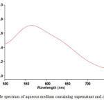 |
Figure 1: UV–visible spectrum of aqueous medium containing supernatant and copper ion (1 mM)
|
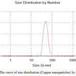 |
Figure 2: The curve of size distribution (Copper nanoparticles) by number
|
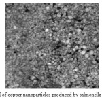 |
Figure 3: SEM of copper nanoparticles produced by salmonella typhimurium Click here to View figure |
Conclusions
Copper nanoparticles in the range of 40-60 nm are synthesized by the supernatant of salmonella typhimurium when copper nitrate is added to it. The time required to complete the copper ions reduction was obtained about 40 min. This methodology could be used to synthesize a number of metallic nanoparticles involving other metals with good size and shape morphology. This study would therefore lead to an easy procedure for producing copper nanoparticles with the added advantage of biosafety.
References
- Parveen, S., Misra, R., Sahoo, SK. (2012). Nanoparticles: a boom to drug delivery, therapeutics diagnostics and imaging. Nanomed-Nanotechnol, 8(2), 147–166.
- Tavakoli, A., Sohrabi, M. & Kargari, A. (2007). A review of methods for synthesis of nanostructured metals with emphasis on iron compounds, Chem Pap 61(3) 151–170.
- Park, B. K., Jeong, S., Kim, D., Moon, J., Lim, S. & Kim, J. S. (2007). Synthesis and size control of monodisperse copper nanoparticles by polyol method, J Colloid Interf Sci, 311(2), 417-424.
- ohanpuria, P., Rana, K. N. & Yadav, S. K. (2008). Biosynthesis of nanoparticles: technological concepts and future applications, J Nanopart Res, 10(3), 507–517.
- Ghorbani, H.R., Safekordi, A.A., Attar, H. & Rezayat Sorkhabadi, S. M. (2011). Biological and Non-biological Methods for Silver Nanoparticles Synthesis, Chem Biochem Eng Q, 25(3) 317–326.
- Majumber ,D.R.,(2012). Bioremediation: copper Nanoparticles from electronic-waste. Inter J Eng Sci Technol, 4,4380–4389.
- Lee, H.J., Lee, G., Jang,N.R., Yun,J.H., Song, J.Y. , Kim, B.S. (2011). Biological synthesis of copper nanoparticles using plant extract. Nanotech. 1(1), 371-374.
- Kuber Valodkar, M., Jadeja,R.N., Thounaojam,M.C., Devkar, R.V. , Thakore,S. (2011). Biocompatible synthesis of peptide capped copper nanoparticles and their biological effect on tumor cells. Mater Chem. Phys., 128(1-2), 83-89.
- Varshney, R., Bhadauria, S., Gaur, M.S. & Pasricha, R. (2010). Characterization of Copper Nanoparticles Synthesized by a Novel Microbiological Method, J Met, 62(12), 100-102.
- Ramanathan, R., Bhargava, S.K. & Bansal, V. (2011). Biological synthesis of Copper/Copper Oxide nanoparticles, In Rose Amal (ed.) Chemeca Conference. Eng A Better World, Australia, 1-8.

This work is licensed under a Creative Commons Attribution 4.0 International License.
![]()
A New Edition of Web of Science
Journal Impact Factor
2022: 0.5
Five Year: 0.8
Journal is Indexed in

Cabells Whitelist
![]()


