Synthesis and Evaluation of the Antimicrobial Activity of Dodecyl Trimethyl Ammonium and N-(N, N-Dimethylamino) Propyl Glucosylamine
Théoneste Muhizi
University of Rwanda, School of Pure and Applied Science, Department of Chemistry, University avenue 117 Butare, Rwanda.
DOI : http://dx.doi.org/10.13005/ojc/300239
Article Received on :
Article Accepted on :
Article Published : 14 Apr 2014
In this study Dodecyl trimethyl ammonium sulphate (DTA) was obtained through quaternization reaction between dodecylamine and dimethyl sulfate (yield: 51.5%) while Dimethylamino propyl glucosylamine (DAPGA) was successively synthesized from D-glucose and 3-Dimethylamino propylamine (yield: 93.6%). Both compounds were analyzed through NMR spectroscopy. Evaluation of their biological activity against Salmonella Typhimurium, Staphylococcus aureus and Fusarium oxysporum cubens indicated that at a concentration greater than 0.001 10-4 mole mL-1, DTA significantly inhibited growth of Salmonella Typhimurium and Staphylococcus aureus while DAPGA was not effective. Furthermore, these two compounds showed a pronounced antifungal activity against a toxicogen fungal, Fusarium oxysporum cubens.
KEYWORDS:Dodecyl trimethyl ammonium; Dimethylamino propyl glucosylamine; Salm. Typhimurium; Staph. aureus; F. oxysporum cubens; biocides
Download this article as:| Copy the following to cite this article: Muhizi T. Synthesis and Evaluation of the Antimicrobial Activity of Dodecyl Trimethyl Ammonium and N-(N, N-Dimethylamino) Propyl Glucosylamine. Orient J Chem 2014;30(2). |
| Copy the following to cite this URL: Muhizi T. Synthesis and Evaluation of the Antimicrobial Activity of Dodecyl Trimethyl Ammonium and N-(N, N-Dimethylamino) Propyl Glucosylamine. Orient J Chem 2014;30(2). Available from: http://www.orientjchem.org/?p=3005 |
INTRODUCTION
Development of pathogen bacteria and mycotoxicogen fungi in food constitute a danger for humanity. These harmful microorganisms can be major source of food toxi-infection leading to different human diseases. For instance Salmonella and Listeria species, very resistant mircroorganisms at different conditions used for food preservation, can lead to salmonellose and listeriose, two known human fatal diseases 1-6. Furthermore, fungi such us Aspergillus, Penicillium and Fusarium species essentially found on cereal are responsible of teratogenic and carcinogenic toxins which seriously affect human life7-14. Due to the diversity and ravages of these harmful microorganisms, human prefer to use antimicrobials to fight against them. Nevertheless, even if the importance of these chemicals is no longer demonstrable, some of them crop up serious eco-toxicological problems and therefore were interdicted or limited from the markets15, 16. These decisions come up very rapidly and left questions on the means to be used for replacing them17. Furthermore, future effectiveness of antimicrobial therapy is somewhat in doubt since various microorganisms are becoming more resistant to the existing antimicrobial agents1-6. Therefore, it is the responsibility of scientists to work hard and to find out environment friendly active compounds which can be used in this struggling. Previous reports indicated that amino sugars and ammonium compounds could be one of the good candidates to reach the goal18-24. This study is our contribution in this scientists’ task and was enabled to synthesize two molecules with promised results.
EXPERIMENTAL
Chemicals such us glucose (Sigma Aldrich), dodecylamine 98% (Sigma Aldrich), 3-dimethylamino propylamine (Sigma Aldrich), dimethyl sulphate 99% (Aldrich), Sodium sulphate 99% (Aldrich), chloroform (Prolabo), ethanol (Schorlau) and methanol (Labosi) were used with out any further purification. Culture media Mueller Hinton agar (Biomérieux), Nutrient broth (Difco) and Potato dextrose Agar (Himedia RM 007) were used to assess biological activity. Azithromycin (Alice Pharma PVT, LTD, India) was used as positive reference.
General methods
1H NMR spectra of DTA and DAPGA were recorded at 300 MHZ on a Bruker Avance 300 spectrometer and chemical shifts are given in ppm. The melting point of DTA was determined by an Electrothermal 9100 Digital Melting Point Apparatus IA 9100.
Synthesis of DTA
1-Dodecylamine (0.0539 mole) was dissolved in 20 mL of chloroform. The mixture was refluxed at 50°C and under stirring a small quantity of sodium sulphate was added. After 30 min of reaction, 0.2158 mole of dimethyl sulphate was carefully added to the mixture and reflux was maintained for 6 hours. Solvent was then removed under reduced pressure and residue was co-evaporated four times in methanol to recuperate a white solid which was finally recristallized in ethanol to obtain 9 grams of DTA (Figure 1).
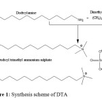 |
Figure 1: Synthesis scheme of DTA Click here to View Figure |
Synthesis of DAPGA
Reported method of Petit (1999)25 was adapted to synthesize DAPGA (Figure 2). Ten grams of glucose (0.0555 mole) and 5.7 g (0.0558 mole) of 3-dimethylamino propylamine were dissolved in 66 mL of methanol. The reaction mixture was refluxed at 40°C under stirring for 4 hours. The solvent was removed under reduced pressure using a rotary evaporator and a viscous yellowish liquid of 9.3 g was obtained.
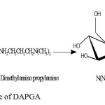 |
Figure 2: Synthesis scheme of DAPGA Click here to View Figure |
Microorganisms
The fungal strain, Fusarium oxysporum cubens, was isolated and characterized in the laboratory of University of Rwanda, College of Science and Technology. Two bacteria Salmonella Typhimurium (Institut Pasteur 5858) and Staphylococcus aureus (Institut Pateur 25923) were obtained from the Laboratory of US2B, Université Bordeaux 1, France.
Antifungal activity assessment
Potato dextrose agar (PDA) medium was used to isolate, identify and revivify F. oxysporum cubens. The radial growth method17 was used to assess the antifungal activity of DTA and DAPGA against F. oxysporum cubens. Concentrations of 0.4 x 10⁻⁴ and 1.5 x 10⁻⁴ mole mL-1 for both compounds were evaluated. Distilled water and methanol were used to dissolve DTA and DAPGA, respectively. Each essay was done in triplicate and control experiments which contain either distilled water or methanol were done in parallel. The percentage of inhibition from the two products was calculated after 8 days of incubation using the formula previously reported26. In addition, the diameter of fungi was daily measured during 8 days of incubation to evaluate the effect of the two products on the kinetic growth of fungi.
Antibacterial activity assessment on agar media
Mueller-Hinton culture medium was used to assess biological activity of DTA and DAPGA against Salm. Typhimurium and Staph. aureus. Disc diffusion method was used to conduct the test17, 27.
Assessment of antibacterial efficacy of DTA and DAPGA in liquid medium
Nine milliliters of liquid medium, nutrient broth, in test tubes were separately contaminated by 1 mL of 18h revivified bacteria strains, Salm. Typhimurium and Staph. aureus, separately. Then a given quantity of DTA or DAPGA was added prior to have a final concentration of 0.08.10-4 mole mL-1 of these products. The obtained tubes were then incubated at 37°C for 24 hours. Control tubes without drugs were concurrently incubated. After 24 hours of incubation, 1 mL of this microbial suspension was previously 106 times diluted and 100 mL were deposited on the Mueller-Hinton agar medium and gently spread onto the surface of this prior to incubation at 37°C for 24h. Forming colony unities were then counted and number recorded. The experiment was conducted in triplicate.
Statistical analysis
Results from antibacterial assays were statistically analyzed using one-way analysis of variance (ANOVA). The significant difference between activities of DTA and DAPGA or between these drugs and control experiments was obtained when the probability (p) found was higher than the significance threshold of 0.05.
RESULTS AND DISCUSSION
Chemical Synthesis
In this study, both DTA and DAPGA were successively synthesized and characterized by 1H NMR spectroscopy.
DTA
Yield: 51.5%, melting point: 75.1°C, 1H NMR (300 MHz, D2O, Figure 3): dH 3.4 (2H, t, -NCH2-), 3.19 (9H, s, -N+(CH3)3), 1.7-1.9 (2H, br, -CH2CH3), 1.4-1.5 (18H, -CH2-), 0.9-1 (3H, t, -CH3). The total synthesis of DTA was indicated by a signal in the form of singulet at 3.19 ppm representing +N(CH3) protons (Figure 3).
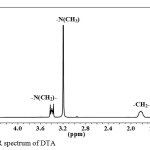 |
Figure 3: 1H NMR spectrum of DTA Click here to View Figure |
Our result was confirmed by previous reports21, 24, 26, 28 in which quaternary ammonium protons were characterized by signals found at 3.1, 3.33 and 3.67 ppm respectively, depending on the type of chemical groups surrounding them.
DAPGA
Yield: 93.6%, 1H NMR (300 MHz, D2O): dH 3.9-4.0 (1H, d, H-1), 3.8-3.9 (1H, dd, H-6a), 3.6-3.7 (1H, dd, H-6b), 3.4-3.5 (1H, t, H-3), 3.3-3.4 (2H, complex, H-4 and H-5), 3.1-3.2 (1H, t, H-2), 2.78-2.86 (2H, mm, -NHCH2-), 2.35 (2H, t, -CH2N=), 2.15 (6H, s, -N(CH3)2), 1.54-1.65 (2H, m, -CH2-). The synthesis of DAPGA was confirmed by H-1 signal in the form of doublet found at lower chemical shift of 3.9-4.0 ppm compared to H-1a and H-1b of glucose which were reported to be around 5.23 and 4.65 ppm29, respectively, and this indicated the replacement of OH-1 group by -NH- group in glucosylamine. The same observation was reported in various previous works17, 27, 29, 30.
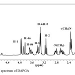 |
Figure 4: 1H NMR spectrum of DAPGA Click here to View Figure |
Antifungal activity of DTA and DAPGA on F. oxysporum cubens
Some fungi are harmful either to animals or plants by intoxicating them. Among these fungi we chose to work with one toxicogen fungus called Fusarium oxysporum cubens. Fusarium species are known not only to be implicated in the annually worldwide harvest lost12 but also to produce a variety of toxins such as fumonisins, zearalenone and trichothecenes in food mainly cereal, and thus exhibiting chronic toxicity7,8,10,14,31. In this study, both two products DTA and DAPGA were effective against the growth of Fusarium oxysporum cubens (Figure 5 and Table 1). At all concentrations used, 0.4 x 10⁻⁴ and 1.5 x 10⁻⁴ mole mL-1, DTA completely inhibited the growth of Fusarium oxysporum cubens while DAPGA exhibited a significant antifungal activity (percentage of inhibition of 81.5 %) when a concentration of 0.4 x 10⁻⁴ mole mL-1 was tested and completely inhibited the growth of fungal at a concentration of 1.5 x 10⁻⁴ mole mL-1. Methanol and water used as negative control exhibited non significant antifungal activity.
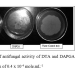 |
Figure 5: Illustration of antifungal activity of DTA and DAPGA on Fusarium oxysporum cubens at a concentration of 0.4 x 10⁻⁴ mole.mL-1 Click here to View Figure |
Table 1: Inhibition of DTA, methanol and DAPGA on the radial growth of F.oxysporum cubens at the 8th day of incubation
| Biocides | Concentration (mol ml-1)10-4 |
Percentage of inhibition ±SEM |
| DTA |
0.4 |
100.0 ± 0.0 |
|
1.5 |
100.0 ± 0.0 |
|
| DAPGA |
0.4 |
81.5 ± 1.2 |
|
1.5 |
100.0 ± 0.0 |
|
| Distilled water |
1 mL |
0.0 ± 0.0 |
| Methanol |
1 mL |
11.5 ± 1.4 |
Furthermore, the antifungal activity of these two compounds was observed during a week to visualize well what was happened during incubation of fungus. At concentration of 0.4 x 10⁻⁴ mole mL-1, DTA completely inhibited the growth of fungi while DAPGA significantly affected the kinetic growth of fungus by strongly changing the shape of its kinetic growth. Methanol used as negative control did not show significant effect on the kinetic growth of fungal but slightly delayed it. All these observations were done in comparison with the effect of distilled water used as control (Figure 6).
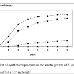 |
Figure 6: The effect of synthesized products on the kinetic growth of F. oxysporum cubens at the concentration of 0.4 x 10⁻⁴ mole mL-1 Click here to View Figure |
Antibacterial activity of DTA and DAPGA (Figure 7)
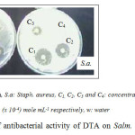 |
Figure 7: Illustration of antibacterial activity of DTA on Salm. Typhimurium and Staph. Click here to View Figure |
Note: S.t.: Salm. Typhimurium, S.a: Staph. aureus, C1, C2, C3 and C4: concentrations of drugs which are equal to: 0.04, 0.02, 0.01 and 0.001 (x 10-4) mole mL-1 respectively, w: water
At all concentrations used DAPGA did not affect the growth of both bacteria while DTA showed its efficacy in inhibiting these microbes. At the concentration of 0.08 x 10⁻⁴ mole mL-1, DTA exhibited higher activity against both bacteria. It inhibited the growth of Staph. aureus and Salm. Typhimurium with inhibition zones of 24 and 15 mm respectively. These inhibition zones are too sufficient to consider DTA as effective against both bacteria 32. In this study, at a concentration previously quoted, DTA led to an inhibition diameter which was closed to 16 mm (viz 15.3 ± 0.9) for Salm. Typhimurium and thus can be used as the minimal effective concentration against that bacterium. Furthermore, a concentration of around 0.02 mole mL-1 can be taken as minimal effective one for Staph. aureus. As shown in the table 2, DTA with a concentration greater than 0.001.10-4 mole mL-1 had higher antibacterial activity on Staph. aureus, Gram positive bacterium, than the antibacterial activity it had on a Gram negative bacterium, Salm. Typhimurium. This may partially due to the chemical composition of their cell-wall structures which are different and thus conduct to their different permeability towards drugs33, 34. In addition, at a concentration of 0.08 mole mL-1, Azithromycin, an antibiotic used as positive control, exhibited comparable antibacterial activity with DTA. DTA indicated inhibition zones of 24.3 ± 0.3 and 15.3 ± 0.9 for Staph. aureus and Salm. Typhimurium (Table 2), respectively, while for Azithromycin the inhibition zones were found to be 26.7 ± 1.2 and 20.3 ± 1.8 mm respectively for Staph. aureus and Salm. Typhimurium. Comparison of these results indicated that efficiency of DTA is closely comparable to that from know antibiotic, Azithromycin. At concentrations lower than or equal to 0.08 x 10⁻⁴ mole mL-1, DAPGA did not show any significant antibacterial activity.
Table 2: Inhibition diameter values of DTA on Staph. aureus and Salm. Typhimurium
|
Inhibition diameter ± SEM (in mm) |
|||||
| Bacteria | Concentrations (mol mL-1)10-4 | ||||
|
0.08 |
0.04 |
0.02 |
0.01 |
0.001 |
|
| Staph. aureus | 24.3 ± 0.3 | 20.3 ± 0.5 | 18.3 ± 0.7 | 9.2 ± 0.8 | 0.0 ± 0.0 |
| Salm. Typhimurium | 15.3 ± 0.9 | 9.7 ± 0.3 | 8.0 ± 0.0 | 2.1 ± 0.5 | 0.0 ± 0.0 |
To verify the reason of difference between biological activities remarked between DTA and DAPGA, the efficacy of these two drugs were also assessed in liquid media. DTA completely inhibited the growth of both bacteria while DAPGA didn’t show any significant inhibition compared to control experiments (Table 3).
Table 3: Antibacterial activity of DTA and DAPGA in water
| Drugs |
Antibacterial activity (UFC ± SD) |
|
| Salm. Typhimurium | Staph. aureus | |
| Control experiment | 374 ± 13 | 299 ± 23 |
| DTA | 0 ± 0 | 0 ± 0 |
| DAPGA | 362 ± 17 | 277 ± 20 |
Having these observations, decomposition of DAPGA in D2O was followed using 1H-NMR. Both disappearance of –NH(CH2)- and decreasing intensity of H-1 signals were served as references (Figure 8). We chose to work with D2O as solvent, because of its quite same properties, in hydrolysis process, with H2O used to prepare liquid medium in which both compounds was tested. After 24 h in D2O, the initial DAPGA decreased of 50% while at 48 h, only 37.5% of DAPGA was remained in the medium without any decomposition (Figure 8).
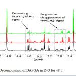 |
Figure 8: Decomposition of DAPGA in D2O for 48 h Click here to View Figure |
This remarked rapid decomposition of DAPGA could be the reason of its poor antibacterial activity. As previously reported17, the lack of activity of glucosylamines against bacteria, may be partially explained by their rapid hydrolysis during the first 24 hours. To verify this assumption, the antibacterial activity of DAPGA was compared to that of Dodecyl glucosylamine (DoGA), a molecule which was previously synthesized and found to be not highly hydrolyzed17. At a concentration of 0.5 x 10⁻⁴ mole mL-1, both Salm. Typhimurium and Staph. aureus were exactly sensible to DoGA but not to DAPGA (Table 4).
Table 4: Inhibition diameter values of DAPGA, DoGA and 3-DMAP on Staph. aureus and Salm. Typhimurium
|
Biocides |
Inhibition diameter ± SEM (in mm) |
||
|
Concentration (mole mL-1) x 10-4 |
Staph. aureus |
Salm. Typhimurium |
|
|
DoGA |
0.5 |
16.0 ± 0.5 |
10.6 ± 0.5 |
|
DAPGA |
0.5 |
0.0 ± 0.0 |
0.0 ± 0.0 |
|
2.0 |
6.0 ± 0.3 |
8.0 ± 0.6 |
|
DAPGA started to exhibit antibacterial activity at higher concentration of 2.0 x 10⁻⁴ mole mL-1 compared to DoGA and this observed activity was even not significant.
In conclusion, results revealed that at all concentrations used, DTA and DAPGA were effectiveness to inhibit the growth of F. oxysporum cubens. Furthermore, Dodecyl trimethyl ammonium (DTA) showed significant antibacterial activity against the growth of Salm. Typhimurium and Staph. aureus. The lack of significant antibacterial of N-(N,N-Dimethylamino) propyl glycosylamine against Staph. aureus and Salm. Typhimurium could be due to its remarked decomposition in D2O and research on its possible stabilization should be of interest. Through our findings, the reported antibacterial efficacy of ammonium compounds21, 24, 28 was confirmed: satifying antimicrobial activity was obtained from small biodegradable molecule, dodecyl trimethyl ammonium sulphate. It is however to note that the toxicity assessment of this compound should be carefully realized before any application of this compound in various domains.
Acknowledgment
We thank the University of Rwanda-Swedish International Development Cooperation Agency (UR-SIDA) Project and UR Research Commission for funding this work.
References
- Sabharwal E.R., Indian Journal of Pathological Microbiology, 53 (2): 389 (2010).
- Sarker A., Saha S.K., Islam M., Hossain M.A., Dhaka University Journal of Biological Science 19 (2): 137 (2010).
- Dermatin M.A., Gaborieau V., Song Y., Roumagnac P., Marchou B., Achtman M., Weil F.X., Emerg. Infect. Dis. 17 (6): 1 (2011).
- Tajibakhsh M., Hendriksen R.S., Nochi Z., Zari Z.M., Aerestrup F.M., Garcia-Migura L., Folia Microbiologica 57 (2): 91 (2012).
- Granier S.A., Moubarack C., Colaneri C., Lemire A., Roussel S., Dao T., Courvalin P., Brisabois A., Appl. Environ. microbiol. 78: 2043 (2012).
- Collins B. Guinane C.M., Cotter P.D., Hill C., Ross R.P., Appl. Environ. microbiol. 78(8): 2923 (2012).
- Smith T. K., McMillan E. G., Castillo J. B., Journal of Animal Science 75: 2184 (1997).
- Gouze M. E., Oswald I. P., Journées de la Recherche Porcine en France 33: 277 (2001).
- Magan N.; Hope R.; Colleate A.; Bacter E. S. Eur. J. Plant Pathol. 108: 685 (2002).
- Yiannikouris A., Jouany J.P., INRA Production Animale 15 (1): 3 (2002).
- Molinié, A.; Pfohl-Leszkowicz, A. Note ASEDIS-SO no spécial Mycotoxines, Laboratoire de Toxicologie et sécurité alimentaire, Auzeville-Tolosane, 9 (2003).
- Bi Z., Wang Z., Xu L., Acta botanica Sinica 46 (1): 124 (2004).
- Hibar K., Daami-Remadi M., Khiareddine H., El Mahjoub M., Biotechnol. Agron. Soc. Envir. 9 (3): 163 (2005).
- Cavret S., Lecoeur S., Annuel de Médécine Vétérinaire 150: 43 (2006).
- INERIS. Fiche de données toxicologiques et environnementales des substances chimiques, Dieldrine, Ineris, Paris, 119 (2008a).
- INERIS. Fiche de données toxicologiques et environnementales des substances chimiques, Chlordane, Ineris, Paris, 113 (2008b).
- Muhizi, T. Synthèse d’aminosucres conduisant à des biocides d’origine naturelle, Thèse, Université Bordeaux 1, 188 (2008).
- Kim C.H., Choi J.W., Chun H.J., Choi, K.S., Polym. Bull. 38: 387 (1997).
- Jia Z., Shen D., Xu W., Carbohydr. Res. 333: 1 (2001).
- Avadi M.R., Sadeghi A.M.M., Tahzibi, A., Bayati K., Pouladzadeh M., Zohuriaan-Mehr M.J., Rafiee-Tehrani M., Eur. Polym. J. 40: 1355 (2004).
- Huang R., Du Y., Zheng L., Liu H., Fan L., React. Funct. Polym. 59: 41 (2004).
- Matejuk J.Z., Czaczyk, K. Wood Sci. Technol. 40: 461 (2006).
- Kenawy E.R., Abdel-Hay F.I., El-Magd A.A., Mahmoud Y., React. Funct. Polym. 66: 419 (2006).
- Belalia R., Grelier S., Benaissa M., Coma V. J. Agric. Food Chem. 56: 1582 (2008).
- Petit S. Brevet no CA2243510, 12 p (1999).
- Muhizi T., Coma V., Grelier S., Pest management Science 67 (3): 287 (2011),.
- Muhizi T., Grelier S., Coma V., J. Agric. Food Chem. 57 (23): 11092 (2009).
- Holappa J., Nevalainen T., Savolainen J., Soininen P., Elomaa M., Safin R., Suvanto S., Pakkanen T., Másson M., Loftsson T., Järvinen T., Macromolecules 37: 2784 (2004).
- Blasko A., Bunton A.B., Bunel S., Ibarra C., Moraga E., Carbohydr. Res., 298: 163 (1997).
- Muhizi T., Coma V., Grelier S. Carbohydrate research: 343: 2369 (2008).
- Fandohan P., Hell K., Marasas W.F.O., Wingfield M.J., African Journal of Biotechnology 2 (12): 570 (2003).
- Johnson and Case, Laboratory Experiments in Microbiology, 4th ed.; Benjamin Cummings Publishing Co: Redwood City, CA, 445 (1995).
- Derevitskaya V.A., Molodtsov N.V., Koehetkov N.K., UDC: 542-547 (2000).
- Sun L., Du Y., Fan L., Chen X., Yang J., Polymer 47: 1796 (2006).

This work is licensed under a Creative Commons Attribution 4.0 International License.
![]()
A New Edition of Web of Science
Journal Impact Factor
2022: 0.5
Five Year: 0.8
Journal is Indexed in

Cabells Whitelist
![]()


