Vibrational Spectroscopic Investigations of 4-Nitropyrocatechol
Y. Sheena Mary1, C.Yohannan Panicker2* and Hema Tresa Varghese1
1Department of Physics, Fatima Mata National College, Kollam, Kerala (India). 2Department of Physics, TKM College of Arts and Science, Kollam, Kerala (India).
The IR and Raman spectra of 4-nitropyrocatechol have been recorded and analyzed. The harmonic vibrational wavenumbers were calculated theoretically using Gaussian03 software. Calculations were performed by the DFT level using the standard 6-31G* basis. The calculated wavenumbers agree well with the observed wavenumbers. The data obtained from vibrational wavenumber calculations are used to assign vibrational bands found in the IR and Raman spectra of the title compound. The predicted infrared intensities and Raman activities are reported.
KEYWORDS:IR; Raman; DFT; Nitropyrocatechol
Download this article as:| Copy the following to cite this article: Mary Y. S, Panicker C. Y, Varghese H. T. Vibrational Spectroscopic Investigations of 4-Nitropyrocatechol. Orient J Chem 2012;28(2). |
| Copy the following to cite this URL: Mary Y. S, Panicker C. Y, Varghese H. T. Vibrational Spectroscopic Investigations of 4-Nitropyrocatechol. Available from: http://www.orientjchem.org/?p=23489 |
Introduction
Catechol, also known as pyrocatechol or 1,2-dihydroxybenzene, is an organic compound with the molecular formula C6H4(OH)2. It is the ortho isomer of the three isomeric benzenediols. This colorless compound occurs naturally in trace amounts. Catechol occurs as feathery white crystals that are very rapidly soluble in water. Catechol occurs in free form naturally in kino and in beechwood tar; its sulfonic acid has been detected in the urine of horse and humans1. Small amounts of catechol occur naturally in fruits and vegetables, along with the enzyme polyphenol oxidase (also known as catecholase, or catechol oxidase). Upon mixing the enzyme with the substrate and exposure to oxygen (as when a potato or apple is cut and left out), the colorless catechol oxidizes to reddish-brown melanoid pigments, derivatives of benzoquinone. The enzyme is inactivated by adding an acid, such as lemon juice, and slowed with cooling. Excluding oxygen also prevents the browning reaction. Benzoquinone is said to be antimicrobial, which slows the spoilage of wounded fruits and other plant parts. Catechol moieties are also found widely within the natural world. Arthropod cuticle consists of chitin linked by a catechol moiety to protein. The cuticle may be strengthened by cross-linking, in particular, in insects, and of course by biomineralization2. Catechols are produced through the metabolism of cholesterol by bacteria such as mycobacterium tuberculosis3. Urushiols are naturally existing organic compounds that have the catechol skeleton structure and diphenol functionality but with alkyl groups substituted onto the aromatic ring. Urushiols are the skin-irritating poisons found in plants like poison ivy, etc. Catecholamines are biochemically significant hormones/neurotransmitters that are phenethylamines in which the phenyl group has a catechol skeleton structure. Parts of a molecule of catechin, another natural compound presents in tea, has the catechol skeleton structure in it. Approximately 50% of synthetic catechol is consumed in the production of pesticides, the remainder being used as a precursor to fine chemicals such as perfumes and pharmaceuticals1. It is a common building block in organic synthesis4. Several industrially significant flavors and fragrances are prepared starting from catechol. Guaiacol is prepared by methylation of catechol and is then converted to vanillin. The related monoethyl ether of catechol, guethol, is converted to ethylvanillin, a component of chocolate confectioneries. 3-Trans-Isocamphylcyclohexanol, widely used as a replacement for sandalwood oil, is prepared from catechol via guaiacol and camphor. Piperonal, a flowery scent, is prepared from the methylene diether of catechol followed by condensation with glyoxal and decarboxylation5. Catechol is used as a black-and-white photographic developer, but, except for some special purpose applications, its use until recently was largely historical. Research shows that catechol and catechols with electron donating substituents, specially in acidic and neutral media, show one anodic peak and a corresponding cathodic one, which correspond to the transformation of catechol to o-benzoquinone and vice versa. In the present study the FT-IR and FT-Raman and theoretical calculations of the wavenumbers of the title compound are reported.
Experimental
The FT-IR spectrum was recorded on a DR/Jasco FT-IR 6300 spectrometer in KBr pellets, number of scans 16, resolution 2 cm-1. The FT-Raman spectrum was obtained on a BRUKER RFS 100/S, Germany. For excitation of the spectrum the emission of a Nd:YAG laser was used, excitation wavelength 1064 nm, maximal power 150 mW. One thousand scans were accumulated with a total registration time of about 30 min. The spectral resolution after apodization was 2 cm-1.
Computational Details
Calculations of the title compound were carried out with Gaussian03 software program6 using the B3LYP/6-31G* basis sets to predict the molecular structure and vibrational wavenumbers. The DFT hybrid B3LYP functional tends also to overestimate the fundamental modes; therefore scaling factors have to be used for obtaining a considerably better agreement with experimental data. Therefore, a scaling factor of 0.9613 was uniformly applied to the DFT calculated wavenumbers7. The assignment of the calculated wavenumbers is aided by the animation option of MOLEKEL program, which gives a visual presentation of the vibrational modes8,9.
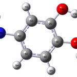 |
Scheme 1 Click here to View scheme |
Results and Discussion
The observed IR, Raman and calculated (scaled) wavenumbers and the assignments are given in Table 1. The DFT calculations give the OH stretching bands at 3618, 3573 cm-1 and strong bands are observed in the IR spectrum at 3388, 3300 cm-1. The in-plane deformation10 is expected in the region 1400± 40 cm-1 and the bands at 1444, 1380 cm-1 in the IR spectrum and at 1446, 1382 cm-1 in the Raman spectrum are assigned as the in-plane deformation of OH band. The calculated values are 1451 and 1372 cm-1. The stretching mode of the hydroxyl group with respect to the phenyl moiety C-O appears at 1251, 1185 cm-1 in the IR spectrum, 1184 cm-1 in the Raman spectrum and the calculated value are 1232, 1179 cm-1. This band is expected in the region11, 12 1220 ± 40 cm-1. El-Shahway et al.13 reported C-O stretching at 1240 cm-1.
In nitro compounds, υasNO2 are located in the region 1580 ± 80 cm-1. The nitro benzene derivatives show υasNO2 in the region10 1535 ± 30cm-1 and the 3-nitropyridine at 1530 ± 20cm-1. In the present case, υasNO2 are obtained at 1601 cm-1 theoretically (DFT) and at 1592 cm-1 in IR and at 1597 cm-1 in Raman spectrum. In nitro compounds, υsNO2 are located in the region 1300-1360 cm-1. The nitrobenzene derivatives show υsNO2 in 1345 ± 30 cm-1 and the 3-nitropyridines in 1350 ± 20cm-1. For the title compound, υsNO2 is observed at 1341 cm-1 (DFT), 1327 cm-1 (IR) and 1327 cm-1 (Raman). In the aromatic nitro compounds, bands are usually seen at 855 ± 40cm-1 (NO2 scissors deformation δNO2), 760 ± 30cm-1 (NO2 out of plane wag ωNO2), 540 ± 30cm-1 (NO2 in-plane rock ρNO2) and 70 ± 20cm-1 (tNO2). In the present case, the observed values for δNO2 are 901 cm-1 (DFT); ωNO2 is 784 cm-1 (DFT); ρNO2 are 549 cm-1 (DFT), 546 cm-1 (IR) and 551 cm-1 (Raman) and tNO2 is at 61cm-1 (DFT). Panicker et al.14 reported the NO2 deformation bands at 800,724,534 cm-1 (theoretically) and 809,727,717,524 cm-1 (experimentally). Sundaraganesan et al.15 reported the NO2 deformation bands at 839,744 and 398cm-1 experimentally and 812, 716,703 and 327 cm-1 theoretically.
The existence of one or more aromatic rings in a structure is normally readily determined from the C–H and C=C–C related vibrations. The C–H stretching occurs above 3000 cm-1 and is typically exhibited as a multiplicity of weak to moderate bands, compared with the aliphatic C–H stretch16. In the present case, the DFT calculations give the υCH modes in the range 3137– 3062 cm-1. The band observed at 3067 cm-1 in the IR spectrum is assigned as υCH mode of the benzene ring. The benzene ring possesses six ring stretching vibrations, of which the four with the highest wavenumbers (occurring near 1600, 1580, 1490 and 1440 cm-1) are good group vibrations10. In the absence of ring conjugation, the band near 1580 cm-1 is usually weaker than that at 1600 cm-1. The fifth ring stretching vibration which is active near 1335 ± 35 cm-1 a region which overlaps strongly with that of the CH in-plane deformation and the intensity is in general, low or medium high10. The sixth ring stretching vibration or ring breathing mode appears as a weak band near 1000 cm-1 in mono, 1,3-di and 1,3,5-trisubstituted benzenes. In the other wise substituted benzene, however, this vibration is substituent sensitive and difficult to distinguish from the ring in-plane deformation. For tri-substituted phenyl ring the υPh modes10 are seen in the region 1640 – 1110 cm-1 and these modes are observed at 1628, 1511, 1284, 1121 cm-1 in the IR spectrum, 1505, 1279, 1129 cm-1 in the Raman spectrum and at 1614, 1564, 1498, 1281, 1139 cm-1 (DFT) theoretically. In asymmetric tri-substituted benzenes, when all the three substituents are light, the wavenumber interval of the breathing mode11 is between 500 and 600 cm-1. When all the three substituents are heavy, the wavenumber appears above 1100 cm-1. In the case of mixed substituents, the wavenumber is expected11 to appear between 600 and 750 cm-1. For the title compound the phenyl ring breathing mode is observed at 653 cm-1 in the IR spectrum, 656 cm-1 in the Raman spectrum and at 666 cm-1 theoretically. Mary et al.17 reported the ring breathing mode of tri-substituted benzene ring at 733 cm-1 in the IR spectrum and at 738 cm-1 theoretically. The in-plane bending δCH modes10 of the phenyl ring are expected above 1000 cm-1. For the tri-substituted benzene ring these modes are observed at 1185, 1081 cm-1 in IR, 1184, 1081 cm-1 in Raman and at 1179, 1099, 1057 cm-1 theoretically. The CH out-of-plane deformations10 are observed between 1000 and 700 cm-1. Generally the CH out-of-plane deformations with the highest wavenumbers have a weaker intensity than those absorbing at lower wavenumbers. These γCH modes are observed at 947, 788 (IR), 935, 794 (Raman), 929, 876, 795 cm-1 (DFT). The in-plane and out-of-plane substituent modes of the phenyl ring are also identified and assigned (Table 1).
Conclusion
The IR and Raman spectra of 4-nitropyrocatechol have been recorded and analyzed. The harmonic vibrational wavenumbers were calculated theoretically using Gaussian03 software. Calculations were performed by the DFT level using the standard 6-31G* basis. The calculated wavenumbers agree well with the observed wavenumbers. The data obtained from vibrational wavenumber calculations are used to assign vibratinal bands found in IR and Raman spectra of the studied molecule.
Table 1: Calculated (Scaled) wavenumbers, IR, Raman bands and assignments
| B3LYP/6-31G* υ(cm-1) | IR intensity | Raman activity | IRυ(cm-1) | Ramanυ(cm-1) | Assignments |
| 3618 | 92.31 | 162.63 | 3388 | υOH | |
| 3573 | 90.16 | 69.54 | 3300 | υOH | |
| 3137 | 5.80 | 53.13 | υCH | ||
| 3135 | 2.45 | 62.87 | υCH | ||
| 3062 | 12.01 | 102.69 | 3067 | υCH | |
| 1614 | 4.07 | 2.96 | 1628 | υPh | |
| 1601 | 44.90 | 103.87 | 1592 | 1597 | υasNO2 |
| 1564 | 265.60 | 7.93 | υPh | ||
| 1498 | 114.85 | 1.87 | 1511 | 1505 | υPh |
| 1451 | 26.73 | 7.34 | 1444 | 1446 | δOH |
| 1372 | 1.21 | 1.83 | 1380 | 1382 | δOH |
| 1341 | 328.11 | 281.00 | 1327 | 1327 | υsNO2 |
| 1307 | 221.65 | 20.73 | 1308 | υCN | |
| 1281 | 287.39 | 39.09 | 1284 | 1279 | υPh |
| 1232 | 39.54 | 9.99 | 1251 | υCO | |
| 1179 | 45.63 | 6.06 | 1185 | 1184 | υCO, δCH |
| 1139 | 63.75 | 2.53 | 1121 | 1129 | υPh |
| 1099 | 50.48 | 1.61 | 1081 | 1081 | δCH |
| 1057 | 92.15 | 20.72 | δCH | ||
| 929 | 31.50 | 5.18 | 947 | 935 | γCH |
| 901 | 1.55 | 1.98 | δNO2 | ||
| 876 | 19.50 | 1.20 | 871 | γCH | |
| 795 | 16.09 | 5.67 | 788 | 794 | γCH |
| 784 | 14.54 | 3.15 | ωNO2 | ||
| 773 | 37.71 | 22.90 | 746 | 750 | γOH |
| 721 | 35.31 | 2.35 | 722 | γPh | |
| 666 | 0.90 | 0.18 | 653 | 656 | υPh |
| 632 | 23.78 | 2.24 | 632 | δPh(X) | |
| 574 | 7.82 | 6.76 | γOH | ||
| 549 | 0.06 | 0.21 | 546 | 551 | ρNO2 |
| 532 | 1.86 | 0.64 | γPh(X) | ||
| 442 | 5.65 | 0.91 | 448 | γPh(X) | |
| 416 | 3.46 | 3.36 | δPh(X) | ||
| 412 | 56.47 | 1.11 | δPh(X) | ||
| 347 | 0.14 | 2.42 | 351 | δCX(X) | |
| 333 | 2.22 | 1.60 | δCX(X) | ||
| 302 | 5.41 | 0.33 | δCX(X) | ||
| 282 | 189.45 | 2.76 | γCX(X) | ||
| 208 | 0.86 | 0.33 | 210 | γCX(X) | |
| 207 | 2.49 | 2.25 | γCX(X) | ||
| 114 | 0.38 | 0.48 | 111 | tPh | |
| 61 | 0.25 | 0.08 | tNO2 |
υ-stretching; δ-in-plane deformation; γ-out-of-plane deformation; ω-wagging; ρ-rocking; t-torsion; as-asymmetric; s-symmetric; Ph-phenyl ring; X-substituent sensitive
References
- Fiegel, H., Voges, H.W., Hamamoto, T., Umemura, S., Iwata, T., Miki, H., Fujita, Y., Buysch, H., Garbe, D., and Paulus, W., Ullmann’s Encyclopedia of Industrial Chemistry, Wiley-VCH, Weinheim (2002).
- Briggs, D.E.G., Philos. Trans.Royal Soc. B: Biol. Sci. 354: 7 (1999).
- Yam, K.C., D’Angelo, I., Kalscheuer, R., Zhu, H., Wang, J.X., Snieckus, V., Ly, L.H., Converse, P.J., Jacobs, W.R., Strynadka, N., and Eltis, L.D., PLoS Pathog. 5, e1000344(2009).
- Barner, B.A., Encyclopedia of Reagents for Organic Synthesis (Ed:Paquette,L.), Wiley and Sons, New York (2004).
- Fahlbusch, K., Hammerschmidt, F., Panten, J., Pickenhagen, W., Schatkowski, D., Bauer, K., Garbe, D., and Surburg, H., Ullmann’s Encyclopedia of Industrial Chemistry, Wiley-VCH, Weinheim (2005).
- Frisch, M.J., et al., Gaussian 03, Revision C.02 Gaussian, Inc., Wallingford CT (2004).
- Foresman, J.B., in: Frisch, E., (Ed.), Exploring Chemistry with Electronic Structure Methods: A Guide to Using Gaussian”, Pittsburg, PA (1996).
- Flukiger, P., Luthi, H.P., Portmann, S., and Weber, J., MOLEKEL 4.3, Swiss Centre for Scientific Computing, Manno, Switzerland (2000-2002).
- Portmann, S., and Luthi, H.P., Chimia, 54: 766 (2000).
- Roeges, N.P.G., A Guide to the Complete Interpretation of the Infrared spectra of organic structures, Wiley, New York (1994).
- Varsanyi, G., Assignments for Vibrational Spectra of Seven Hundred Benzene Derivatives, New York, Wiley (1974).
- Colthup, N.B., Daly, L.H., and Wiberly, S.E., Introduction to Infrared and Raman Spectroscopy. Ed. , Academic Press, Boston (1990).
- El-Shahaway, A.S., Ahmed, S.M., and Sayed, N.K., Spectrochim. Acta 66A: 143 (2007).
- Panicker, C.Y., Varghese, H.T., Ushakumari, L., Ertan, T., Yildiz, I., Granadeiro, C.M., Nogueira, H.I.S., and Mary, Y.S., J. Raman Spectrosc. 41: 381 (2010).
- Sundaraganesan, N., Ayyappan, S., Umamaheshawari, H., and Joshua, B.D., Spectrochim. Acta 66A: 17 (2007).
- Coates, J., and Meyers, R.A., Introduction to Infrared Spectrum, A Practical Approach, Chichester, John Wiley and Sons Ltd., (2000).
- Mary, Y.S., Varghese, H.T., Panicker, C.Y., Ertan, T., Yildiz, I., and Arpaci, O.T., Spectrochim. Acta 71A: 566 (2008).

This work is licensed under a Creative Commons Attribution 4.0 International License.
![]()
A New Edition of Web of Science
Journal Impact Factor
2022: 0.5
Five Year: 0.8
Journal is Indexed in

Cabells Whitelist
![]()


