Transforming Molecules, Transforming Medicine: A Comprehensive Review of Bioisosterism in Drug Design
 and Vipul M. Vaghela2
1Department of Pharmacy, Sumandeep Vidyapeeth Deemed to be University, Piparia, Waghodia, Vadodara, Gujarat, India.
2A. R. college of pharmacy and G. H. Patel institute of pharmacy, Vallabh Vidyanagar, Anand, Gujarat, India.
Corresponding Author E-mail:nbp9171@gmail.com
Download this article as:
ABSTRACT:Bioisosterism has transformed from empirical group replacement into a precision-guided cornerstone of drug discovery, integrating computational intelligence with chemical intuition. This review critically examines the evolution from classical monovalent through tetravalent substitutions to AI-driven, context-aware bioisosteric design.
Modern computational frameworks—molecular electrostatic potential mapping, density functional theory, fragment molecular orbital analysis, and molecular dynamics—now predict electronic and structural equivalence with unprecedented accuracy. Graph neural networks, generative models, and reinforcement learning algorithms have revolutionized bioisostere identification, enabling rapid exploration of previously inaccessible chemical space.
Contemporary successes—venetoclax's BCL-2 selectivity, elexacaftor's CFTR modulation, sotorasib's covalent KRAS targeting—exemplify bioisosterism's power in addressing selectivity, metabolic vulnerability, and undruggable targets. Yet critical challenges persist: predictive model limitations, synthetic tractability barriers, and extension to peptide/biologic modalities demand continued innovation.
As personalized medicine and multi-target therapeutics reshape pharmaceutical landscapes, bioisosterism stands uniquely positioned at the convergence of computational prediction and medicinal chemistry creativity, driving next-generation therapeutic breakthroughs.
KEYWORDS:Artificial Intelligence; Bioisosterism; Computational modelling; Drug design; Medicinal chemistry; Pharmacophore; Scaffold hopping
Introduction
Historical Background
The Genesis of Isosterism: Langmuir’s Electronic Theory
The conceptual foundation of bioisosterism traces its intellectual lineage to an unexpected discovery. In 1919, Irving Langmuir confronted a paradox that would reshape molecular design: why do N₂ and CO—chemically distinct entities—behave as functional twins? His answer challenged prevailing dogma. Langmuir revealed that physicochemical behavior emerges not from atomic identity per se, but from electronic architecture.1,2 Molecules sharing identical valence electron configurations exhibit convergent properties despite compositional divergence—a principle that seemed almost alchemical in its implications.3–5
Grimm’s Hydride Displacement Law: Expanding Isosteric Relationships
Grimm’s 1925 intervention proved transformative, yet conceptually audacious. His Hydride Displacement Law didn’t merely catalog isosteric relationships—it imposed electronic logic onto the periodic table itself. Grimm reconceptualized neighboring elements as valence-shifted variants of carbon: rightward elements behaving as electron-enriched carbon analogues, leftward as electron-depleted surrogates. The resulting equivalence series—CH≈NH≈O≈F—was elegant, almost suspiciously so.6,7 Could atomic identity truly be reduced to electron counting? Grimm’s framework succeeded as predictive theory but failed as biological prophecy. While physicochemical properties aligned beautifully across his isosteric series, he operated in a vacuum—literally devoid of enzymes, receptors, or living systems. His carbon-centric view anticipated molecular interchangeability,2,8 yet the critical question remained unanswered: would biology honor these electronic substitutions? Grimm provided the mathematical scaffold; medicinal chemistry would later test whether nature respected his electron-based equivalences or demanded something more nuanced. The gap between chemical elegance and biological reality had yet to be confronted.
Erlenmeyer’s Systematic Classification: Birth of Classical Bioisosterism
Erlenmeyer Sr. delivered the critical refinement in 1932, crystallizing isosterism into operational precision. His redefinition—”atoms, ions, or molecules with identical peripheral electron layers”—seemed narrow, yet unlocked systematic prediction.6 But Erlenmeyer’s genius lay not in classification, but in recognizing its inherent incompleteness.
His dual taxonomy appeared straightforward:
- Class I (Classical): Exact valence electron matching (N₂/CO, CO₂/N₃⁻, O₂/S₂)
- Class II (Non-Classical): Columnar equivalents with electronic flexibility (F/Cl/Br/I, NH₂/OH/SH)
Yet Erlenmeyer observed something profoundly destabilizing: biological systems sometimes accepted substitutions that violated strict isosteric rules. Certain molecular swaps preserved activity despite electronic or steric deviations—a paradox that pure chemistry couldn’t explain. This wasn’t theoretical weakness; it was prescient biology. Erlenmeyer had inadvertently discovered that living systems operate on functional mimicry, not electronic formalism. He named no such concept, but his observation foreshadowed bioisosterism itself: biology rewards molecular impersonation, not merely electron accounting.1
Modern Definition aand Significance in Drug Discovery
The Evolution of Bioisosterism: From Structure to Function
Friedman’s 1951 coinage of “bioisostere” marked chemistry’s capitulation to biology.9 The term itself was revolutionary—not for defining what isosteres are, but for what they do. Friedman severed the dogmatic link between electronic identity and functional equivalence, liberating medicinal chemists from Grimm’s electron-counting formalism.10 Biology, it turned out, didn’t care about perfect electronic mimicry; it cared about performance.
“Atoms, molecules, or functional groups possessing similar physicochemical properties that produce comparable biological effects when substituted within a bioactive molecule.”10
This definition is deceptively permissive. “Similar” properties span:
- Electronic architecture (charge distribution, polarization, dipole topology)
- Spatial geometry (shape, volume, conformational dynamics)
- Hydrogen bonding signatures (donor/acceptor motifs)
- Solvation equilibria (lipophilicity/hydrophilicity partitioning)
- Metabolic destiny (liability vs. resistance)
Contemporary bioisosterism is not structural cosplay—it is molecular engineering. The goal isn’t mimicry; it’s optimization. Replacements succeed when they preserve target engagement while reshaping ADME liabilities, metabolic soft spots, or off-target promiscuity. Friedman’s legacy endures precisely because he recognized what Erlenmeyer suspected: biology rewards functional equivalence, not chemical purity.
Strategic Applications in Modern Drug Development
Bioisosterism has evolved from academic curiosity to industrial imperative. Contemporary drug optimization operates on four strategic axes, each wielding bioisosteric substitution as precision instrumentation:
Pharmacokinetic Sculpting
- Metabolic armor: shielding labile sites from enzymatic attack
- Absorption control: lipophilicity tuning for barrier negotiation
- Distribution steering: tissue-selective penetration
- Half-life extension: clearance pathway obstruction
Efficacy Amplification
- Binding thermodynamics: affinity refinement through optimized contacts
- Selectivity sharpening: off-target silencing via structural divergence
- Residence time modulation: kinetic tuning for sustained engagement
- Resistance circumvention: exploiting alternate binding modes
Safety Engineering
- Toxicophore elimination: excising structural liabilities
- Metabolic detoxification: blocking reactive intermediate pathways
- Interaction minimization: CYP and transporter evasion
- Alert neutralization: removing genotoxic or idiosyncratic triggers
Intellectual Property Warfare
- Freedom-to-operate: patent landscape navigation through structural novelty
- Lifecycle extension: bioisosteric successors as commercial hedges
- Competitive differentiation: therapeutic equivalence with legal distinction
The transformation is complete: medicinal chemistry has migrated from empirical substitution to rational molecular engineering.11 Bioisosterism no longer asks “Can we replace this?” but “What performance metric improves when we do?“.1 As computational prediction converges with structural insight, bioisosteric design operates at the intersection of chemical imagination and biological constraint—where therapeutic innovation becomes systematically achievable rather than serendipitously discovered.
Classification of Bioisosteres
Bioisosteric classification isn’t mere taxonomy—it’s strategic architecture. Organized frameworks enable medicinal chemists to navigate molecular modification systematically rather than empirically, transforming structural redesign from art into engineering.12
Classical Bioisosteres
Classical bioisosteres operate on a deceptively simple promise: electronic equivalence guarantees functional equivalence. Atoms or groups sharing identical valence electron counts and comparable steric profiles theoretically produce predictable biological outcomes.13–15 Reality, as always, proves more nuanced. While these replacements maintain molecular geometry and charge distribution with reasonable fidelity, their biological performance depends critically on context—binding site microenvironments, desolvation penalties, and off-target promiscuity often disrupt neat electronic predictions.
Monovalent Bioisosteres
Monovalent substitutions represent molecular scalpel work—small changes, disproportionate consequences. Their power derives not from steric mimicry (they’re often geometrically mismatched) but from strategic manipulation of electronics, metabolism, and binding complementarity. Consider the hydrogen-to-fluorine swap: nearly isosteric yet profoundly transformative. Fluorine’s extreme electronegativity polarizes adjacent bonds, blocks oxidative metabolism, and alters pKa—all while occupying barely more space than hydrogen. Conversely, chlorine or methyl introductions amplify lipophilicity and membrane transit but risk metabolic liabilities and promiscuous hydrophobic contacts.
Polar monovalent groups—hydroxyls, amines—operate differently. They strengthen target engagement through hydrogen bonding yet exact pharmacokinetic penalties: reduced permeability, accelerated clearance, metabolic vulnerability. The monovalent design challenge isn’t choosing structurally similar groups; it’s orchestrating electronic and physicochemical trade-offs within three-dimensional binding constraints. Representative monovalent bioisosteres and their key properties are summarized in Table 1.16
Table 1: Representative monovalent bioisosteric substitutions illustrating their influence on electronic properties, lipophilicity, hydrogen-bonding capacity, and metabolic stability in medicinal chemistry design.
|
Bioisostere |
Structure / Group | Key Properties |
| Hydrogen | –H |
Baseline reference; smallest substituent; minimal steric impact |
|
Fluorine |
–F | Similar size to H (1.47 Å vs. 1.20 Å); highly electronegative; blocks oxidative metabolism; enhances metabolic stability |
| Chlorine | –Cl |
Larger than F; increases lipophilicity; contributes to binding affinity via halogen bonding |
|
Bromine |
–Br | Even larger halogen; increases lipophilicity and polarizability |
| Hydroxyl | –OH |
Acts as both H-bond donor and acceptor; increases polarity and solubility |
|
Thiol |
–SH | Weaker H-bond donor than –OH; more nucleophilic; lower electronegativity |
| Amino | –NH₂ |
Basic group; good H-bond donor; increases solubility and potential for ionic interactions |
|
Methyl |
–CH₃ | Increases lipophilicity; minimal electronic effect; enhances membrane permeability |
| Azido | –N₃ |
Linear geometry; electron-rich; used in click chemistry and molecular labeling |
Divalent Bioisosteres
Divalent bioisosteres are deceptive—they appear as simple linkers but function as molecular rheostats. Their true influence extends beyond connectivity to conformational control, electronic relay, and hydrogen-bonding topology. Selection of a divalent bioisostere governs flexibility versus rigidity, polarity versus lipophilicity, and ultimately, whether a molecule adopts its bioactive conformation or collapses into inactive ensembles.
Oxygen versus sulfur illustrates this principle: both link molecular fragments, yet oxygen imparts polarity and planarity-inducing lone pairs, while sulfur expands the linker, reduces electronegativity, and dampens hydrogen bonding. Carbonyl versus thiocarbonyl substitutions further demonstrate electronic tuning—carbonyl’s planarity and strong acceptor character contrast sharply with thiocarbonyl’s lipophilicity and weakened polarization. Effective divalent bioisosterism prioritizes conformational preorganization over simple linkage equivalence.16 Representative divalent bioisosteres and their design implications are summarized in Table 2.
Table 2: Representative divalent bioisosteres highlighting their effects on molecular geometry, polarity, and electronic communication relevant to medicinal chemistry design.
|
Bioisostere |
Structure / Group |
Key Properties |
|
Methylene |
–CH₂– | Simplest divalent linker; imparts conformational flexibility and non-polar character |
| Oxygen | –O– |
Hydrogen bond acceptor; increases polarity and solubility; common in ethers and esters |
|
Sulfur |
–S– | Larger, less electronegative than oxygen; more lipophilic; limited H-bonding |
| Selenium | –Se– |
Heavier chalcogen; offers redox potential; larger atomic radius |
|
Amino |
–NH– | Hydrogen bond donor; basic; introduces sites for protonation or tautomerism |
| Carbonyl | –C=O |
Planar geometry; strong H-bond acceptor; central in amides, ketones, and esters |
|
Thiocarbonyl |
–C=S | Sulfur analog of carbonyl; increases lipophilicity; modulates electronic properties |
| Imine | –C=N– |
Planar and conjugated; H-bond acceptor; enhances rigidity and π-character |
|
Sulfoxide |
–SO– |
Highly polar; tetrahedral geometry; contributes to dipole-driven interactions |
In practice, divalent bioisosteres are most effective when conformational control rather than simple linkage replacement is the primary design objective.
Trivalent Bioisosteres
Trivalent centers don’t replace—they reorganize. Unlike lower-valency substitutions that preserve directionality, trivalent bioisosteres function as geometric hubs, controlling substituent orientation and three-dimensional topology. Their medicinal chemistry value lies in redirecting binding vectors without disrupting pharmacophoric anchors.
Carbon-to-nitrogen replacement at trivalent centers introduces lone-pair electronics and hydrogen-bond accepting capability while maintaining planarity. Phosphorus, arsenic, and antimony offer progressively larger atomic radii and polarizability—useful when steric expansion or electronic diversification is required without sacrificing tetrahedral geometry. Yet these are not functional mimics; they are scaffold modulators. Trivalent replacements excel when optimizing target engagement geometry, not when attempting direct group equivalence.16:
Table 3: Trivalent bioisosteres illustrating their impact on molecular geometry, electronic properties, and scaffold behavior in drug design.
|
Bioisostere |
Structure | Key Properties |
| Methine | =CH– |
Carbon-based trivalent center; serves as the standard reference group |
|
Nitrogen |
=N– | More electronegative; provides hydrogen bond acceptor capability; planar geometry |
| Phosphorus | =P– |
Larger than nitrogen; capable of expanded valency; introduces electronic diversity |
|
Arsenic |
=As– | Heavier Group 15 element; increased polarizability and size |
| Antimony | =Sb– |
Largest stable trivalent Group 15 element used in drug design; more lipophilic |
Trivalent replacements are therefore better viewed as tools for reorganizing binding vectors than as direct functional mimics.
Tetravalent Bioisosteres
Tetrahedral centers define molecular architecture, and their bioisosteric replacement rewrites three-dimensional logic. Carbon provides the reference geometry, but alternative tetravalent elements—silicon, germanium, quaternary nitrogen, phosphorus—introduce bond-length expansion, charge introduction, or hypervalency that profoundly alter recognition landscapes.
Silicon-for-carbon substitution lengthens bonds (Si–C: 1.87 Å vs. C–C: 1.54 Å) and increases lipophilicity—subtle changes that can enhance metabolic stability or disrupt binding geometry depending on pocket tolerance. Quaternary nitrogen introduces permanent positive charge, transforming hydrophobic cores into cationic anchors. Sulfone groups impose rigidity and strong dipoles, useful for organizing hydrogen-bonding networks. The challenge with tetravalent bioisosterism isn’t preserving geometry—it’s exploiting geometry changes to access new interaction modes while maintaining sufficient overlap with the parent scaffold.16:
Table 4: Representative tetravalent bioisosteres illustrating how substitution at tetrahedral centers modulates molecular geometry, electronic properties, and target recognition in medicinal chemistry.
|
Bioisostere |
Structure |
Key Properties |
|
Carbon |
–C– | The standard tetrahedral center in organic compounds; basis for comparison |
| Silicon | –Si– |
Larger atomic radius; longer bonds and altered angles; increases lipophilicity |
|
Germanium |
–Ge– | Heavier Group 14 analog; maintains tetrahedral geometry with enhanced polarizability |
| Quaternary Nitrogen | –N⁺– |
Tetrahedral when fully substituted; introduces positive charge and ionic character |
|
Phosphorus |
–P– | Enables hypervalency and diverse substitution; electronic and steric versatility |
| Sulfone | –S(O)₂– |
Strongly polar tetrahedral group; contributes to hydrogen bonding and rigidity |
These substitutions are particularly valuable in late-stage optimization, where subtle changes in geometry or polarity can disproportionately affect pharmacokinetics.
Ring Equivalents
Ring bioisosterism operates at the intersection of electronics and topology, where cyclic framework replacement modulates both π-character and three-dimensional occupancy. This is not simple aromatic exchange—it’s strategic heteroatom placement to tune basicity, hydrogen bonding, desolvation energy, and metabolic soft spots while preserving spatial recognition.
Benzene-to-pyridine replacement introduces a basic nitrogen, enhancing solubility but risking protonation-dependent activity loss. Pyrimidine further increases polarity and nucleobase mimicry. Five-membered heterocycles—thiophene, furan, pyrrole, imidazole—offer planarity with varying heteroatom electronics: sulfur provides lipophilicity, oxygen polarity, nitrogen donor/acceptor versatility. Non-aromatic rings like cyclopropyl mimic π-systems through Walsh orbital character while introducing metabolic resistance via strain energy.
Modern ring bioisosterism integrates heteroatom positioning with metabolic liability prediction and desolvation thermodynamics—aromaticity alone is insufficient.6,17,18
Table 5: Representative ring bioisosteres illustrating how heteroatom substitution, ring size, and aromaticity modulate polarity, hydrogen bonding, and metabolic stability while preserving spatial recognition elements.
|
Ring System |
Type | Key Properties |
| Benzene | Aromatic (6-membered) |
Prototypical planar aromatic ring; hydrophobic; used as a reference scaffold |
|
Pyridine |
Aromatic (6-membered) | One nitrogen atom; introduces basicity and hydrogen bond acceptor capacity |
| Pyrimidine | Aromatic (6-membered) |
Two nitrogen atoms; increased polarity; useful in nucleobase mimicry |
|
Thiophene |
Aromatic (5-membered) | Sulfur-containing; planar and lipophilic; π-system preserved |
| Furan | Aromatic (5-membered) |
Oxygen-containing; more polar; reduced aromatic stability |
|
Pyrrole |
Aromatic (5-membered) | Nitrogen-containing; provides hydrogen bond donor functionality |
| Imidazole | Aromatic (5-membered) |
Two nitrogen atoms; both donor and acceptor properties; polar and amphoteric |
|
Oxazole/Thiazole |
Aromatic (5-membered) | Mixed heteroatoms (O/N or S/N); altered electron density; versatile pharmacophore |
| Cyclopropyl | Non-aromatic (3-membered) |
Strained, rigid; can mimic π-systems; increases metabolic stability |
|
Cyclobutyl |
Non-aromatic (4-membered) |
Moderate strain; lipophilic; retains compact spatial footprint |
Successful ring bioisosterism increasingly demands computational validation of desolvation penalties and CYP liability—structural similarity is necessary but insufficient.While classical bioisosteres rely on valence and electronic similarity, their utility becomes limited as molecular complexity and target selectivity increase. This limitation has driven the widespread adoption of non-classical bioisosteres, which prioritize functional mimicry and interaction patterns over strict structural equivalence.
Non-Classical Bioisosteres
Non-classical bioisosteres abandon electronic formalism entirely. These replacements prioritize functional mimicry over valence equivalence—atoms need not match, electrons need not align, yet biological outcomes converge. The paradigm shift is profound: structure becomes negotiable; function becomes absolute6.
Carboxylic Acid Bioisosteres
Carboxylic acids dominate bioactive molecules through hydrogen bonding and ionic anchoring—yet they’re pharmacokinetically catastrophic. Poor membrane permeability, rapid glucuronidation, and metabolic lability demand alternatives that preserve acidity while circumventing ADME failures.19,20 Tetrazole represents the archetype: pKa ~4.5–5.0 mimics carboxylate ionization, lipophilicity increases membrane transit, and metabolic resistance extends half-life—functional equivalence without structural identity. Sulfonic and phosphonic acids offer stronger acidity but sacrifice permeability; hydroxamic acids moderate pKa (~8–9) while enabling metalloprotease chelation. Acylsulfonamides elongate geometry yet maintain acidity (pKa ~5–6) with enhanced metabolic armor. Boronic acids exploit reversible covalent bonding—Lewis acidity replaces Brønsted acidity, enabling serine protease inhibition through transient adduct formation.
The design principle: carboxylic acid bioisosterism optimizes charge distribution and metabolic vulnerability, not carbon–oxygen bond preservation.
Table 6: Carboxylic Acid Bioisosteres
|
Bioisostere |
Key Properties |
| Tetrazole |
pKa ~4.5–5.0; mimics acidity and binding of carboxylic acid; enhanced metabolic and chemical stability |
|
Sulfonic Acid (–SO₃H) |
Strong acid (pKa ~0–2); extended geometry; high polarity and water solubility |
| Phosphonic Acid (–PO₃H₂) |
Diprotic acid; mimics phosphate groups; strong metal chelation; often used in bone-targeting agents |
|
Hydroxamic Acid (–CONHOH) |
Moderate acidity (pKa ~8–9); excellent hydrogen bonding; commonly found in metalloprotease inhibitors |
| Acylsulfonamide (–CONHSO₂R) |
Acidic (pKa ~5–6); elongated structure; multiple H-bonding sites; improved metabolic resistance |
|
Oxadiazolone |
Aromatic heterocycle with acidic proton; charge delocalization improves stability and solubility |
| Sulfonylurea |
Combines urea and sulfonamide motifs; distinct H-bonding pattern; known from antidiabetic drugs |
|
Boronic Acid (–B(OH)₂) |
Lewis acidic; forms reversible covalent bonds with nucleophiles (e.g., serine); used in protease inhibitors |
Amide Bioisosteres
Amides are structurally ubiquitous yet metabolically fragile. Planarity and hydrogen bonding make them pharmacophorically ideal; susceptibility to peptidases makes them pharmacokinetically disastrous. Amide bioisosterism therefore targets enzymatic resistance while preserving geometry and interaction topology.4,21 Sulfonamides replace carbonyl with sulfone—tetrahedral geometry shifts subtly, but enzymatic recognition collapses. Ureas and thioureas maintain planarity while modulating hydrogen-bonding strength and lipophilicity. Heterocyclic replacements—1,2,4-oxadiazoles, triazoles, imidazoles—rigidify geometry while introducing aromatic stability that peptidases cannot process. Retroamides invert connectivity, reversing dipole orientation without disrupting binding. Hydroxyethylene isosteres mimic the tetrahedral intermediate of amide hydrolysis, creating transition-state analogs that bind proteases irreversibly yet resist cleavage.
Effective amide bioisosterism exploits enzyme selectivity: preserve target recognition, evade metabolic machinery.\
Table 7: Amide Bioisosteres
|
Bioisosteres |
Key Properties |
| Sulfonamide (–SO₂NH–) |
Replaces carbonyl with sulfone; enhanced resistance to enzymatic hydrolysis; preserves H-bonding pattern |
|
Urea (–NHCONH–) |
Offers additional H-bonding potential; similar planarity and geometry to amide |
| Thiourea (–NHCSNH–) |
Sulfur replaces oxygen; increases lipophilicity; maintains donor/acceptor properties |
|
Retroamide (–NHCO–) |
Reverses connectivity; alters dipole orientation and H-bonding features |
| 1,2,4-Oxadiazole |
Rigid, planar heterocycle; mimics trans-amide geometry; improved metabolic stability |
|
1,2,4-Triazole |
Aromatic heterocycle with multiple H-bond sites; enhanced rigidity and metabolic resistance |
| Imidazole |
Planar ring system with donor/acceptor capability; mimics amide-like binding interactions |
|
Carbamate (–OCONH–) |
Retains hydrogen bonding features; less susceptible to enzymatic cleavage than amides |
| Hydroxyethylene (–CH(OH)CH₂–) |
Non-hydrolyzable moiety; mimics tetrahedral intermediate of amide hydrolysis; used in protease inhibitors |
Carbonyl Bioisosteres
Carbonyl groups anchor hydrogen-bonding networks, yet their electrophilic reactivity invites metabolic attack. Bioisosteric replacements maintain acceptor capacity while modulating polarity, geometry, and chemical liability21
Sulfones impose tetrahedral geometry with enhanced polarity—stronger hydrogen bonding but reduced lipophilicity. Sulfoximines combine sulfone and imine electronics, creating unique dipole profiles. Difluoromethylene offers non-bonding mimicry: steric and electronic similarity to C=O without hydrogen-bond capability—useful when acceptor elimination is desired. Thioamides preserve planarity while increasing lipophilicity; phosphonates introduce tetrahedral geometry with strong acidity, enabling phosphate ester mimicry in kinase inhibitors.
Carbonyl bioisosterism balances electronic mimicry against metabolic liability—geometry preservation is secondary to interaction optimization.
Table 8: Carbonyl Bioisosteres
|
Bioisostere |
Structure | Key Features |
| Sulfone (–SO₂–) | Tetrahedral |
Strong hydrogen bond acceptor; increased polarity; mimics carbonyl geometry |
|
Sulfoximine (–S(=O)(=NH)–) |
Tetrahedral with NH | Unique electronic profile; combines sulfone and imine behavior; modulates polarity |
| Sulfonamide (–SO₂N–) | Tetrahedral with N |
Dual H-bond donor/acceptor; enhanced metabolic stability and solubility |
|
Ketone Hydrate Equivalent (e.g., gem-diols) |
Tetrahedral | Mimics hydrated ketones; important in enzyme-target interactions involving carbonyl hydrates |
| Thioamide (–CSNH–) | Planar or near-planar |
Sulfur replaces oxygen; increased lipophilicity; retains H-bond acceptor capacity |
|
Hydroxylamine (–C(=NOH)–) |
Planar with donor/acceptor | Additional H-bond donor; modulates polarity and electronic properties |
| Difluoromethylene (–CF₂–) | Planar |
Non-hydrogen bonding; mimics dipole and sterics of C=O; improves metabolic stability |
|
Dicyanomethylene (–C(CN)₂–) |
Planar | Strong electron-withdrawing group; alters electronic distribution and π-character |
| Phosphonate (–PO(OR)₂–) | Tetrahedral |
Strong H-bond acceptor; mimics phosphate esters; useful in enzyme inhibition |
Emerging Classes of Bioisosteres
Traditional classifications constrain innovation. Emerging bioisosteric strategies exploit unconventional elements and complex motifs previously dismissed as synthetically intractable or biologically irrelevant.
Silicon-Based Bioisosteres
The strategic replacement of carbon with silicon (sila-substitution) has gained attention due to silicon’s unique properties. Silicon-for-carbon substitution violates classical isosterism yet succeeds functionally. Larger atomic radius (Si: 1.11 Å vs. C: 0.76 Å), lower electronegativity (1.9 vs. 2.5), and hypervalency potential create distinct electronic landscapes. Sila-substitution lengthens bonds, alters geometry, and reshapes metabolic trajectories—CYP enzymes evolved for carbon oxidation often fail against silicon analogs.rties22 Examples include silaproline (proline replacement with altered ring pucker), silacyclopentane scaffolds (conformational rigidification), and silafluorene derivatives (extended π-systems with silicon bridges).
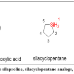 |
Figure 1: Examples include silaproline, silacyclopentane analogs, and silafluorene derivatives. Click here to View Figure |
Fluorinated Bioisosteres
Fluorine transcends monovalent substitution when deployed as complex motifs. Trifluoromethyl groups mimic methyl, chlorine, or methoxy depending on context—lipophilicity without oxidative liability. Difluoromethyl functions as a lipophilic hydrogen-bond donor, exploiting fluorine’s electronegativity to activate the remaining C–H. Pentafluorosulfanyl (–SF₅) delivers extreme lipophilicity and electronegativity simultaneously. Trifluoroborate complexes replace carboxylates with Lewis-acidic boron centers, enabling reversible receptor coordination.23
Fluorinated bioisosterism weaponizes electronegativity and metabolic resistance—structural similarity is coincidental.
Peptide Bond Mimetics
Peptide therapeutics demand metabolic invisibility. Standard amide bonds invite proteolytic destruction; non-hydrolyzable mimetics—reduced amides (–CH₂NH–), ketomethylenes (–COCH₂–), hydroxyethylenes (–CHOHCH₂–)—preserve geometry while eliminating scissile bonds. Thioamides introduce sulfur’s lower electronegativity, modulating hydrogen bonding without sacrificing planarity. Methyleneamines and aminomethylenes adjust rigidity and basicity.24,25
Peptide bond bioisosterism prioritizes proteolytic evasion over structural fidelity—recognition persists, cleavage does not.
Physicochemical properties Affecting Bioisosteric Replacements
Bioisosteric success demands physicochemical precision across electronegativity, lipophilicity, steric geometry, and hydrogen bonding—parameters governing potency, ADME profiles, and safety. These properties rarely align cooperatively; effective design requires strategic trade-offs, not isolated optimization.
Electronegativity and pKa
Electronegativity governs electron distribution, reshaping dipoles, charge localization, and binding interactions. Bioisosteric replacements altering electronegativity modulate enzyme recognition, receptor affinity, and transporter selectivity.26 Hydroxyl-to-fluorine substitution illustrates electronegativity-driven optimization: the substantial difference in electron-withdrawing capacity between oxygen and fluorine redistributes charge density across neighboring bonds, simultaneously conferring resistance to oxidative degradation and strengthening target interactions—achieved within a near-identical steric envelope 27.
pKa determines ionization at physiological pH, controlling solubility, permeability, and target engagement.28 Acidic and basic groups engage charged binding-site residues, yet ionization impedes membrane transit. Lowering pKa increases ionization (enhanced solubility, reduced permeability); raising pKa favors neutral forms (improved lipophilicity, diminished aqueous solubility).29
The angiotensin receptor blocker (ARB) class demonstrates pKa-guided bioisosteric design: tetrazole’s ionization profile (pKa ~4.5–5.0) ensures equivalent charge-state population at physiological pH while simultaneously improving the lipophilicity-driven oral absorption that limited earlier carboxylate-containing candidates—a dual optimization impossible through pKa matching alone.30
Lipophilicity
Lipophilicity, quantified through logP and pH-dependent logD, serves as the central variable connecting passive membrane diffusion, hydrophobic binding pocket engagement, and enzymatic susceptibility—three outcomes that often require opposing optimization directions.31
Hydroxyl-to-fluorine substitution increases lipophilicity while maintaining size, improving BBB penetration for CNS drugs.32,33 Hydrophobic pockets reward increased lipophilicity through strengthened van der Waals contacts.34 Hydrogen-to-trifluoromethyl replacement in steroids deepens hydrophobic engagement, enhancing receptor selectivity.35,36
Yet lipophilicity paradoxically threatens stability. Paradoxically, the same lipophilicity that facilitates membrane transit also increases cytochrome P450 (CYP450) recognition, accelerating first-pass oxidation and systemic clearance.37 Methyl-to-chlorine substitution blocks oxidation sites while modestly increasing lipophilicity, extending half-life without permeability loss.38 Effective bioisosterism navigates the permeability-solubility-metabolism inflection point.32
Molecular Size and Shape
Steric geometry determines binding complementarity and conformational preorganization.
Binding pockets enforce dimensional tolerance windows: substituents exceeding the pocket’s van der Waals boundary by even 0.5 Å trigger steric rejection, while undersized replacements leave hydrophobic voids that extract desolvation penalties without compensating contacts.34 Methyl-to-tert-butyl substitution blocks enzymatic approach, preventing off-target interactions or metabolism.39 Conversely, undersized replacements fail to fill hydrophobic voids, reducing affinity.
While ring bioisosteres achieve rigidification through cyclic constraints (discussed in Section 3), open-chain modifications employ a complementary strategy: single-bond-to-double-bond conversion restricts torsional freedom, enforcing bioactive geometry by eliminating inactive rotameric populations and reducing the entropic penalty of target engagement.40
PSA correlates inversely with passive permeability; values below 140 Ų favor oral bioavailability.41 Carboxyl-to-sulfonamide replacement increases PSA, reducing passive permeation while promoting renal excretion—useful when limiting CNS exposure is desired.
Hydrogen Bonding Capability
Hydrogen bonding anchors molecular recognition yet constrains membrane permeability.
Polar donors (–OH, –NH₂) and acceptors (–C=O, –N=) establish directional contacts essential for aqueous solvation and protein-ligand complementarity, yet each additional hydrogen bond imposes a desolvation penalty of approximately 2–5 kJ/mol during membrane transit—a thermodynamic cost that compounds rapidly with increasing donor/acceptor count.42 Ketone-to-thiocarbonyl substitution weakens acceptance (sulfur’s lower electronegativity reduces lone-pair availability), increasing lipophilicity.11
Increased hydrogen bonding enhances water solubility but impedes bilayer transit.43 Amide-to-cyano replacement eliminates donation, increasing permeability while sacrificing solubility—effective when absorption, not solubility, limits bioavailability.6
Hydrogen bonds govern enzyme recognition and receptor specificity.34
Hydroxyl-to-thiol substitution in metalloproteinase inhibitors weakens hydrogen bonding but enhances metal coordination through softer Lewis basicity, shifting mechanisms from hydrogen networks to chelation.44 Intramolecular hydrogen bonds mask polar groups, improving permeation.34 Prodrugs exploit this: internal bonds shield functionalities during absorption; metabolic cleavage liberates active forms post-permeation.45
These physicochemical parameters operate interdependently, often antagonistically. Optimizing lipophilicity may compromise solubility; enhancing hydrogen bonding may block permeability; increasing size may improve selectivity yet hinder absorption. Effective bioisosteric design demands context-driven, multi-parameter optimization—not isolated property maximization.
Late-stage failures trace to disproportionate emphasis on single parameters without accounting for emergent effects on metabolism, permeability, and engagement. Modern computational and AI-driven strategies enable simultaneous evaluation across biologically and pharmacokinetically relevant constraint spaces. Single-property optimization is obsolete; multi-dimensional assessment is requisite.
Mechanistic insights into Bioisosteric Modifications
Bioisosteric success is mechanistically complex, governed not by structural mimicry but by coordinated modulation of electronic distribution, steric geometry, binding kinetics, and metabolic fate. Effective replacements balance multiple competing parameters—a challenge that computational integration increasingly addresses.
Electronic Considerations
Electronic equivalence determines molecular recognition; deviations collapse binding.
Resonance effects preserve π-electron delocalization across conjugated systems, maintaining charge distribution essential for aromatic stacking and electrostatic anchoring.46,47 Inductive effects through electronegative substituents modulate local charge densities, preserving dipole-driven interactions without disrupting binding geometry.48,49 Polarizability matching ensures van der Waals interactions survive replacement—critical when induced-dipole forces dominate recognition.11
These are not independent variables. Altering one electronic parameter reshapes others; resonance stabilization may weaken inductive withdrawal, or enhanced polarizability may disrupt dipole alignment. Effective bioisosterism orchestrates these effects simultaneously.
Steric Considerations
Spatial geometry governs binding pocket occupancy and conformational preorganization.
Volumetric matching between isosteres, assessed through van der Waals surface overlap calculations, predicts spatial complementarity—but shape congruence alone proves insufficient without concurrent evaluation of desolvation thermodynamics and induced-fit adaptation.50 Yet volume alone is insufficient—shape matters. Linear versus bent geometries alter binding trajectories; planar versus tetrahedral centers reorient hydrogen-bonding vectors.
Conformational control through rigidification or flexibility introduction profoundly impacts binding entropy.40 Restricting rotation reduces entropic penalties during binding (fewer inactive conformers to depopulate); introducing flexibility enables induced-fit adaptation. The design choice depends on target plasticity: rigid binding sites reward preorganization; flexible pockets favor adaptability.
Advanced Structure-Activity Relationships (SAR)
Quantitative Structure-Activity Relationships (QSAR)
Modern QSAR integrates bioisosteric replacements with machine-learning algorithms, predicting activity from molecular descriptors.51 Key parameters—Hammett constants (σ) for electronic effects, Hansch-Fujita constants (π) for lipophilicity, Taft steric parameters (Es) for bulk—now operate within multidimensional models that capture non-linear property interactions impossible to intuit manually.
Fragment-Based Drug Design (FBDD)
Bioisosterism drives FBDD optimization: initial fragments undergo systematic bioisosteric exploration, linker regions are refined for geometry and electronics, and optimized fragments assemble into high-affinity leads.52 This modular approach decouples pharmacophore preservation from property tuning, enabling independent optimization of binding and ADME.
Influence on Pharmacodynamics: Detailed Mechanisms
Modulation of Target Binding Kinetics
Bioisosteric modifications alter association (kon) and dissociation (koff) rates independently, reshaping residence time and therapeutic windows.53 Long residence times extend pharmacological action beyond plasma concentrations—critical for kinetically driven efficacy.
Allosteric Modulation
Strategic replacements induce conformational shifts, accessing allosteric sites or modulating protein-protein interfaces.54 This transcends orthosteric binding: bioisosteres can convert competitive inhibitors into allosteric modulators through geometry-driven perturbations.
Effect on Pharmacokinetics (ADME): Advanced Considerations
Metabolic Soft Spots
Strategic bioisosteric intervention at identified metabolic soft spots disrupts CYP450 substrate recognition through altered electronic density or steric shielding, converting vulnerable sites into metabolically inert positions without compromising target affinity.55 Metabolic tuning is bidirectional: reducing liability or deliberately introducing clearance mechanisms.
Transporter Interactions
Replacements modulate P-glycoprotein, OATP, and other transporter recognition, governing absorption, distribution, and efflux.56 Small structural changes disproportionately affect transporter affinity—a chlorine-for-methyl swap can convert a substrate into non-substrate.
Bioisosteric success emerges from coordinated electronic, steric, kinetic, and metabolic optimization—not isolated parameter improvements. Enhancing binding affinity may introduce transporter liabilities; improving metabolic stability may compromise solubility. This inherent trade-off demands integrated SAR, fragment-based strategies, and computational modeling to identify bioisosteres balancing pharmacodynamic potency with pharmacokinetic robustness. Single-parameter optimization fails; multi-dimensional design succeeds.
Case Studies Illustrating SAR Relationships
HIV protease inhibitor evolution demonstrates bioisosteric refinement across three generations, each addressing predecessor failures through strategic replacement.
First-generation inhibitors (saquinavir) employed peptide-like scaffolds—structurally logical yet pharmacokinetically catastrophic due to poor oral bioavailability and rapid proteolytic degradation. Second-generation designs (lopinavir) introduced non-peptidic bioisosteres, replacing hydrolyzable amides with metabolically stable mimetics, dramatically improving absorption and half-life. Latest-generation inhibitors (darunavir) feature optimized bioisosteric replacements that enhance potency while suppressing resistance emergence through optimized binding geometry and reduced mutational escape pathways.57,58
Each iteration preserved HIV protease recognition while systematically eliminating ADME liabilities—bioisosterism as progressive pharmacokinetic problem-solving.
Kinase Inhibitor Optimization
Selective kinase inhibition demands bioisosteric precision across three structural regions, each governing distinct selectivity and ADME parameters.59,60
Hinge-binding region: Adenine-mimetic core modifications modulate selectivity across kinase families—subtle heteroatom placement differentiates isoform recognition through altered hydrogen-bonding geometry.
Solvent-exposed regions: Solubilizing group introduction improves pharmacokinetics without disrupting target engagement—polarity tuning occurs distant from binding determinants, decoupling ADME optimization from affinity.
DFG-out binders: Type II inhibitor bioisosteric replacements optimize allosteric pocket interactions, exploiting conformational plasticity unavailable to ATP-competitive (type I) designs.
Kinase bioisosterism operates regioselectively: binding affinity, selectivity, and pharmacokinetics each map to distinct molecular sectors, enabling independent optimization.
Applications of Bioisosteres in Drug Design
Case Studies: Successful Bioisosteric Replacements
Applications of Bioisosterism in Drug Design: Selected Case Studies
Table 9: Applications of Bioisosterism in Drug Design: Selected Case Studies
|
Drug/Case (Approval/Discovery Year) |
Key Bioisosteric Strategy | Improvements Achieved | Mechanistic/Clinical Impact | Ref. |
| Losartan (1995) | Carboxylic acid → tetrazole | ↑ Oral bioavailability, ↑ metabolic stability, ↓ clearance | Tetrazole preserved ionic interaction with Lys199, avoided glucuronidation; established “sartan” antihypertensives (multi-billion sales) |
30,61 |
|
Cimetidine → Ranitidine (1981) |
Imidazole → furan; side-chain tuning | 5–10× potency, ↓ CYP450 inhibition, fewer DDIs, lower dosing | Maintained binding electronics while reducing CYP interactions; Ranitidine became first blockbuster H₂ antagonist | 62 |
| Serotonin → Sumatriptan (1991) | Indole core preserved; sulfonamide + amine modification | ↑ Selectivity (5-HT1B/1D), ↓ BBB penetration, improved PK, ↓ ergot-like side effects | Indole maintained essential binding; modifications conferred receptor subtype selectivity; pioneered triptan class for migraine |
63,64 |
|
Ezetimibe (2002) |
Fluorine substitution (C–F as C–H bioisostere) | ↑ Metabolic stability, ↑ NPC1L1 binding, ↑ bioavailability, ↑ half-life | Fluorine blocked oxidative metabolism with minimal steric change; established fluorine as a standard design tool (20% of drugs) | 65,66 |
| Celecoxib (1998) | Carboxylic acid → sulfonamide; pyrazole scaffold | 1000× COX-2 selectivity, ↓ GI side effects, preserved efficacy | Sulfonamide exploited COX-2 side pocket; founded “coxib” class of selective NSAIDs |
67,68 |
|
HIV Protease Inhibitors (1995–) |
Peptide bond → hydroxyethylene / hydroxyethylamine | ↑ Protease resistance, ↑ bioavailability, ↑ half-life | Hydroxyethylene mimicked tetrahedral intermediate; led to potent protease inhibitors (e.g., Saquinavir), transforming HIV into chronic condition | 69–71 |
| Morphine → Fentanyl (1960s) | Phenol scaffold → piperidine; anilide substitution | 50–100× potency, ↑ BBB penetration, faster onset, shorter duration | Piperidine N maintained ionic receptor interaction; synthetic opioids revolutionized anesthesia and pain management |
72,73 |
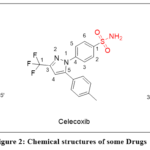 |
Figure 2: Chemical structures of some Drugs Click here to View Figure |
Impact on Drug Discovery
Integration into Modern Medicinal Chemistry
Structure-Based Bioisosterism
Computational algorithms now identify bioisosteric replacements through electrostatic potential mapping and molecular field analysis, enabling rational design beyond empirical substitution.74
Fragment-based libraries accelerate SAR exploration by systematically varying bioisosteric motifs,.75 while protein-ligand interaction mapping guides replacements that preserve binding geometry while optimizing pharmacological properties.76,77
High-Throughput Bioisosteric Screening
Automated parallel synthesis generates bioisosteric libraries at scale.78,79Positional scanning systematically replaces functional groups, identifying optimal bioisosteres through exhaustive exploration.80Machine learning algorithms predict successful replacements from historical datasets, transforming bioisosterism from iterative experimentation into predictive design.74
Property-Based Design
Bioisosteres strategically address ADME deficiencies,7replace toxic moieties with safer alternatives,81and enhance solubility while maintaining target engagement.4 This represents bioisosterism as precision tool—not for creating new pharmacophores, but for optimizing existing ones.
Emerging Trends and Current Applications
Ring Transformations in Drug Design
Scaffold hopping and ring walking expand chemical space while modulating properties. Scaffold hopping replaces central ring systems, accessing new IP and altering physicochemical profiles.82 Ring walking repositions heteroatoms within rings, fine-tuning hydrogen bonding, dipole orientation, and metabolic vulnerability.
SGLT2 inhibitor development from phlorizin to dapagliflozin exemplifies this: heterocyclic replacements improved oral bioavailability, metabolic stability, and target affinity—transforming a natural product into a clinical therapeutic through systematic ring bioisosterism.83 These changes not only improved oral bioavailability but also enhanced metabolic resilience and target affinity, showcasing the power of ring-focused bioisosteric strategies.
Halogen Bonding in Drug Design
Halogens transcend lipophilicity and metabolic blocking—they form directional halogen bonds. Iodine and bromine’s σ-holes interact with electron-rich acceptors (carbonyl oxygens, aromatic π-systems), enhancing binding affinity and selectivity.84
Thyroid hormone receptor modulators exploit iodine-mediated halogen bonds with ligand-binding domain residues, improving receptor specificity and pharmacodynamics.85 This elevates halogens from passive substituents to active binding elements.
Peptidomimetics and Beyond
Peptidomimetics employ bioisosterism to overcome peptide liabilities—proteolytic vulnerability, poor permeability, conformational flexibility.
Transition-state mimics resemble enzymatic reaction intermediates, enhancing inhibitory potency. Secondary structure mimetics constrain geometry, replicating β-turns or α-helices critical for protein-protein interactions.86
BACE1 inhibitors for Alzheimer’s employ hydroxyethylene isosteres simulating the enzyme’s transition state,87 achieving potency and selectivity impossible with hydrolyzable peptides. Bioisosterism thus enables peptide-like recognition with small-molecule-like stability.
These trends demonstrate bioisosterism’s evolution from substitution strategy to integrated design framework—combining computational prediction, high-throughput validation, and mechanistic understanding to systematically optimize pharmacological profiles.
Integration with Fragment-Based Drug Discovery
In FBDD, bioisosterism evolves low-molecular-weight fragments into potent leads. Fragment growing employs isosteric substitutions to enhance affinity or solubility without disrupting binding interactions. Fragment linking utilizes bioisosteric spacers connecting fragments while preserving conformations and binding vectors.88
Venetoclax (Bcl-2 inhibitor) exemplifies this: fragment screening integrated with bioisosteric design generated a molecule with high affinity, optimized pharmacokinetics, and oral bioavailability.89
Applications in Targeted Covalent Inhibitors
Bioisosterism fine-tunes electrophilic warhead reactivity for selective nucleophile targeting. Covalent engagement of cysteine residues demands precise warhead placement and reactivity control—isosteric modifications balance irreversible binding with safety and selectivity.90
BTK inhibitors demonstrate this: strategic covalent group optimization enabled selective, irreversible BTK engagement with minimal off-target activity, advancing B-cell malignancy therapies.91
Future Directions
Artificial Intelligence and Bioisosterism
AI convergence with bioisosterism revolutionizes molecular design. Trained models predict novel replacements with desired properties, accelerating optimization. Generative models propose chemically feasible, non-traditional bioisosteres beyond human intuition.92
Closed-loop design systems—AI suggestion, automated synthesis, immediate biological evaluation—enable iterative, real-time optimization, redefining drug discovery pace and precision.93
Expanding Beyond Traditional Medicinal Chemistry
Bioisosterism extends into peptide therapeutics through unnatural amino acids and peptide bond surrogates, improving stability, permeability, and proteolytic resistance. Biologics—antibodies, protein scaffolds—incorporate bioisosteres to modulate affinity and half-life.94
Beyond therapeutics, bioisosteric principles influence materials science: molecular replacements tune polymer properties, sensors, and drug delivery systems.95 This interdisciplinary expansion validates bioisosterism’s conceptual adaptability.
Precision Medicine Applications
- Patient-specific bioisosteres: modifications tailored to genetic polymorphisms
- Phenotypic optimization: replacements guided by disease-specific cellular responses
- Targeted delivery systems: bioisostere incorporation enhancing tissue-specific distribution
Bioisosterism evolves from structural modification strategy into predictive, interdisciplinary framework driving precision therapeutics.
Classical Applications of Bioisosteric Strategies in Successful Drug Design
The following cases weren’t lucky accidents—they were strategic strikes. Each bioisosteric modification solved a specific problem: poor selectivity, unacceptable toxicity, failed pharmacokinetics. These weren’t chemists randomly swapping groups; they were deliberate interventions guided by nascent SAR understanding. Their success proved bioisosterism works as rational design, not guesswork. The lessons still drive modern computational drug discovery.
The summarized details are presented as below:
Table 10: Classical Applications of Bioisosteric Strategies in Successful Drug Design.
|
Drug Evolution |
Key Bioisosteric Strategy | Benefits Achieved | Clinical/Mechanistic Insight | Reference |
| Aspirin → Celecoxib (1899–1998) | Carboxylic acid → sulfonamide; pyrazole core | ↑ COX-2 selectivity, ↓ GI side effects, improved compliance | Sulfonamide exploits COX-2 side pocket inaccessible to COX-1 |
5,96,97 |
|
Chlorpromazine → Clozapine (1950s–1971) |
Phenothiazine → dibenzodiazepine; side-chain tuning | ↓ Extrapyramidal effects, broader receptor profile, efficacy in resistant cases | Multireceptor engagement critical for complex symptom control | 98–100 |
| Salicylic Acid → Probenecid (Natural product → 1951) | Scaffold change: salicylate → benzoic acid; sulfonamide addition | ↑ Uricosuric effect, ↓ penicillin competition, ↑ half-life | Maintains acidic group while enhancing OAT1/3 transporter binding |
101–104 |
Note: Probenecid is not a classical atom- or group-level bioisostere of salicylic acid; rather, it represents scaffold and functional evolution guided by bioisosteric principles through preservation of the acidic pharmacophore.
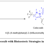 |
Figure 3: Aspirin to Celecoxib with Bioisosteric Strategies in Successful Drug Design Click here to View Figure |
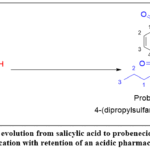 |
Figure 4: Structural evolution from salicylic acid to probenecid illustrating scaffold modification with retention of an acidic pharmacophore. Click here to View Figure |
Recent Case Studies
Recent breakthroughs in drug discovery have demonstrated how advanced bioisosteric strategies can transform lead compounds into clinically successful therapeutics. By fine-tuning electronic, steric, and metabolic properties, modern bioisosteric design has enabled improvements in potency, selectivity, and pharmacokinetics across diverse therapeutic areas. These innovations underscore bioisosterism’s role in overcoming long-standing drug development challenges. The key examples are summarized as below:
Table 11: Recent Case Studies of Bioisosteric Strategies in Drug Development.
|
Drug (Approval Year) |
Key Bioisosteric Strategy | Benefits Achieved | Clinical Impact | Reference |
| Sitagliptin (2006) | Trifluoromethyl as methyl bioisostere; fluorinated triazolopiperazine | ↑ Metabolic stability, ↓ oxidation, ↑ DPP-4 binding | First-in-class DPP-4 inhibitor for T2D; >$6B sales |
105–107 |
|
Venetoclax (2016) |
Chlorophenylpyrrole for naphthalene; azaindole for indole; halogen tuning | ↑ Potency (Ki < 0.01 nM), ↑ selectivity, oral bioavailability | Breakthrough in Bcl-2 inhibition for CLL/AML | 108–112 |
| Baricitinib (2018) | Pyrazole for pyrrole; azetidine as amine bioisostere | ↑ JAK1/2 selectivity, ↓ hERG risk, QD dosing | Used in RA, alopecia, COVID-19 |
113–115 |
|
Elexacaftor (2019) |
Trifluoromethoxy for methoxy; pyrazole for phenyl | ↑ CFTR binding, ↑ metabolic stability, broader coverage | Part of Trikafta; called “functional cure” for CF | 116–118 |
| Sotorasib (2021) | Piperazine as acrylamide bioisostere; pyrimidine for phenyl | ↑ Selectivity for KRAS(G12C), oral stability | First approved KRAS inhibitor |
119–122 |
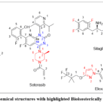 |
Figure 5: Chemical structures with highlighted Bioisosterically modification Click here to View Figure |
Modern Computational Approaches to Bioisosterism
Computational methods have transformed bioisosterism from retrospective rationalization into prospective design. Advanced tools now predict, validate, and optimize replacements before synthesis, accelerating drug development while reducing empirical screening.6,123
Structure-Based Bioisosteric Design
Structure-based approaches decode ligand-target interactions, revealing which molecular features are negotiable and which are sacred.
Three-dimensional pharmacophore modeling identifies spatial arrangements of chemical features essential for activity, generating hypotheses that guide bioisosteric selection toward functionally equivalent yet structurally distinct groups.124,125
HIV integrase inhibitor development exemplifies this: pharmacophore models identified the diketo acid motif as critical for Mg²⁺ chelation in the catalytic site. This insight directed medicinal chemists toward hydroxytriazolines and tetrazoles as bioisosteric replacements, culminating in raltegravir and dolutegravir—clinically successful agents that preserve metal coordination through non-obvious structural mimicry.126,127
Molecular electrostatic potential (MEP) analysis quantifies electronic surface similarity, identifying bioisosteres that preserve charge distribution despite structural divergence.128 Factor Xa inhibitor design demonstrates this power: MEP analysis revealed that certain heterocycles mimic amidine electron distribution. Oxazolidinone incorporation in rivaroxaban—guided by MEP equivalence—yielded an orally bioavailable anticoagulant with superior drug-like properties.129
Dynamic and Quantum Mechanical Modeling
Static models fail when dynamics matter. Binding sites flex, water networks reorganize, ligands adopt multiple conformations—phenomena invisible to rigid structural analysis.
Molecular dynamics (MD) simulations capture time-dependent behavior, evaluating water-mediated interactions, protein plasticity, and conformational adaptability of bioisosteric replacements.5 JAK inhibitor optimization illustrates this: MD simulations revealed that alternative heterocycles disrupted conserved water bridges, modulating selectivity and potency. These insights enabled rational tofacitinib derivative design with enhanced isoform discrimination.130
Density functional theory (DFT) provides quantum-level accuracy for electronic structure, reactivity, charge distribution, dipole moments, and pKa prediction—parameters governing bioisosteric functional equivalence. BACE1 inhibitor development leveraged DFT modeling to design hydroxyethylamine moieties mimicking the tetrahedral intermediate of amide hydrolysis, yielding potent inhibitors with improved metabolic profiles.131,132
Fragment molecular orbital (FMO) analysis decomposes ligand-receptor systems into fragments, quantifying individual contributions to binding affinity.133 Applied to Hsp90 inhibitors, FMO identified the resorcinol moiety’s hydrogen-bonding criticality within the ATP-binding site. Guided by this energetic dissection, heterocyclic bioisosteres preserved key interactions while improving metabolic stability—achieving comparable potency with enhanced drug-like properties.134
Advanced Docking and Scoring Techniques
Modern docking transcends geometry matching to model chemical mechanisms and solvent thermodynamics.
Covalent docking simulates irreversible bond formation between electrophilic ligands and nucleophilic residues, essential for covalent inhibitor design.135 EGFR T790M inhibitor development demonstrates this: covalent docking optimized acrylamide warheads, enabling osimertinib’s clinical success against resistance mutations in non-small cell lung cancer.136
Water-aware docking integrates solvent thermodynamics, exploiting energetically unfavorable water molecules as displacement opportunities.137 Not all binding-site waters stabilize complexes—some exact entropic penalties or form weak interactions. Well-positioned ligand groups can displace these high-energy waters, enhancing affinity through favorable enthalpy-entropy exchange.
CDK4/6 inhibitor development applied this principle: water-aware docking identified heterocyclic bioisosteres capable of displacing interfacial waters with unfavorable energetics. This strategy contributed to abemaciclib’s design, where selective water displacement enhanced binding affinity and isoform selectivity.138
Machine Learning and AI-Driven Approaches
Artificial intelligence has fundamentally recoded bioisosterism—from pattern recognition by human experts to pattern extraction by trained algorithms.
Graph neural networks (GNNs) model molecules as graphs, learning from vast datasets to identify non-intuitive bioisosteric replacements. The Novartis NIBR Bioisostere Finder exemplifies this: GNNs trained on curated medicinal chemistry transformations propose functionally equivalent substitutions invisible to traditional structure-activity heuristics.139,140 The paradigm shift: algorithms now detect bioisosteric relationships humans miss.
Generative models—transformers, variational autoencoders—transcend screening to actively design novel bioisosteres. Rather than selecting from known replacements, these models generate context-specific structures tailored to scaffolds and binding environments.141 AstraZeneca’s MolBART, trained on chemical transformation corpora, produces synthetically tractable bioisosteres that expand accessible chemical space beyond historical precedent.142
Deep learning frameworks predict how bioisosteric modifications cascade through pharmacokinetic and physicochemical property landscapes.143 DeepChem and similar platforms employ multi-parameter models assessing solubility, permeability, metabolic stability simultaneously—enabling holistic optimization rather than sequential property tuning.144 The advantage: coordinated multi-objective design replaces iterative single-parameter refinement.
Integrated AI Platforms and Iterative Optimization
Bioisosteric design divorced from synthetic feasibility is intellectually satisfying but practically useless. Modern platforms integrate retrosynthetic planning with bioisostere suggestion, prioritizing replacements by both biological relevance and synthetic accessibility.
IBM RXN for Chemistry, coupled with bioisostere engines, generates optimized molecules alongside feasible retrosynthetic routes.145 his integration eliminates synthetically intractable substitutions before they waste resources, streamlining lead optimization and accelerating development timeline.146 Design and synthesis now operate in dialogue, not isolation.
Reinforcement learning (RL) introduces dynamic optimization: algorithms iteratively improve bioisosteric replacements based on experimental feedback, learning which modifications succeed across potency, selectivity, and ADME objectives. Exscientia’s Centaur Chemist platform demonstrates this closed-loop approach, achieving fivefold acceleration in hit-to-lead optimization compared to traditional methods.147,148 RL transforms bioisosterism into an adaptive process—each experiment informs the next design cycle.
Natural Language Processing and Chemical Language Models
Medicinal chemistry literature harbors decades of bioisosteric experiments—successes, failures, contextual nuances. Natural language processing platforms like SciBite’s TERMite mine this corpus, extracting reported bioisosteric pairs with usage contexts and outcomes. This structured knowledge feeds machine learning models, improving predictive accuracy through historical wisdom.149,150
Chemical language models—MolGPT and analogues—treat molecules as linguistic sequences, learning syntactic and semantic rules of chemical transformations. Fragment-to-bioisostere replacement becomes molecular translation: input a fragment, output contextually appropriate alternatives based on learned chemical grammar.151,152 Chemistry becomes language; bioisosterism becomes translation.
Integrative Perspective: From Bio isosteric Principles to Predictive Design
These computational methodologies collectively represent bioisosterism’s evolution from empirical art to predictive science. Classical bioisosteric decisions relied on physicochemical analogy and SAR intuition—educated guesses refined through synthesis and testing. Contemporary approaches—structure-based modeling, quantum mechanics, AI—enable systematic evaluation within electronic, conformational, solvation, and synthetic constraint spaces.
Critically, these methods operate synergistically, not independently. Structure-based modeling and molecular dynamics reveal binding modes and flexibility requirements; quantum methods rationalize electronic equivalence; machine learning prioritizes viable candidates across multi-dimensional optimization criteria. Successful bioisosteric outcomes emerge from coordinated design decisions, not isolated substitutions.
This integration explains why modern bioisosterism succeeds systematically rather than sporadically. Replacements are predicted computationally, validated experimentally, refined iteratively—each method compensating for others’ limitations. Structure provides geometry; dynamics add temporal dimension; quantum mechanics ensure electronic fidelity; AI navigates combinatorial space; synthesis constrains reality. Together, they transform bioisosterism from retrospective rationalization into prospective engineering—a central, rational pillar of contemporary medicinal chemistry.
Emerging Horizons and Future Directions in Bioisosterism
Bioisosterism has outgrown its origins. What began as atomic substitution arithmetic has evolved into a predictive, multi-dimensional design paradigm governing therapeutics, agrochemicals, and functional materials simultaneously.13,153 The question is no longer what can be replaced—but what replacement optimally reshapes molecular destiny across binding, metabolism, selectivity, and beyond.
Data-Driven Discovery and AI Integration
AI hasn’t merely accelerated bioisosterism—it has fundamentally altered what’s discoverable.
Algorithms trained on vast molecular datasets predict isosteric replacement impacts on pharmacokinetics, target engagement, and off-target profiles with increasing accuracy.
.154,155 GNNs, generative models, and quantum machine learning identify bioisosteres with optimal spatial and electronic congruence—replacements human intuition would never propose.140
3D electrostatic potential matching, HOMO-LUMO descriptors, and MD simulations now generate isosteric scaffolds with improved ADMET profiles computationally, before synthetic investment.156,157 The critical insight: AI doesn’t optimize known bioisosteres—it discovers previously inconceivable ones, expanding chemical space beyond historical precedent.
Expanding Molecular Targets and Modalities
Classical bioisosterism targeted enzymes and receptors—well-defined pockets with predictable pharmacophores. Modern bioisosterism confronts fundamentally different challenges: protein-protein interactions, RNA structures, intrinsically disordered proteins, and allosteric sites that defy conventional design logic.7,81 These demanding interfaces require unconventional solutions: rigidified linkers enforce binding geometry against flat PPI surfaces; macrocyclic constraints pre-organize conformations reducing entropic binding penalties; sp³-rich bioisosteres improve three-dimensional complementarity with complex binding architectures.
Triazole, tetrazole, and oxadiazole isosteres in RNA-targeted ligands, covalent warheads, and stapled peptides demonstrate bioisosterism’s modality expansion.158 PROTAC linker optimization, molecular glue design, and nucleic acid mimetics represent emerging frontiers where bioisosteric principles govern efficacy.159 The critical limitation: predictive models trained on classical targets perform poorly against these novel modalities—new training datasets are urgently required.
Green Chemistry and Sustainable Innovation
Biological activity optimized at environmental cost is an incomplete victory. Green bioisosterism integrates ecological impact, synthetic tractability, and resource efficiency alongside pharmacological performance.160
Biocatalysis, photoredox catalysis, and late-stage C–H functionalization now install bioisosteric groups under mild, chemoselective conditions—eliminating harsh reagents and reducing synthetic steps.161 The critical insight: green bioisosterism reframes replacement selection as simultaneously pharmacological and ecological—sustainability becomes a design parameter, not an afterthought. This imperative intensifies in agrochemicals and materials science, where regulatory constraints demand environmental accountability.
System-Level Modeling and Digital Twins
Single-target optimization produces potent molecules that fail systemically. Complex diseases—cancer, neurodegeneration, metabolic disorders—demand molecules that perform coherently across entire biological networks.
Digital molecular twins coupled with systems pharmacology and multi-omics data simulate cascading biological consequences of structural modifications across cellular networks.162–164 A bioisosteric substitution can now be evaluated for therapeutic efficacy, polypharmacology, and toxicity simultaneously—before synthesis occurs.
The critical transformation: bioisosteric design shifts from molecular to systemic thinking. Replacements aren’t evaluated in binding-site isolation but against whole-organism biological coherence. This is bioisosterism’s most ambitious frontier—designing molecules that don’t merely bind targets but harmonize with biological complexity.
Conclusion
Bioisosterism has transitioned from a heuristic principle into a scientifically rigorous and predictive framework central to modern drug design. Through strategic replacement of functional groups and scaffolds, bioisosteres have consistently enhanced pharmacokinetics, safety, and therapeutic efficacy, with clinical successes ranging from classical NSAIDs to novel targeted inhibitors. Recent advances in computational modeling and AI have further redefined bioisosterism, transforming it into a data-driven approach capable of generating non-obvious replacements and accelerating lead optimization. Despite challenges in synthetic feasibility and predictive robustness, the continued integration of bioisosterism with machine learning, fragment-based design, and precision medicine offers a powerful path toward addressing complex diseases and unmet therapeutic needs. As the field evolves, bioisosterism will remain a guiding principle of medicinal chemistry—reshaping molecules, redefining mechanisms, and expanding the boundaries of therapeutic innovation.
Acknowledgement
The authors gratefully acknowledge the Almighty for guidance and strength, their family for unwavering support, and their institution for providing the necessary facilities that enabled the successful completion of this work.
AI Disclosure
Artificial intelligence tools (ChatGPT, GPT-5, OpenAI, USA) were used only for language refinement and grammar correction. All scientific content and interpretations are solely those of the authors
Funding Sources
The author has independently prepared this paper for publication based on their own research and efforts.
Conflicts of Interest
Our research and findings are driven solely by scientific merit and integrity, without any competing interests.
Data Availability Statement
All data generated and analyzed for this study have been included in the research article.
Ethics Statement
As this study did not involve animals or humans, no ethical clearance was required.
Author Contributions
The authors of this paper have adhered to the guidelines of the International Committee of Medical Journal Editors (ICMJE), contributed significantly to the study, and approved the final manuscript for submission and publication.
References
- Lima, L.; Barreiro, E. Bioisosterism: A Useful Strategy for Molecular Modification and Drug Design. Curr. Med. Chem. 2005, 12 (1), 23–49. https://doi.org/10.2174/0929867053363540.
CrossRef - Meanwell, N. A. Applications of Bioisosteres in the Design of Biologically Active Compounds. J. Agric. Food Chem. 2023, 71 (47), 18087–18122. https://doi.org/10.1021/ACS.JAFC.3C00765/ASSET/IMAGES/MEDIUM/JF3C00765_0071.GIF.
CrossRef - Ahmad, S.; Abdul Qadir, M.; Ahmed, M.; Imran, M.; Yousaf, N.; Wani, T. A.; Zargar, S.; Ali, I.; Muddassar, M. Exploring the Potential of Propanamide-Sulfonamide Based Drug Conjugates as Dual Inhibitors of Urease and Cyclooxygenase-2: Biological and Their in Silico Studies. Front. Chem. 2023, 11, 1206380. https://doi.org/10.3389/FCHEM.2023.1206380/BIBTEX.
CrossRef - Meanwell, N. A. The Influence of Bioisosteres in Drug Design: Tactical Applications to Address Developability Problems. Top. Med. Chem. 2014, 9, 283–382. https://doi.org/10.1007/7355_2013_29/FIGURES/40.
CrossRef - Osman, A. M. A.; Arabi, A. A. Quantum and Classical Evaluations of Carboxylic Acid Bioisosteres: From Capped Moieties to a Drug Molecule. ACS Omega 2023, 8 (1), 588–598. https://doi.org/10.1021/ACSOMEGA.2C05708/ASSET/ IMAGES/LARGE/AO2C05708_ 0008.JPEG.
CrossRef - Patani, G. A.; LaVoie, E. J. Bioisosterism: A Rational Approach in Drug Design. Chem. Rev. 1996, 96 (8), 3147–3176. https://doi.org/10.1021/CR950066Q.
CrossRef - Meanwell, N. A. Applications of Bioisosteres in the Design of Biologically Active Compounds. J. Agric. Food Chem. 2023, 71 (47), 18087–18122. https://doi.org/10.1021/ACS.JAFC.3C00765/ASSET/IMAGES/MEDIUM/JF3C00765_0071.GIF.
CrossRef - Brown, D. G.; Boström, J. Analysis of Past and Present Synthetic Methodologies on Medicinal Chemistry: Where Have All the New Reactions Gone? J. Med. Chem. 2016, 59 (10), 4443–4458. https://doi.org/10.1021/ACS.JMEDCHEM.5B01409/ ASSET/IMAGES/ LARGE/ JM-2015-01409Z_0006.JPEG.
CrossRef - First Symposium on Chemical-Biological Correlation, May 26-27, 1950; National Academies Press, 1951. https://doi.org/10.17226/18474.
CrossRef - Huang, S.; Wang, S.; Dong, J.; Xu, M.; Yuan, S. NeBULA: A Web-Based Novel Drug Design Platform for up-to-Date Bioisosteric Replacement. Med. Drug Discov. 2025, 28, 100231. https://doi.org/10.1016/J.MEDIDD.2025.100231.
CrossRef - Meanwell, N. A. Synopsis of Some Recent Tactical Application of Bioisosteres in Drug Design. J. Med. Chem. 2011, 54 (8), 2529–2591. https://doi.org/10.1021/JM1013693.
CrossRef - Chatzopoulou, M. Bioisosterism in Drug Discovery. Bioorganic Med. Chem. 2024, 108. https://doi.org/10.1016/j.bmc.2024.117758.
CrossRef - Wermuth, C. G.; Ganellin, C. R.; Lindberg, P.; Mitscher, L. A. Chapter 36. Glossary of Terms Used in Medicinal Chemistry (IUPAC Recommendations 1997. Annu. Rep. Med. Chem. 1998, 33 (C), 385–395. https://doi.org/10.1016/S0065-7743(08)61101-X.
CrossRef - Lima, L. M.; Barreiro, E. J. Beyond Bioisosterism: New Concepts in Drug Discovery. Compr. Med. Chem. III 2017, 1–8, 186–210. https://doi.org/10.1016/B978-0-12-409547-2.12290-5.
CrossRef - Dunker, C.; Schlegel, K.; Junker, A. Phenol (Bio)Isosteres in Drug Design and Development. Arch. Pharm. (Weinheim). 2025, 358 (1). https://doi.org/10.1002/ARDP.202400700.
CrossRef - Silverman, R. B.; Holladay, M. W. The Organic Chemistry of Drug Design and Drug Action: Third Edition. Org. Chem. Drug Des. Drug Action Third Ed. 2015, 1–517. https://doi.org/10.1016/C2009-0-64537-2.
CrossRef - Subbaiah, M. A. M.; Meanwell, N. A. Bioisosteres of the Phenyl Ring: Recent Strategic Applications in Lead Optimization and Drug Design. J. Med. Chem. 2021, 64 (19), 14046–14128. https://doi.org/10.1021/ACS.JMEDCHEM.1C01215/ASSET/ IMAGES/MEDIUM/ JM1C01215_0140.GIF.
CrossRef - Bauer, M. R.; Di Fruscia, P.; Lucas, S. C. C.; Michaelides, I. N.; Nelson, J. E.; Storer, R. I.; Whitehurst, B. C. Put a Ring on It: Application of Small Aliphatic Rings in Medicinal Chemistry. RSC Med. Chem. 2021, 12 (4), 448. https://doi.org/10.1039/D0MD00370K.
CrossRef - Ballatore, C.; Huryn, D. M.; Smith, A. B. Carboxylic Acid (Bio)Isosteres in Drug Design. ChemMedChem 2013, 8 (3), 385–395. https://doi.org/10.1002/cmdc.201200585.
CrossRef - Lassalas, P.; Gay, B.; Lasfargeas, C.; James, M. J.; Tran, V.; Vijayendran, K. G.; Brunden, K. R.; Kozlowski, M. C.; Thomas, C. J.; Smith, A. B.; Huryn, D. M.; Ballatore, C. Structure Property Relationships of Carboxylic Acid Isosteres. J. Med. Chem. 2016, 59 (7), 3183–3203. https://doi.org/10.1021/ACS.JMEDCHEM.5B01963.
CrossRef - Meanwell, N. A. Improving Drug Candidates by Design: A Focus on Physicochemical Properties as a Means of Improving Compound Disposition and Safety. Chem. Res. Toxicol. 2011, 24 (9), 1420–1456.https://doi.org/10.1021/TX200211V/ASSET/IMAGES/MEDIUM/ TX-2011-00211V_0041.GIF.
CrossRef - Franz, A. K.; Wilson, S. O. Organosilicon Molecules with Medicinal Applications. J. Med. Chem. 2013, 56 (2), 388–405. https://doi.org/10.1021/JM3010114.
CrossRef - Gillis, E. P.; Eastman, K. J.; Hill, M. D.; Donnelly, D. J.; Meanwell, N. A. Applications of Fluorine in Medicinal Chemistry. J. Med. Chem. 2015, 58 (21), 8315–8359. https://doi.org/10.1021/ACS.JMEDCHEM.5B00258.
CrossRef - Lenci, E.; Trabocchi, A. Peptidomimetic Toolbox for Drug Discovery. Chem. Soc. Rev. 2020, 49 (11), 3262–3277. https://doi.org/10.1039/D0CS00102C.
CrossRef - Estiarte, M. A.; Rich, D. H. Peptidomimetics for Drug Design. Burger’s Med. Chem. Drug Discov. 2003, 633–685. https://doi.org/10.1002/0471266949.BMC014.
CrossRef - Lipinski, C. A. Drug-like Properties and the Causes of Poor Solubility and Poor Permeability. J. Pharmacol. Toxicol. Methods 2000, 44 (1), 235–249. https://doi.org/10.1016/S1056-8719(00)00107-6.
CrossRef - Sheikhi, N.; Bahraminejad, M.; Saeedi, M.; Mirfazli, S. S. A Review: FDA-Approved Fluorine-Containing Small Molecules from 2015 to 2022. Eur. J. Med. Chem. 2023, 260. https://doi.org/10.1016/j.ejmech.2023.115758.
CrossRef - Manallack, D. T.; Prankerd, R. J.; Yuriev, E.; Oprea, T. I.; Chalmers, D. K. The Significance of Acid/Base Properties in Drug Discovery. Chem. Soc. Rev. 2012, 42 (2), 485–496. https://doi.org/10.1039/C2CS35348B.
CrossRef - Reijenga, J.; van Hoof, A.; van Loon, A.; Teunissen, B. Development of Methods for the Determination of PKa Values. Anal. Chem. Insights 2013, 8 (1), 53–71. https://doi.org/10.4137/ACI.S12304.
CrossRef - Herr, R. J. 5-Substituted-1H-Tetrazoles as Carboxylic Acid Isosteres: Medicinal Chemistry and Synthetic Methods. Bioorganic Med. Chem. 2002, 10 (11), 3379–3393. https://doi.org/10.1016/S0968-0896(02)00239-0.
CrossRef - Waring, M. J. Lipophilicity in Drug Discovery. Expert Opin. Drug Discov. 2010, 5 (3), 235–248. https://doi.org/10.1517/17460441003605098.
CrossRef - Arnott, J. A.; Planey, S. L. The Influence of Lipophilicity in Drug Discovery and Design. Expert Opin. Drug Discov. 2012, 7 (10), 863–875. https://doi.org/10.1517/17460441.2012.714363.
CrossRef - Park, B. K.; Kitteringham, N. R.; O’Neill, P. M. Metabolism of Fluorine-Containing Drugs. Annu. Rev. Pharmacol. Toxicol. 2001, 41, 443–470. https://doi.org/10.1146/ANNUREV.PHARMTOX.41.1.443.
CrossRef - Bissantz, C.; Kuhn, B.; Stahl, M. A Medicinal Chemist’s Guide to Molecular Interactions. J. Med. Chem. 2010, 53 (14), 5061–5084. https://doi.org/10.1021/JM100112J.
CrossRef - Kirk, K. L. Fluorine in Medicinal Chemistry: Recent Therapeutic Applications of Fluorinated Small Molecules. J. Fluor. Chem. 2006, 127 (8), 1013–1029. https://doi.org/10.1016/J.JFLUCHEM.2006.06.007.
CrossRef - Zhou, Y.; Wang, J.; Gu, Z.; Wang, S.; Zhu, W.; Acenã, J. L.; Soloshonok, V. A.; Izawa, K.; Liu, H. Next Generation of Fluorine-Containing Pharmaceuticals, Compounds Currently in Phase II-III Clinical Trials of Major Pharmaceutical Companies: New Structural Trends and Therapeutic Areas. Chem. Rev. 2016, 116 (2), 422–518. https://doi.org/10.1021/ACS.CHEMREV.5B00392.
CrossRef - Zhang, Z.; Tang, W. Drug Metabolism in Drug Discovery and Development. Acta Pharm. Sin. B 2018, 8 (5), 721–732. https://doi.org/10.1016/j.apsb.2018.04.003.
CrossRef - Zhao, M.; Ma, J.; Li, M.; Zhang, Y.; Jiang, B.; Zhao, X.; Huai, C.; Shen, L.; Zhang, N.; He, L.; Qin, S. Cytochrome P450 Enzymes and Drug Metabolism in Humans. Int. J. Mol. Sci. 2021, 22 (23), 12808. https://doi.org/10.3390/IJMS222312808/S1.
CrossRef - Obach, R. S.; Walsky, R. L.; Venkatakrishnan, K.; Gaman, E. A.; Houston, J. B.; Tremaine, L. M. The Utility of in Vitro Cytochrome P450 Inhibition Data in the Prediction of Drug-Drug Interactions. J. Pharmacol. Exp. Ther. 2006, 316 (1), 336–348. https://doi.org/10.1124/JPET.105.093229.
CrossRef - Fang, Z.; Song, Y.; Zhan, P.; Zhang, Q.; Liu, X. Conformational Restriction: An Effective Tactic in ’Follow-on’-Based Drug Discovery. Future Med. Chem. 2014, 6 (8), 885–901. https://doi.org/10.4155/FMC.14.50.
CrossRef - Shultz, M. D. Two Decades under the Influence of the Rule of Five and the Changing Properties of Approved Oral Drugs. J. Med. Chem. 2019, 62 (4), 1701–1714. https://doi.org/10.1021/ACS.JMEDCHEM.8B00686.
CrossRef - Kenny, P. W. Hydrogen Bonding, Electrostatic Potential, and Molecular Design. J. Chem. Inf. Model. 2009, 49 (5), 1234–1244. https://doi.org/10.1021/CI9000234.
CrossRef - Ritchie, T. J.; Macdonald, S. J. F. The Impact of Aromatic Ring Count on Compound Developability–Are Too Many Aromatic Rings a Liability in Drug Design? Drug Discov. Today 2009, 14 (21–22), 1011–1020. https://doi.org/10.1016/J.DRUDIS.2009.07.014.
CrossRef - Jacobsen, F. E.; Lewis, J. A.; Cohen, S. M. The Design of Inhibitors for Medicinally Relevant Metalloproteins. ChemMedChem 2007, 2 (2), 152–171. https://doi.org/10.1002/CMDC.200600204.
CrossRef - Rautio, J.; Kumpulainen, H.; Heimbach, T.; Oliyai, R.; Oh, D.; Järvinen, T.; Savolainen, J. Prodrugs: Design and Clinical Applications. Nat. Rev. Drug Discov. 2008, 7 (3), 255–270. https://doi.org/10.1038/NRD2468.
CrossRef - Hernández-Lladó, P.; Meanwell, N. A.; Russell, A. J. A Data-Driven Perspective on Bioisostere Evaluation: Mapping the Benzene Bioisostere Landscape with BioSTAR. J. Med. Chem. 2025, 68 (16), 16921. https://doi.org/10.1021/ACS.JMEDCHEM.5C01641.
CrossRef - Patrick, G. . An Introduction to Medicinal Chemistry, 5th editio.; Oxford University Press, 2016.
- Taylor, R. D.; Maccoss, M.; Lawson, A. D. G. Rings in Drugs. J. Med. Chem. 2014, 57 (14), 5845–5859. https://doi.org/10.1021/JM4017625.
CrossRef - Verma, A.; Waiker, D. K.; Gupta, P. S.; Kumar, M.; Shrivastava, S. K. Bioisosterism: An Approach to Develop New Anti-Cancer Agents. Eur. J. Med. Chem. 2025, 300. https://doi.org/10.1016/J.EJMECH.2025.118166.
CrossRef - Arabi, A. A. Routes to Drug Design via Bioisosterism of Carboxyl and Sulfonamide Groups. Future Med. Chem. 2017, 9 (18), 2167–2180. https://doi.org/10.4155/FMC-2017-0136.
CrossRef - Cherkasov, A.; Muratov, E. N.; Fourches, D.; Varnek, A.; Baskin, I. I.; Cronin, M.; Dearden, J.; Gramatica, P.; Martin, Y. C.; Todeschini, R.; Consonni, V.; Kuz’Min, V. E.; Cramer, R.; Benigni, R.; Yang, C.; Rathman, J.; Terfloth, L.; Gasteiger, J.; Richard, A.; Tropsha, A. QSAR Modeling: Where Have You Been? Where Are You Going To? J. Med. Chem. 2014, 57 (12), 4977–5010.
https://doi.org/10.1021/JM4004285/ASSET/IMAGES/MEDIUM/JM-2013-004285_0009.GIF.
CrossRef - Erlanson, D. A.; Fesik, S. W.; Hubbard, R. E.; Jahnke, W.; Jhoti, H. Twenty Years on: The Impact of Fragments on Drug Discovery. Nat. Rev. Drug Discov. 2016, 15 (9), 605–619. https://doi.org/10.1038/NRD.2016.109.
CrossRef - Copeland, R. A.; Pompliano, D. L.; Meek, T. D. Drug-Target Residence Time and Its Implications for Lead Optimization. Nat. Rev. Drug Discov. 2006, 5 (9), 730–739. https://doi.org/10.1038/NRD2082.
CrossRef - Lu, S.; Zhang, J. Small Molecule Allosteric Modulators of G-Protein-Coupled Receptors: Drug-Target Interactions. J. Med. Chem. 2019, 62 (1), 24–45. https://doi.org/10.1021/ACS.JMEDCHEM.7B01844.
CrossRef - Zhang, D.; Zhu, M.; Humphreys, W. G. Drug Metabolism in Drug Design and Development: Basic Concepts and Practice. Drug Metab. Drug Des. Dev. Basic Concepts Pract. 2007, 1–609. https://doi.org/10.1002/9780470191699.
CrossRef - Montanari, F.; Ecker, G. F. Prediction of Drug–ABC-Transporter Interaction — Recent Advances and Future Challenges. Adv. Drug Deliv. Rev. 2015, 86, 17–26. https://doi.org/10.1016/J.ADDR.2015.03.001.
CrossRef - Ghosh, A. K.; Osswald, H. L.; Prato, G. Recent Progress in the Development of HIV-1 Protease Inhibitors for the Treatment of HIV/AIDS. J. Med. Chem. 2016, 59 (11), 5172–5208. https://doi.org/10.1021/ACS.JMEDCHEM.5B01697.
CrossRef - Voshavar, C. Protease Inhibitors for the Treatment of HIV/AIDS: Recent Advances and Future Challenges. Curr. Top. Med. Chem. 2019, 19 (18), 1571–1598. https://doi.org/10.2174/1568026619666190619115243.
CrossRef - Ferguson, F. M.; Gray, N. S. Kinase Inhibitors: The Road Ahead. Nat. Rev. Drug Discov. 2018, 17 (5), 353–376. https://doi.org/10.1038/NRD.2018.21.
CrossRef - Parikh, P. K.; Ghate, M. D. Recent Advances in the Discovery of Small Molecule C-Met Kinase Inhibitors. Eur. J. Med. Chem. 2018, 143, 1103–1138. https://doi.org/10.1016/j.ejmech.2017.08.044.
CrossRef - Bredael, K.; Geurs, S.; Clarisse, D.; De Bosscher, K.; D’hooghe, M. Carboxylic Acid Bioisosteres in Medicinal Chemistry: Synthesis and Properties. J. Chem. 2022, 2022. https://doi.org/10.1155/2022/2164558.
CrossRef - Ma, Y. S.; Xin, R.; Yang, X. L.; Shi, Y.; Zhang, D. D.; Wang, H. M.; Wang, P. Y.; Liu, J. Bin; Chu, K. J.; Fu, D. Paving the Way for Small-Molecule Drug Discovery. American Journal of Translational Research. 2021, pp 853–870. https://pubmed.ncbi.nlm.nih.gov/ 33841626/ (accessed 2025-11-14).
- Humphrey, P. P. A. The Discovery and Development of the Triptans, a Major Therapeutic Breakthrough. Headache 2008, 48 (5), 685–687. https://doi.org/10.1111/j.1526-4610.2008.01097.x.
CrossRef - Blumenfeld, A.; Tepper, S. J.; Khanna, R.; Doty, E.; Vincent, M.; Miller, S. I. Serotonin Syndrome in the Acute Treatment Landscape of Migraine: The Lasmiditan Experience. Front. Neurol. 2023, 14. https://doi.org/10.3389/FNEUR.2023.1291102.
CrossRef - Gillis, E. P.; Eastman, K. J.; Hill, M. D.; Donnelly, D. J.; Meanwell, N. A. Applications of Fluorine in Medicinal Chemistry. J. Med. Chem. 2015, 58 (21), 8315–8359. https://doi.org/10.1021/ACS.JMEDCHEM.5B00258.
CrossRef - Henary, E.; Casa, S.; Dost, T. L.; Sloop, J. C.; Henary, M. The Role of Small Molecules Containing Fluorine Atoms in Medicine and Imaging Applications. Pharm. 2024, Vol. 17, Page 281 2024, 17 (3), 281. https://doi.org/10.3390/PH17030281.
CrossRef - Jiang, B.; Zeng, Y.; Li, M. J.; Xu, J. Y.; Zhang, Y. N.; Wang, Q. J.; Sun, N. Y.; Lu, T.; Wu, X. M. Design, Synthesis, and Biological Evaluation of 1,5-Diaryl-1,2,4-Triazole Derivatives as Selective Cyclooxygenase-2 Inhibitors. Arch. Pharm. (Weinheim). 2010, 343 (9), 500–508. https://doi.org/10.1002/ARDP.200900227.
CrossRef - Chahal, S.; Rani, P.; Kiran; Sindhu, J.; Joshi, G.; Ganesan, A.; Kalyaanamoorthy, S.; Mayank; Kumar, P.; Singh, R.; Negi, A. Design and Development of COX-II Inhibitors: Current Scenario and Future Perspective. ACS Omega 2023, 8 (20), 17446–17498. https://doi.org/10.1021/ACSOMEGA.3C00692.
CrossRef - Roberts, N. A.; Martin, J. A.; Kinchington, D.; Broadhurst, A. V.; Craig, J. C.; Duncan, I. B.; Galpin, S. A.; Handa, B. K.; Kay, J.; Kröhn, A.; Lambert, R. W.; Merrett, J. H.; Mills, J. S.; Parkes, K. E. B.; Redshaw, S.; Ritchie, A. J.; Taylor, D. L.; Thomas, G. J.; Machin, P. J. Rational Design of Peptide-Based HIV Proteinase Inhibitors. Science 1990, 248 (4953), 358–361. https://doi.org/10.1126/ SCIENCE. 2183354.
CrossRef - Ghosh, A. K.; Nyalapatla, R. P.; Kovela, S.; Rao, K. V.; Brindisi, M.; Osswald, H. L.; Amano, M.; Aoki, M.; Agniswamy, J.; Wang, Y. F.; Weber, I. T.; Mitsuya, H. Design and Synthesis of Highly Potent HIV-1 Protease Inhibitors Containing Tricyclic Fused Ring Systems as Novel P2 Ligands: Structure-Activity Studies, Biological and X-Ray Structural Analysis. J. Med. Chem. 2018, 61 (10), 4561–4577. https://doi.org/10.1021/acs.jmedchem.8b00298.
CrossRef - Funicello, M.; Chiummiento, L.; Santarsiere, A.; Poggio, F.; Lupattelli, P. Recent Advances in Heterocyclic HIV Protease Inhibitors. Int. J. Mol. Sci. 2025, Vol. 26, Page 9023 2025, 26 (18), 9023. https://doi.org/10.3390/IJMS26189023.
CrossRef - Vardanyan, R. S.; Hruby, V. J. Fentanyl-Related Compounds and Derivatives: Current Status and Future Prospects for Pharmaceutical Applications. Future Med. Chem. 2014, 6 (4), 385–412. https://doi.org/10.4155/FMC.13.215.
CrossRef - Zarin, M. K. Z.; Dehaen, W.; Salehi, P.; Asl, A. A. B. Synthesis and Modification of Morphine and Codeine, Leading to Diverse Libraries with Improved Pain Relief Properties. Pharmaceutics 2023, 15 (6), 1779. https://doi.org/10.3390/PHARMACEUTICS15061779.
CrossRef - Zhang, T.; Sun, S.; Wang, R.; Li, T.; Gan, B.; Zhang, Y. BioisoIdentifier: An Online Free Tool to Investigate Local Structural Replacements from PDB. J. Cheminform. 2024, 16 (1), 1–17. https://doi.org/10.1186/S13321-024-00801-8/FIGURES/12.
CrossRef - Wirth, M.; Zoete, V.; Michielin, O.; Sauer, W. H. B. SwissBioisostere: A Database of Molecular Replacements for Ligand Design. Nucleic Acids Res. 2013, 41 (D1). https://doi.org/10.1093/NAR/GKS1059.
CrossRef - Seddon, M. P.; Cosgrove, D. A.; Gillet, V. J. Bioisosteric Replacements Extracted from High-Quality Structures in the Protein Databank. ChemMedChem 2018, 13 (6), 607–613. https://doi.org/10.1002/CMDC.201700679,.
CrossRef - Adasme, M. F.; Linnemann, K. L.; Bolz, S. N.; Kaiser, F.; Salentin, S.; Haupt, V. J.; Schroeder, M. PLIP 2021: Expanding the Scope of the Protein-Ligand Interaction Profiler to DNA and RNA. Nucleic Acids Res. 2021, 49 (W1), W530–W534. https://doi.org/10.1093/NAR/GKAB294,.
CrossRef - Dombrowski, A. W.; Aguirre, A. L.; Shrestha, A.; Sarris, K. A.; Wang, Y. The Chosen Few: Parallel Library Reaction Methodologies for Drug Discovery. J. Org. Chem. 2022, 87 (4), 1880–1897. https://doi.org/10.1021/acs.joc.1c01427.
CrossRef - Duléry, B. D.; Verne-Mismer, J.; Wolf, E.; Kugel, C.; Van Hijfte, L. Analyses of Compound Libraries Obtained by High-Throughput Parallel Synthesis: Strategy of Quality Control by High-Performance Liquid Chromatography, Mass Spectrometry and Nuclear Magnetic Resonance Techniques. J. Chromatogr. B Biomed. Sci. Appl. 1999, 725 (1), 39–47. https://doi.org/10.1016/S0378-4347(98)00570-2.
CrossRef - Sampaio-Dias, I. E.; Reis-Mendes, A.; Costa, V. M.; García-Mera, X.; Brea, J.; Loza, M. I.; Pires-Lima, B. L.; Alcoholado, C.; Algarra, M.; Rodríguez-Borges, J. E. Discovery of New Potent Positive Allosteric Modulators of Dopamine D2Receptors: Insights into the Bioisosteric Replacement of Proline to 3-Furoic Acid in the Melanostatin Neuropeptide. J. Med. Chem. 2021, 64 (9), 6209–6220. https://doi.org/10.1021/acs.jmedchem.1c00252.
CrossRef - Jayashree, B. S.; Nikhil, P. S.; Paul, S. Bioisosterism in Drug Discovery and Development – An Overview. Med. Chem. (Los. Angeles). 2022, 18 (9), 915–925. https://doi.org/10.2174/1573406418666220127124228,.
CrossRef - Wang, S.; Zhang, R.; Li, X.; Cai, F.; Ma, X.; Tang, Y.; Xu, C.; Wang, L.; Ren, P.; Liu, L.; Wu, S.; Qian, Q.; Fang Bai, &. Recent Advances in Molecular Representation Methods and Their Applications in Scaffold Hopping. npj Drug Discov. 2025 21 2025, 2 (1), 14-. https://doi.org/10.1038/s44386-025-00017-2.
CrossRef - Maccari, R.; Ottanà, R. Sodium-Glucose Cotransporter Inhibitors as Antidiabetic Drugs: Current Development and Future Perspectives. J. Med. Chem. 2022, 65 (16), 10848–10881. https://doi.org/10.1021/ACS.JMEDCHEM.2C00867.
CrossRef - Ali, S.; Tian, X.; Meccia, S. A.; Zhou, J. Highlights on U.S. FDA-Approved Halogen-Containing Drugs in 2024. Eur. J. Med. Chem. 2025, 287. https://doi.org/10.1016/j.ejmech.2025.117380.
CrossRef - Verteramo, M. L.; Ignjatović, M. M.; Kumar, R.; Wernersson, S.; Ekberg, V.; Wallerstein, J.; Carlström, G.; Chadimová, V.; Leffler, H.; Zetterberg, F.; Logan, D. T.; Ryde, U.; Akke, M.; Nilsson, U. J. Interplay of Halogen Bonding and Solvation in Protein–Ligand Binding. iScience 2024, 27 (4), 109636. https://doi.org/10.1016/J.ISCI.2024.109636.
CrossRef - Romagnoli, A.; Rexha, J.; Perta, N.; Di Cristofano, S.; Borgognoni, N.; Venturini, G.; Pignotti, F.; Raimondo, D.; Borsello, T.; Di Marino, D. Peptidomimetics Design and Characterization: Bridging Experimental and Computer-Based Approaches. Prog. Mol. Biol. Transl. Sci. 2025, 212, 279–327. https://doi.org/10.1016/bs.pmbts.2024.07.002.
CrossRef - Ghosh, A. K. BACE1 Inhibitor Drugs for the Treatment of Alzheimer’s Disease: Lessons Learned, Challenges to Overcome, and Future Prospects†. Glob. Heal. Med. 2024, 6 (3), 164–169. https://doi.org/10.35772/GHM.2024.01033.
CrossRef - Sternicki, L. M.; Poulsen, S. A. Fragment-Based Drug Discovery Campaigns Guided by Native Mass Spectrometry. RSC Med. Chem. 2024, 15 (7), 2270–2285. https://doi.org/10.1039/D4MD00273C.
CrossRef - Croce, C. M.; Vaux, D.; Strasser, A.; Opferman, J. T.; Czabotar, P. E.; Fesik, S. W. The BCL-2 Protein Family: From Discovery to Drug Development. Cell Death Differ. 2025 328 2025, 32 (8), 1369–1381. https://doi.org/10.1038/s41418-025-01481-z.
CrossRef - Hillebrand, L.; Liang, X. J.; Serafim, R. A. M.; Gehringer, M. Emerging and Re-Emerging Warheads for Targeted Covalent Inhibitors: An Update. J. Med. Chem. 2024, 67 (10), 7668–7758. https://doi.org/10.1021/ACS.JMEDCHEM.3C01825.
CrossRef - Broccoli, A.; Del Re, M.; Danesi, R.; Zinzani, P. L. Covalent Bruton Tyrosine Kinase Inhibitors across Generations: A Focus on Zanubrutinib. J. Cell. Mol. Med. 2025, 29 (3). https://doi.org/10.1111/JCMM.70170.
CrossRef - Hernández-Lladó, P.; Meanwell, N. A.; Russell, A. J. A Data-Driven Perspective on Bioisostere Evaluation: Mapping the Benzene Bioisostere Landscape with BioSTAR. J. Med. Chem. 2025, 68 (16), 16921–16939. https://doi.org/10.1021/ACS.JMEDCHEM.5C01641.
CrossRef - Tom, G.; Schmid, S. P.; Baird, S. G.; Cao, Y.; Darvish, K.; Hao, H.; Lo, S.; Pablo-García, S.; Rajaonson, E. M.; Skreta, M.; Yoshikawa, N.; Corapi, S.; Akkoc, G. D.; Strieth-Kalthoff, F.; Seifrid, M.; Aspuru-Guzik, A. Self-Driving Laboratories for Chemistry and Materials Science. Chem. Rev. 2024, 124 (16), 9633–9732. https://doi.org/10.1021/ACS.CHEMREV.4C00055.
CrossRef - Pereira, A. J.; de Campos, L. J.; Xing, H.; Conda-Sheridan, M. Peptide-Based Therapeutics: Challenges and Solutions. Med. Chem. Res. 2024 338 2024, 33 (8), 1275–1280. https://doi.org/10.1007/S00044-024-03269-1.
CrossRef - Lombardi, L.; Li, J.; Williams, D. R. Peptide-Based Biomaterials for Combatting Infections and Improving Drug Delivery. Pharm. 2024, Vol. 16, Page 1468 2024, 16 (11), 1468. https://doi.org/10.3390/PHARMACEUTICS16111468.
CrossRef - Ahmadi, M.; Bekeschus, S.; Weltmann, K. D.; von Woedtke, T.; Wende, K. Non-Steroidal Anti-Inflammatory Drugs: Recent Advances in the Use of Synthetic COX-2 Inhibitors. RSC Med. Chem. 2022, 13 (5), 471–496. https://doi.org/10.1039/D1MD00280E.
CrossRef - Nzerue, C. M. The Coxibs, Selective Inhibitors of Cyclooxygenase-2. N. Engl. J. Med. 2001, 345 (23), 1708–1709. https://doi.org/10.1056/NEJM200112063452314.
CrossRef - Miyamoto, S.; Duncan, G. E.; Marx, C. E.; Lieberman, J. A. Treatments for Schizophrenia: A Critical Review of Pharmacology and Mechanisms of Action of Antipsychotic Drugs. Mol. Psychiatry 2005, 10 (1), 79–104. https://doi.org/10.1038/SJ.MP.4001556,.
CrossRef - Morrison, P. D.; Jauhar, S.; Young, A. H. The Mechanism of Action of Clozapine. J. Psychopharmacol. 2025, 39 (4), 297. https://doi.org/10.1177/02698811251319458.
CrossRef - Gammon, D.; Cheng, C.; Volkovinskaia, A.; Baker, G. B.; Dursun, S. M. Clozapine: Why Is It So Uniquely Effective in the Treatment of a Range of Neuropsychiatric Disorders? Biomolecules 2021, 11 (7), 1030. https://doi.org/10.3390/BIOM11071030.
CrossRef - Hamada, T.; Ichida, K.; Hosoyamada, M.; Mizuta, E.; Yanagihara, K.; Sonoyama, K.; Sugihara, S.; Igawa, O.; Hosoya, T.; Ohtahara, A.; Shigamasa, C.; Yamamoto, Y.; Ninomiya, H.; Hisatome, I. Uricosuric Action of Losartan via the Inhibition of Urate Transporter 1 (URAT 1) in Hypertensive Patients. Am. J. Hypertens. 2008, 21 (10), 1157–1162. https://doi.org/10.1038/AJH.2008.245,.
CrossRef - Otani, N.; Ouchi, M.; Hayashi, K.; Jutabha, P.; Anzai, N. Roles of Organic Anion Transporters (OATs) in Renal Proximal Tubules and Their Localization. Anat. Sci. Int. 2017, 92 (2), 200–206. https://doi.org/10.1007/S12565-016-0369-3,.
CrossRef - MARSON, F. G. Sodium Salicylate and Probenecid in the Treatment of Chronic Gout; Assessment of Their Relative Effects in Lowering Serum Uric Acid Levels. Ann. Rheum. Dis. 1954, 13 (3), 233–245. https://doi.org/10.1136/ARD.13.3.233,.
CrossRef - Wright, S. H.; Dantzler, W. H. Molecular and Cellular Physiology of Renal Organic Cation and Anion Transport. Physiol. Rev. 2004, 84 (3), 987–1049. https://doi.org/10.1152/PHYSREV.00040.2003,.
CrossRef - Xu, J.; Liang, G. Medicinal Chemistry and Process Development of the Type 2 Diabetes Drug Sitagliptin. Med. Chem. Drug Dev. 2025, 1–20. https://doi.org/10.1016/B978-0-443-27402-2.00019-X.
CrossRef - Mathur, V.; Alam, O.; Siddiqui, N.; Jha, M.; Manaithiya, A.; Bawa, S.; Sharma, N.; Alshehri, S.; Alam, P.; Shakeel, F. Insight into Structure Activity Relationship of DPP-4 Inhibitors for Development of Antidiabetic Agents. Molecules 2023, 28 (15), 5860. https://doi.org/10.3390/MOLECULES28155860.
CrossRef - Singhal, S.; Manikrao Patil, V.; Verma, S.; Masand, N. Recent Advances and Structure-Activity Relationship Studies of DPP-4 Inhibitors as Anti-Diabetic Agents. Bioorg. Chem. 2024, 146, 107277. https://doi.org/10.1016/J.BIOORG.2024.107277.
CrossRef - Fairbrother, W. J.; Leverson, J. D.; Sampath, D.; Souers, A. J. Discovery and Development of Venetoclax, a Selective Antagonist of BCL-2. Success. Drug Discov. 2019, 4, 225–245. https://doi.org/10.1002/9783527814695.CH9.
CrossRef - Birkinshaw, R. W.; Gong, J. nan; Luo, C. S.; Lio, D.; White, C. A.; Anderson, M. A.; Blombery, P.; Lessene, G.; Majewski, I. J.; Thijssen, R.; Roberts, A. W.; Huang, D. C. S.; Colman, P. M.; Czabotar, P. E. Structures of BCL-2 in Complex with Venetoclax Reveal the Molecular Basis of Resistance Mutations. Nat. Commun. 2019 101 2019, 10 (1), 1–10. https://doi.org/10.1038/s41467-019-10363-1.
CrossRef - Ahmad, N. M.; Li, J. J. Venetoclax (Venclexta): A BCL-2 Antagonist for Treating Chronic Lymphocytic Leukemia. Curr. Drug Synth. 1st Ed. 2022, 143–163. https://doi.org/10.1002/9781119847281.CH8.
CrossRef - Badawi, M.; Chen, X.; Marroum, P.; Suleiman, A. A.; Mensing, S.; Koenigsdorfer, A.; Schiele, J. T.; Palenski, T.; Samineni, D.; Hoffman, D.; Menon, R.; Salem, A. H. Bioavailability Evaluation of Venetoclax Lower-Strength Tablets and Oral Powder Formulations to Establish Interchangeability with the 100 Mg Tablet. Clin. Drug Investig. 2022, 42 (8), 657–668. https://doi.org/10.1007/S40261-022-01172-4,.
CrossRef - Garciaz, S.; Saillard, C.; Hicheri, Y.; Hospital, M. A.; Vey, N. Venetoclax in Acute Myeloid Leukemia. Molecular Basis, Evidences for Preclinical and Clinical Efficacy and Strategies to Target Resistance. Cancers (Basel). 2021, 13 (22), 1–21. https://doi.org/10.3390/cancers13225608.
CrossRef - Mayence, A.; Eynde, J. J. Vanden. Baricitinib: A 2018 Novel FDA-Approved Small Molecule Inhibiting Janus Kinases. Pharmaceuticals 2019, 12 (1). https://doi.org/10.3390/PH12010037,.
CrossRef - Taylor, P. C.; Laedermann, C.; Alten, R.; Feist, E.; Choy, E.; Haladyj, E.; De La Torre, I.; Richette, P.; Finckh, A.; Tanaka, Y. A JAK Inhibitor for Treatment of Rheumatoid Arthritis: The Baricitinib Experience. J. Clin. Med. 2023, 12 (13). https://doi.org/10.3390/JCM12134527,.
CrossRef - Mullard, A. FDA Approves Eli Lilly’s Baricitinib. Nat. Rev. Drug Discov. 2018, 17 (7), 460. https://doi.org/10.1038/NRD.2018.112;SUBJMETA=153,154,251,308,565,631,692,700;KWRD=DRUG+DEVELOPMENT,DRUG+DISCOVERY,IMMUNOTHERAPY.
CrossRef - Van Goor, F.; Hadida, S.; Grootenhuis, P. D. J.; Burton, B.; Stack, J. H.; Straley, K. S.; Decker, C. J.; Miller, M.; McCartney, J.; Olson, E. R.; Wine, J. J.; Frizzell, R. A.; Ashlock, M.; Negulescu, P. A. Correction of the F508del-CFTR Protein Processing Defect in Vitro by the Investigational Drug VX-809. Proc. Natl. Acad. Sci. U. S. A. 2011, 108 (46), 18843–18848. https://doi.org/10.1073/PNAS.1105787108/ -/DCSUPPLEMENTAL/PNAS.201105787SI.PDF.
CrossRef - Keating, D.; Marigowda, G.; Burr, L.; Daines, C.; Mall, M. A.; McKone, E. F.; Ramsey, B. W.; Rowe, S. M.; Sass, L. A.; Tullis, E.; McKee, C. M.; Moskowitz, S. M.; Robertson, S.; Savage, J.; Simard, C.; Van Goor, F.; Waltz, D.; Xuan, F.; Young, T.; Taylor-Cousar, J. L. VX-445–Tezacaftor–Ivacaftor in Patients with Cystic Fibrosis and One or Two Phe508del Alleles. N. Engl. J. Med. 2018, 379 (17), 1612–1620. https://doi.org/10.1056/NEJMOA1807120,.
CrossRef - Lopes-Pacheco, M. CFTR Modulators: The Changing Face of Cystic Fibrosis in the Era of Precision Medicine. Front. Pharmacol. 2020, 10. https://doi.org/10.3389/FPHAR.2019.01662,.
CrossRef - Ganguly, A.; Yoo, E. Sotorasib: A KRASG12C Inhibitor for Non-Small Cell Lung Cancer. Trends Pharmacol. Sci. 2022, 43 (6), 536–537. https://doi.org/10.1016/j.tips.2022.03.011.
CrossRef - Canon, J.; Rex, K.; Saiki, A. Y.; Mohr, C.; Cooke, K.; Bagal, D.; Gaida, K.; Holt, T.; Knutson, C. G.; Koppada, N.; Lanman, B. A.; Werner, J.; Rapaport, A. S.; San Miguel, T.; Ortiz, R.; Osgood, T.; Sun, J. R.; Zhu, X.; McCarter, J. D.; Volak, L. P.; Houk, B. E.; Fakih, M. G.; O’Neil, B. H.; Price, T. J.; Falchook, G. S.; Desai, J.; Kuo, J.; Govindan, R.; Hong, D. S.; Ouyang, W.; Henary, H.; Arvedson, T.; Cee, V. J.; Lipford, J. R. The Clinical KRAS(G12C) Inhibitor AMG 510 Drives Anti-Tumour Immunity. Nature 2019, 575 (7781), 217–223. https://doi.org/10.1038/S41586-019-1694-1,.
CrossRef - Zhang, L.; Griffin, D. J.; Beaver, M. G.; Blue, L. E.; Borths, C. J.; Brown, D. B.; Caille, S.; Chen, Y.; Cherney, A. H.; Cochran, B. M.; Colyer, J. T.; Corbett, M. T.; Correll, T. L.; Crockett, R. D.; Dai, X. J.; Dornan, P. K.; Farrell, R. P.; Hedley, S. J.; Hsieh, H. W.; Huang, L.; Huggins, S.; Liu, M.; Lovette, M. A.; Quasdorf, K.; Powazinik, W.; Reifman, J.; Robinson, J. A.; Sangodkar, R. P.; Sharma, S.; Kumar, S. S.; Smith, A. G.; St-Pierre, G.; Tedrow, J. S.; Thiel, O. R.; Truong, J. V.; Walker, S. D.; Wei, C. S.; Wilsily, A.; Xie, Y.; Yang, N.; Parsons, A. T. Development of a Commercial Manufacturing Process for Sotorasib, a First-in-Class KRASG12CInhibitor. Org. Process Res. Dev. 2022, 26 (11), 3115–3125. https://doi.org/10.1021/ACS.OPRD.2C00249/SUPPL_FILE/OP2C00249_SI_001.PDF.
CrossRef - Lanman, B. A.; Parsons, A. T.; Zech, S. G. Addressing Atropisomerism in the Development of Sotorasib, a Covalent Inhibitor of KRAS G12C: Structural, Analytical, and Synthetic Considerations. Acc. Chem. Res. 2022, 55 (20), 2892–2903. https://doi.org/10.1021/ACS.ACCOUNTS.2C00479/ASSET/IMAGES/LARGE/AR2C00479_0012.JPEG.
CrossRef - Brown, N.; Ertl, P.; Lewis, R.; Luksch, T.; Reker, D.; Schneider, N. Artificial Intelligence in Chemistry and Drug Design. J. Comput. Aided. Mol. Des. 2020, 34 (7), 709–715. https://doi.org/10.1007/S10822-020-00317-X,.
CrossRef - Akram, M.; Kaserer, T.; Schuster, D. Pharmacophore Modeling and Pharmacophore-Based Virtual Screening. Silico Drug Discov. Des. Theory, Methods, Challenges, Appl. 2015, 123–154. https://doi.org/10.1201/B18799-10/PHARMACOPHORE-MODELING-PHARMACOPHORE-BASED-VIRTUAL-SCREENING-MUHAMMAD-AKRAM-TERESA-KASERER-DANIELA-SCHUSTER.
- Yang, S. Y. Pharmacophore Modeling and Applications in Drug Discovery: Challenges and Recent Advances. Drug Discov. Today 2010, 15 (11–12), 444–450. https://doi.org/10.1016/J.DRUDIS.2010.03.013.
CrossRef - Pommier, Y.; Johnson, A. A.; Marchand, C. Integrase Inhibitors to Treat HIV/AIDS. Nat. Rev. Drug Discov. 2005, 4 (3), 236–248. https://doi.org/10.1038/NRD1660;KWRD=BIOMEDICINE.
CrossRef - Summa, V.; Petrocchi, A.; Bonelli, F.; Crescenzi, B.; Donghi, M.; Ferrara, M.; Fiore, F.; Gardelli, C.; Paz, O. G.; Hazuda, D. J.; Jones, P.; Kinzel, O.; Laufer, R.; Monteagudo, E.; Muraglia, E.; Nizi, E.; Orvieto, F.; Pace, P.; Pescatore, G.; Scarpelli, R.; Stillmock, K.; Witmer, M. V.; Rowley, M. Discovery of Raltegravir, a Potent, Selective Orally Bioavailable HIV-Integrase Inhibitor for the Treatment of HIV-AIDS Infection. J. Med. Chem. 2008, 51 (18), 5843–5855. https://doi.org/10.1021/JM800245Z,.
CrossRef - Murray, J. S.; Politzer, P. The Electrostatic Potential: An Overview. Wiley Interdiscip. Rev. Comput. Mol. Sci. 2011, 1 (2), 153–163. https://doi.org/10.1002/WCMS.19;SUBPAGE:STRING:ABSTRACT;WEBSITE:WEBSITE:WIRES;WGROUP:STRING:PUBLICATION.
CrossRef - Perzborn, E.; Roehrig, S.; Straub, A.; Kubitza, D.; Misselwitz, F. The Discovery and Development of Rivaroxaban, an Oral, Direct Factor Xa Inhibitor. Nat. Rev. Drug Discov. 2011 101 2010, 10 (1), 61–75. https://doi.org/10.1038/nrd3185.
CrossRef - Sperti, M.; Malavolta, M.; Ciniero, G.; Borrelli, S.; Cavaglià, M.; Muscat, S.; Tuszynski, J. A.; Afeltra, A.; Margiotta, D. P. E.; Navarini, L. JAK Inhibitors in Immune-Mediated Rheumatic Diseases: From a Molecular Perspective to Clinical Studies. J. Mol. Graph. Model. 2021, 104, 107789. https://doi.org/10.1016/J.JMGM.2020.107789.
CrossRef - Hong, L.; Koelsch, G.; Lin, X.; Wu, S.; Terzyan, S.; Ghosh, A. K.; Zhang, X. C.; Tang, J. Structure of the Protease Domain of Memapsin 2 (β-Secretase) Complexed with Inhibitor. Science (80-. ). 2000, 290 (5489), 150–153. https://doi.org/10.1126/SCIENCE.290.5489.150,.
CrossRef - Parr, R. G.; Weitao, Y. Density-Functional Theory of Atoms and Molecules. Density-Functional Theory Atoms Mol. 1995. https://doi.org/10.1093/OSO/9780195092769.001.0001.
CrossRef - Kitaura, K.; Ikeo, E.; Asada, T.; Nakano, T.; Uebayasi, M. Fragment Molecular Orbital Method: An Approximate Computational Method for Large Molecules. Chem. Phys. Lett. 1999, 313 (3–4), 701–706. https://doi.org/10.1016/S0009-2614(99)00874-X.
CrossRef - Yun, B. G.; Huang, W.; Leach, N.; Hartson, S. D.; Matts, R. L. Novobiocin Induces a Distinct Conformation of Hsp90 and Alters Hsp90-Cochaperone-Client Interactions. Biochemistry 2004, 43 (25), 8217–8229. https://doi.org/10.1021/BI0497998,.
CrossRef - London, N.; Miller, R. M.; Krishnan, S.; Uchida, K.; Irwin, J. J.; Eidam, O.; Gibold, L.; Cimermančič, P.; Bonnet, R.; Shoichet, B. K.; Taunton, J. Covalent Docking of Large Libraries for the Discovery of Chemical Probes. Nat. Chem. Biol. 2014 1012 2014, 10 (12), 1066–1072. https://doi.org/10.1038/nchembio.1666.
CrossRef - Cross, D. A. E.; Ashton, S. E.; Ghiorghiu, S.; Eberlein, C.; Nebhan, C. A.; Spitzler, P. J.; Orme, J. P.; Finlay, M. R. V.; Ward, R. A.; Mellor, M. J.; Hughes, G.; Rahi, A.; Jacobs, V. N.; Brewer, M. R.; Ichihara, E.; Sun, J.; Jin, H.; Ballard, P.; Al-Kadhimi, K.; Rowlinson, R.; Klinowska, T.; Richmond, G. H. P.; Cantarini, M.; Kim, D. W.; Ranson, M. R.; Pao, W. AZD9291, an Irreversible EGFR TKI, Overcomes T790M-Mediated Resistance to EGFR Inhibitors in Lung Cancer. Cancer Discov. 2014, 4 (9), 1046–1061. https://doi.org/10.1158/2159-8290.CD-14-0337,.
CrossRef - Michel, J.; Tirado-Rives, J.; Jorgensen, W. L. Prediction of the Water Content in Protein Binding Sites. J. Phys. Chem. B 2009, 113 (40), 13337–13346. https://doi.org/10.1021/JP9047456/SUPPL_FILE/JP9047456_SI_001.PDF.
CrossRef - O’Leary, B.; Finn, R. S.; Turner, N. C. Treating Cancer with Selective CDK4/6 Inhibitors. Nat. Rev. Clin. Oncol. 2016 137 2016, 13 (7), 417–430. https://doi.org/10.1038/nrclinonc.2016.26.
CrossRef - Jiménez-Luna, J.; Grisoni, F.; Schneider, G. Drug Discovery with Explainable Artificial Intelligence. Nat. Mach. Intell. 2020 210 2020, 2 (10), 573–584. https://doi.org/10.1038/s42256-020-00236-4.
CrossRef - Khemani, B.; Patil, S.; Kotecha, K.; Tanwar, S. A Review of Graph Neural Networks: Concepts, Architectures, Techniques, Challenges, Datasets, Applications, and Future Directions. J. Big Data 2024, 11 (1), 1–43. https://doi.org/10.1186/S40537-023-00876-4/TABLES/11.
CrossRef - Grechishnikova, D. Transformer Neural Network for Protein-Specific de Novo Drug Generation as a Machine Translation Problem. Sci. Rep. 2021, 11 (1), 1–13. https://doi.org/10.1038/S41598-020-79682-4;SUBJMETA=114,154,631;KWRD= COMPUTATIONAL+ BIOLOGY+ AND+BIOINFORMATICS, DRUG+DISCOVERY.
CrossRef - Parvatikar, P. P.; Patil, S.; Khaparkhuntikar, K.; Patil, S.; Singh, P. K.; Sahana, R.; Kulkarni, R. V.; Raghu, A. V. Artificial Intelligence: Machine Learning Approach for Screening Large Database and Drug Discovery. Antiviral Res. 2023, 220, 105740. https://doi.org/10.1016/J.ANTIVIRAL.2023.105740.
CrossRef - Wu, Z.; Ramsundar, B.; Feinberg, E. N.; Gomes, J.; Geniesse, C.; Pappu, A. S.; Leswing, K.; Pande, V. MoleculeNet: A Benchmark for Molecular Machine Learning. Chem. Sci. 2018, 9 (2), 513–530. https://doi.org/10.1039/C7SC02664A.
CrossRef - Ramsundar, B.; Eastman, P.; Walters, P.; Pande, V. Deep Learning for the Life Sciences : Applying Deep Learning to Genomics, Microscopy, Drug Discovery, and More. 2019.
- Schwaller, P.; Petraglia, R.; Zullo, V.; Nair, V. H.; Haeuselmann, R. A.; Pisoni, R.; Bekas, C.; Iuliano, A.; Laino, T. Predicting Retrosynthetic Pathways Using Transformer-Based Models and a Hyper-Graph Exploration Strategy. Chem. Sci. 2020, 11 (12), 3316–3325. https://doi.org/10.1039/C9SC05704H.
CrossRef - Cernak, T.; Dykstra, K. D.; Tyagarajan, S.; Vachal, P.; Krska, S. W. The Medicinal Chemist’s Toolbox for Late Stage Functionalization of Drug-like Molecules. Chem. Soc. Rev. 2016, 45 (3), 546–576. https://doi.org/10.1039/C5CS00628G.
CrossRef - Dara, S.; Dhamercherla, S.; Jadav, S. S.; Babu, C. M.; Ahsan, M. J. Machine Learning in Drug Discovery: A Review. Artif. Intell. Rev. 2022, 55 (3), 1947. https://doi.org/10.1007/S10462-021-10058-4.
CrossRef - Segler, M. H. S.; Kogej, T.; Tyrchan, C.; Waller, M. P. Generating Focused Molecule Libraries for Drug Discovery with Recurrent Neural Networks. ACS Cent. Sci. 2018, 4 (1), 120–131. https://doi.org/10.1021/ACSCENTSCI.7B00512,.
CrossRef - Rebholz-Schuhmann, D.; Oellrich, A.; Hoehndorf, R. Text-Mining Solutions for Biomedical Research: Enabling Integrative Biology. Nat. Rev. Genet. 2012, 13 (12), 829–839. https://doi.org/10.1038/NRG3337;SUBJMETA =114,208,2406,48,553,631; KWRD= BIOINFORMATICS,GENETICS,LITERATURE+MINING,SYSTEMS+BIOLOGY.
CrossRef - King, R. D.; Rowland, J.; Oliver, S. G.; Young, M.; Aubrey, W.; Byrne, E.; Liakata, M.; Markham, M.; Pir, P.; Soldatova, L. N.; Sparkes, A.; Whelan, K. E.; Clare, A. The Automation of Science. Science (80-. ). 2009, 324 (5923), 85–89. https://doi.org/10.1126/ SCIENCE. 1165620.
CrossRef - Bagal, V.; Aggarwal, R.; Vinod, P. K.; Priyakumar, U. D. MolGPT: Molecular Generation Using a Transformer-Decoder Model. J. Chem. Inf. Model. 2022, 62 (9), 2064–2076. https://doi.org/10.1021/ACS.JCIM.1C00600/SUPPL_FILE/CI1C00600_SI_001.PDF.
CrossRef - Lipiński, P. F. J.; Jarończyk, M.; Dobrowolski, J. C.; Sadlej, J. Molecular Dynamics of Fentanyl Bound to μ-Opioid Receptor. J. Mol. Model. 2019 255 2019, 25 (5), 144-. https://doi.org/10.1007/S00894-019-3999-2.
CrossRef - Langdon, S. R.; Ertl, P.; Brown, N. Bioisosteric Replacement and Scaffold Hopping in Lead Generation and Optimization. Mol. Inform. 2010, 29 (5), 366–385. https://doi.org/10.1002/MINF.201000019.
CrossRef - Soori, M.; Arezoo, B.; Dastres, R. Artificial Intelligence, Machine Learning and Deep Learning in Advanced Robotics, a Review. Cogn. Robot. 2023, 3, 54–70. https://doi.org/10.1016/J.COGR.2023.04.001.
CrossRef - Lee, J. The Advent of AI and Its Present and Future Application. Artif. Intell. Int. Law 2022, 5–49. https://doi.org/10.1007/978-981-19-1496-6_2.
CrossRef - Bolcato, G.; Heid, E.; Boström, J. On the Value of Using 3D Shape and Electrostatic Similarities in Deep Generative Methods. Cite This J. Chem. Inf. Model 2022, 2022, 1388–1398. https://doi.org/10.1021/acs.jcim.1c01535.
CrossRef - Zare, F.; Ataollahi, E.; Mardaneh, P.; Sakhteman, A.; Keshavarz, V.; Solhjoo, A.; Emami, L. A Combination of Virtual Screening, Molecular Dynamics Simulation, MM/PBSA, ADMET, and DFT Calculations to Identify a Potential DPP4 Inhibitor. Sci. Rep. 2024, 14 (1), 1–16. https://doi.org/10.1038/S41598-024-58485-X;SUBJMETA=114,154,631; KWRD=COMPUTATIONAL +BIOLOGY+AND+ BIOINFORMATICS,DRUG+DISCOVERY.
CrossRef - Kumar, S.; Khokra, S. L.; Yadav, A. Triazole Analogues as Potential Pharmacological Agents: A Brief Review. Futur. J. Pharm. Sci. 2021 71 2021, 7 (1), 1–22. https://doi.org/10.1186/S43094-021-00241-3.
CrossRef - Zhong, G.; Chang, X.; Xie, W.; Zhou, X. Targeted Protein Degradation: Advances in Drug Discovery and Clinical Practice. Signal Transduct. Target. Ther. 2024, 9 (1). https://doi.org/10.1038/S41392-024-02004-X,.
CrossRef - Hegab, H.; Shaban, I.; Jamil, M.; Khanna, N. Toward Sustainable Future: Strategies, Indicators, and Challenges for Implementing Sustainable Production Systems. Sustain. Mater. Technol. 2023, 36, e00617. https://doi.org/10.1016/J.SUSMAT.2023.E00617.
CrossRef - Sinha, S.; Singh, P. P.; Kanaujia, S.; Singh, P. K.; Srivastava, V. Recent Advances of Photocatalytic Biochemical Transformations. Bioorg. Chem. 2025, 157, 108320. https://doi.org/10.1016/J.BIOORG.2025.108320.
CrossRef - Molla, G.; Bitew, M. Revolutionizing Personalized Medicine: Synergy with Multi-Omics Data Generation, Main Hurdles, and Future Perspectives. Biomedicines 2024, 12 (12), 2750. https://doi.org/10.3390/BIOMEDICINES12122750.
CrossRef - Ocana, A.; Pandiella, A.; Privat, C.; Bravo, I.; Luengo-Oroz, M.; Amir, E.; Gyorffy, B. Integrating Artificial Intelligence in Drug Discovery and Early Drug Development: A Transformative Approach. Biomark. Res. 2025, 13 (1), 1–12. https://doi.org/10.1186/S40364-025-00758-2;TYPE=ARTICLE;KWRD=ARTIFICIAL.
CrossRef - Hassan Ali. Artificial Intelligence in Multi-Omics Data Integration: Advancing Precision Medicine, Biomarker Discovery and Genomic-Driven Disease Interventions. Int. J. Sci. Res. Arch. 2023, 8 (1), 1012–1030. https://doi.org/10.30574/IJSRA.2023.8.1.0189.
CrossRef
Accepted on: 25 Apr 2026
Second Review by: Dr. Asim K. Das
Final Approval by: Dr. Pounraj Thanasekaran


ISSN Online: 2231-5039