Nanoformulated Polyherbal Extract of Momordica charantia for Enhanced Cardioprotective and Antioxidant Activity Against Oxidative Stress
 , Rupali Sontakke2, Pankaj Pradhan3, Shalini Saini4, Kamran Javed Naquvi5, Sushilkumar A. Shinde6, Sarojbala Thokchom7, Dipali M. Khopade8, Rashmi Sharma9 and Rinchi Bora10
1Government Pharmacy College Nalanda, B.M.I.M.S. Campus, Pawapuri, Nalanda, Bihar, India
2Faculty of Pharmacy Medi-Caps University Indore, A.B. Road Pigdamber, Indore, M. P., India.
3Swami Keshvanand Institute of Pharmacy, Jaipur, Rajasthan, India.
4Faculty of Pharmaceutical Sciences-Motherhood University Roorkee, Bhuna, Uttarakhand, India.
5Department of Pharmacy, Tishk International University, Erbil, Iraq
6Cambridge College of Pharmacy, Buldhana Lingade knowledge, Yelgaon Dist... Buldhana, Maharashtra, India
7South East Manipur College, Ningomthong Bazar, Imphal East, Manipur, India.
8Department of Biochemistry, Symbiosis Medical College for Women, Symbiosis International (Deemed University), Pune, Maharashtra, India.
9Shri Vaishnav Institute of Paramedical Sciences, Shri Vaishnav Vidyapeeth Vishwavidyalaya, Indore-Ujjain Road , Gram Baroli, Indore, Madhya Pradesh, India
10Faculty of Pharmaceutical Science, Assam down town University, Sankar Madhab Path, Gandhinagar, Panikhaiti, Guwahati, Assam, India.
Corresponding Author E-mail: piyushchandra001@gmail.com
Download this article as:
ABSTRACT:While oxidative stress is a key pathophysiological mechanism underlying cardiovascular diseases (CVD), conventional cardioprotective treatments are more or less non selective and have some adverse effects, as well as being unable to restore redox balance. Momordica charantia (bitter melon) is rich in other antioxidant phytochemicals, but these are not readily available for clinical application because of the low bioavailability. The purpose of this study is to formulate and evaluate a Nanoformulated polyherbal extract (NPHE) of M. charantia along with its potential cardioprotective activity and antioxidant activity with T. arjuna, W. somnifera and O. sanctum as potential herbs. The PHE was prepared using the Soxhlet and maceration (70% ethanol) and encapsulated into PLGA nanoparticles using double emulsion-solvent evaporation method. The properties of NPHE were determined including the particle size, polydispersity index (PDI), zeta potential, morphology (TEM/SEM), encapsulation efficiency (EE%), drug loading (DL%), in vitro release, antioxidant assay (DPPH, ABTS, SOD, CAT, GPx), and cytoprotection against H9c2 cardiomyoblasts under the oxidative stress of H₂O₂ and doxorubicin (DOX). NPHE exhibited a mean diameter of 184.6 ± 9.2 nm, PDI 0.152, zeta potential −32.7 ± 3.4 mV, EE% 86.3%, and DL% 7.8%. MESMA and TEM/SEM confirmed the morphology to be spherical and non aggregated. The biphasic sustained release (76.4% after 240 h) of NPHE could be best described by Korsmeyer Peppas model (R²=0.994). Compared to unformulated PHE, NPHE demonstrated significantly lower IC₅₀ for DPPH (24.7 vs. 46.3 μg/mL) and ABTS (18.3 vs. 34.9 μg/mL), and higher SOD mimetic (42.8 vs. 28.6 U/mg), CAT (31.2 vs. 18.4 U/mg), and GPx (22.5 vs. 12.7 U/mg) activities (p<0.01). In H9c2 cells, NPHE pre treatment (50 μg/mL) restored viability to 78.6% against H₂O₂ (vs. 48.3% for H₂O₂ alone) and to 72.4% against DOX (vs. 45.6% for DOX alone), with IC₅₀ for protection of 22.4 μg/mL (vs. 41.6 μg/mL for PHE). NPHE (50 mg/kg) significantly reduced the levels of serum CK MB, LDH and cTnI by 62%, 58% and 71% respectively and normalized activities of cardiac SOD, CAT, GPx and GR and the level of serum MDA and increased the level of GSH, when compared with the unformulated PHE in a model of rats induced with isoproterenol. The PLGA nanoformulation of polyherbal extract Momordica charantia has been found to have a greater antioxidant and cardioprotective activity and is a potential nanobiomedicine approach for oxidative stress cardiovascular diseases.
KEYWORDS:Antioxidant; Cardioprotection; H9c2 cells; Isoproterenol; Momordica charantia; Nanoformulation; Oxidative stress; Polyherbal extract; PLGA nanoparticles; Phytochemicals
Introduction
Oxidative Stress, is a basic imbalance of redox balance of the biological systems that occurs as a result of excessive production of reactive oxygen species (ROS) and reactive nitrogen species (RNS) that surpasses the anti-oxidative defenses. The cardiovascular system is particularly vulnerable to oxidative damage due to its high metabolic rate, the presence of large number of mitochondria, and chronic exposure to hemodynamic and environmental factors[1][2]. It is well established that oxidative stress is involved in the pathophysiological mechanism of cardiovascular disease (CVD), and evidence from various studies has suggested that ROS can initiate and induce myocardial injury, endothelial dysfunction and adverse cardiac remodeling. The production of ROS is a prominent feature of the mitochondrial electron transport chain, nicotinamide adenine dinucleotide phosphate (NADPH) oxidases, xanthine oxidase, uncoupled endothelial nitric oxide synthase (eNOS) and inflammatory cells that invade the myocardium, such as macrophages and neutrophils. At low concentrations ROS are essential to signalling processes including the regulation of the tone of the vas, contractility and adaptive response to stress of cardiomyocytes[3][4]. But if ROS production is greater than the capacity of the endogenous antioxidants (such as superoxide dismutase (SOD), catalase (CAT), glutathione peroxidase (GPx), and reduced glutathione (GSH)), a stage of oxidative stress is reached that leads to a cascade of harmful events[5][6]. Oxidative stress is responsible for cardiovascular pathology through several molecular mechanisms which are interwoven. Excesses of superoxide anion (O₂•⁻) and hydrogen peroxide (H₂O₂) can directly damage cellular macromolecules including lipid peroxidation of polyunsaturated fatty acids within cell membranes, protein modification including carbonylation, nitration and generation of disulfide bridges, and the breakage of DNA strands, leading to the activation of poly(ADP-ribose) polymerase (PARP) and depletion of cellular energy reserves. The peroxidation of membrane lipids results in the generation of highly reactive aldehydes (e.g., malondialdehyde [MDA] and 4 hydroxynonenal [4-HNE]) that continue to cause oxidative damage and can form adducts with mitochondrial proteins, thereby impacting cardiac energetics. In the vasculature, oxidative stress can directly scavenge nitric oxide (NO) by generating a potent nitrating and oxidizing species, peroxynitrite (ONOO⁻), which then leads to the uncoupling of the eNOS from NO production to further O₂•⁻ production, creating an uncoupling cycle of eNOS[7][8]. This results in a decreased ability to vasodilate in response to endothelial stimulation, increased vascular tone, increased adhesion of leukocytes, increased platelet aggregation, prothrombotic and pro-inflammatory state, which are associated with atherosclerosis and ischemic events. The restoration of blood flow to an ischemic tissue is an unwanted phenomenon called myocardial ischemia-reperfusion (I/R) injury and is one of the most important mediators. During the ischemic period, hypoxanthine accumulates and xanthine dehydrogenase is converted to xanthine oxidase, which means that there is a high potential for explosive ROS generation during reperfusion[9][10]. When abruptly exposed to oxygen, the xanthine oxidase generates substantial amounts of O₂•⁻ and H₂O₂ while the activated neutrophils generate myeloperoxidase (MPO) and other ROS. This oxidative burst causes the opening of the mitochondrial permeability transition pore (mPTP) which causes loss of membrane potential of the mitochondria, uncoupling of oxidative phosphorylation and the release of pro-apoptotic proteins such as apoptosis inducing factor (AIF) and cytochrome c. Necrotic and apoptotic cell death of the cardiomyocytes produces myocardial stunning (post-ischemic dysfunction) and no reflow phenomenon, and infarct expansion. Chronic oxidative stress is also involved in heart disease processes that include other adverse cardiac remodeling such as heart failure with preserved ejection fraction (HFpEF), diabetic cardiomyopathy and hypertension[11,12]. The persistent production of ROS results in the degradation of the extracellular matrix via production of matrix metalloproteinases (MMPs) leading to thinning of the walls and dilation of the ventricles. Concurrently, oxidative stress promotes the proliferation and differentiation of cardiac fibroblasts to myofibroblasts, causing fibrotic changes and myocardial stiffening and decreased diastolic function. As highlighted by the correlation of high levels of oxidative stress markers such as plasma MDA, ox-LDL, F₂-isoprostanes, and protein carbonyl with the risk of MACE, the clinical significance of oxidative stress is evident. Furthermore, many cardiovascular risk factors, such as hyperlipidaemia, hypertension, diabetes, smoking and obesity, lead to increased oxidative stress. For instance, hyperglycemia is known to cause an increase in the production of mitochondrial ROS in diabetic conditions due to the activation of the polyol pathway, advanced glycation end-product (AGE) and protein kinase C (PKC) pathways. Mechanical stretch and increased angiotensin II signaling results in upregulation of NADPH oxidase activity in vascular smooth muscle and cardiac tissue, which is a characteristic of hypertension. Despite the compelling evidence that oxidative stress plays a role in the development of CVD, antioxidant vitamins have yet to demonstrate any cardiovascular benefits in clinical trials as single agents (vitamin E, beta-carotene, and vitamin C) and suggest the need for more advanced antioxidant strategies to target multiple pathways more effectively and achieve sufficient tissue levels. This has now focused attention on polyphenols and polyherbal preparations that consist of various redox active compounds with synergic effects [14].
Momordica charantia as a Medicinal Plant: Phytochemical Profile
Momordica charantia L. or bitter melon, bitter gourd or karela is a tropical vine of the family Cucurbitaceae. It has been part of traditional medicine systems since ancient times, such as Ayurveda, Traditional Chinese Medicine (TCM), Unani and other indigenous healing practices throughout Asia, Africa and South America. The various medicinal properties it has were empirically recognised in the plant, and thanks to these properties it can be appreciated for treating diabetes, dyslipidaemia, infections, inflammation etc. and recently for cardiovascular disorders. Its rich and diverse phytochemical composition with triterpenoids, cucurbitane-type saponins (charantins), phenolic acids, flavonoids, alkaloids (momordicine and vicine), polypeptides (p-insulin), sterols and volatile oils explains the wide array of its pharmacological activities. The various types of compounds give rise to unique biological properties, which in combination make the plants a potential cardioprotective and antioxidant agent. The major active components of M. charantia are the cucurbitane-type triterpenoids which are characterized by a tetracyclic cucurbitane skeleton. Over 30 such compounds have been isolated such as momordicosides A–K, karavilosides and goyaglycosides. These triterpenoids exert hypoglycemic and hypolipidemic effects that indirectly promote cardiovascular health by its beneficial impact on metabolic risk factors. Importantly, recent studies have shown that cucurbitane triterpenoids have exhibited direct antioxidant activity, including DPPH and ABTS radical scavenging, inhibition of lipid peroxidation and upregulation of endogenous antioxidant enzymes. It is known that the charantins (typical saponins of M. charantia) lower serum cholesterol and triglyceride levels, thus lowering the oxidative stress caused by hyperlipidaemia in the vascular endothelium. Another significant group of antioxidants in the plants are flavonoids. The HPLC and mass spectrometry analyses have revealed the presence of high amounts of quercetin, kaempferol, catechin, epicatechin, rutin and apigenin in various parts of M. charantia, particularly in the fruit and leaves[16][17]. These flavonoids have a direct free radical scavenging effect through the presence of phenolic hydroxyl groups, chelate transition metals (iron and copper) which catalyse Fenton chemistry, and modulate signalling pathways including nuclear factor erythroid 2-related factor 2 (Nrf2), the master regulator of antioxidant response element (ARE)-driven gene expression. Another important fraction of M. charantia’s antioxidative properties is the phenolic acids, which include gallic acid, caffeic acid, chlorogenic acid, ferulic acid and p-coumaric acid. These chemicals are capable of donating hydrogen atoms and electrons to the neutralisation of peroxyl radicals, interrupting lipid peroxidation chain reactions[18]. Gallic acid in particular, has been shown to have cardioprotective effects in animal models of doxorubicin- and isoproterenol-induced cardiotoxicity, which resulted in a decrease in cardiac biomarker levels (CK-MB, LDH, cTnI), restoration of activities of SOD and CAT, and decrease in MDA accumulation. Momordicine is an alkaloid with hypoglycemic property, which also has moderate antioxidant activity. Further, the plant contains lycopene and beta-carotene (carotenoids) which serve as singlet oxygen quenchers and guard against photo-oxidation of cardiac membranes which are more abundant in ripe fruit. The polypeptide p-insulin (also called polypeptide-P) is one of the unique polypeptides of M. charantia and has been investigated for its insulin-mimetic effect, but recent research suggests it could also be beneficial to pancreatic and cardiac tissue: It could play a role in glucose homeostasis and reduce advanced glycation end-products, resulting in reduced oxidative stress. M. charantia has compounds apart from its direct antioxidants, which boost the body’s own defense system. Several cucurbitane glycosides have been demonstrated to induce transcription of enzymes of the Nrf2/ARE pathway including heme oxygenase-1 (HO-1), NAD(P)H quinone oxidoreductase 1 (NQO1) and glutamate-cysteine ligase (GCL), the rate-limiting step in the synthesis of glutathione. This indirect antioxidant mechanism is of particular importance, because it provides a longer lasting and greater protection mechanism than low molecular weight antioxidants which can only have a transient scavenging effect. Furthermore, plant extracts decrease NADPH oxidase activity, which is the main source of production of the superoxide in the vasculature, and increase eNOS activity, thus increasing NO bioavailability[19]. It should be noted that the components of the M. charantia extracts may differ depending on the part of the plant used for extraction (fruit, seed, leaf, stem), plant maturity, extraction solvent (water, ethanol, methanol or combination), and extraction processes. The variability creates a problem in the standardization and creates an important need in the optimal formulation of polyherbal formulations that can be prepared from the various parts of the plant and co-extracted with other complementary herbs to get synergic effects. In terms of cardioprotection, the use of M. charantia along with other antioxidant herbs (e.g., Terminalia arjuna, Withania somnifera, Ocimum sanctum) could overcome the individual limitations of the herbs and could help in the treatment of multifactorial diseases that involve oxidative stress such as cardiovascular disease[20].
Limitations of Conventional Cardioprotective Therapies
Currently available therapeutic options for cardiovascular disease include a variety of drugs, including the HMG-CoA reductase inhibitors (statins), angiotensin-converting enzyme (ACE) inhibitors, angiotensin II receptor blockers (ARBs), beta-blockers, calcium channel blockers, antiplatelet agents (aspirin, clopidogrel), and in the acute setting, thrombolytics and revascularization procedures[21]. All these therapies have clearly reduced the morbidity and mortality of cardiovascular disease like coronary artery disease, heart failure and hypertension, but there are clear limitations showing the need for new therapies, new combinations and therapies that specifically target oxidative stress. Despite optimal medical therapy, there are a number of cardiovascular limitations of conventional cardioprotective therapies, including lack of complete efficacy, adverse effects, and insufficient focus on oxidative stress as a key mechanism, as well as issues with multi-drug regimens in the context of polypharmacy. Though intensive statin therapy has been shown to prevent ischemic events, about 30–40% of patients still have recurrent events, which is believed to reflect the lack of sufficient suppression of inflammatory and oxidative risk factors despite statin therapy. Statins are pleiotropic in their antioxidant and anti-inflammatory properties and their main effect is to lower low density lipoprotein cholesterol (LDL-C). They are unable to directly eliminate ROS or to replace depleted antioxidant levels. Furthermore, statins have no effect if they are used to treat a condition that is not a result of hypercholesterolemia, like diabetic cardiomyopathy, chemotherapy-induced cardiotoxicity (e.g., doxorubicin), or ischemia-reperfusion injury. ACE inhibitors and ARBs are ineffective at addressing the production of superoxide caused by mitochondrial dysfunction, XO or inflammatory cells because they block the activation of NADPH oxidase by angiotensin II. Beta-blockers do not possess any direct radical scavenging capacity, but they do lower the myocardial oxygen demand and lower sympathetic overactivity, thus indirectly lowering the production of ROS, but they also have some side effects of decreasing tolerability and long-term adherence[22][23]. Statins can cause adverse effects such as myalgia, myopathy and sometimes rhabdomyolysis, and a risk for new onset DM. Dry cough is a frequent side effect of ACE inhibitors, affecting 10% to 20% of people, due to elevated levels of a chemical that causes inflammation (bradykinin) and, in rare instances, angioedema may be life-threatening. ARBs are more well tolerated, but still may result in hypotension, hyperkalemia, and renal problems. Beta blockers may aggravate failure to resolve heart failure and precipitate bradyarrhythmias as well as sexual dysfunction and bronchospasm in persons with asthma and can exacerbate heart failure in decompensated heart failure. Antipplatelets make bleeding more likely, such as gastrointestinal and intracranial bleeding. Polypharmacy, the combination of two or more cardiovascular drugs, is common, and often the norm with elderly patients who may have multiple illness coexisting. There is an increased risk of drug-drug interactions, nonadherence, adverse reactions, and health care expenses. Patients will look for complementary and alternative medicines often because they wish to decrease their pill burden or avoid certain side effects [24][25].
Material and Methods
Collection and Authentication of Plant Materials
Momordica charantia (bitter gourd) was harvested from the fruiting stage (June-August) from certified organic farms in the agricultural belt of the region.
Preparation of Polyherbal Extract (PHE)
Selection of Herbal Components
Momordica charantia was chosen as the main herb based on traditional knowledge and the present pharmacological evidence of its antioxidant, hypoglycemic and cardioprotective effects. To get the polyherbal synergy and wider range of antioxidant activity, 3 additional cardioprotective herbs were co-extracted: Terminalia arjuna (bark, rich in arjunolic acid and flavonoids), Withania somnifera (ashwagandha, root, with adaptogenic and antioxidant effect with withanolides), and Ocimum sanctum (tulsi, leaves, rich in eugenol and ursolic acid). A ratio of four herbs – 4:2:2:1 (M. charantia: T. arjuna: W. somnifera: O. sanctum) was optimized using the preliminary screening of total phenolic content and DPPH radical scavenging activity. All plant powders were dried and mixed extensively before extraction, in the defined ratio to ensure homogeneity of the components.
Extraction Procedure (Soxhlet/Maceration)
Modifying extraction methods to maximize recovery of both polar and non-polar bioactive compounds was based on two complementary extraction methods.
Soxhlet extraction: 100 g of polyherbal powder was extracted in 500 mL of 70% ethanol for 6 hours (6 cycles) with the extraction temperature of 60-70 °C. The extraction was done using the ethanol-water mixture which would enable a broad spectrum of phenolics, flavonoids, and triterpenoids to be extracted.
Maceration: 50g of powder was macerated in 250ml of 50% EtOH for 72 hours at room temperature with shaking (100 rpm). Both extracts were filtered through Whatman No. 1 filter paper and then mixed and concentrated at low pressure under 40°C by rotary evaporator at the speed of 120 rpm to eliminate the solvent. The concentrated aqueous extract was then lyophilized (freeze dried for 48 hours at -80°C) to get the dry extract (PHE) of the polyherbals. The extraction yield was calculated using the gravimetric method which is shown as (weight of dry extract/weight of dry plant powder) × 100. The extraction yield of this polyherbal combination was around 12-18% in general.
Phytochemical Screening and Quantification
The presence of the alkaloids, flavonoids, tannins, saponins, terpenoids, glycosides and phenolics were qualitatively confirmed by using the standard chemical tests, namely Dragendorff’s reagent, AlCl3 test, FeCl3 test, foam test, Salkowski test, Keller-Killiani test and ferric chloride test respectively. The method of Folin-Ciocalteu was used for quantification of the content of total phenolics (TPC) with gallic acid as standard. The absorbance of the mixture (Briefly the extract (1mg/mL) + 2.5mL of 0.2N Folin-Ciocalteu reagent + 2mL of 7.5% Na₂CO₃) was read after 30 minutes at room temperature (25°C). The results are reported as mg gallic acid equivalents (GAE) per gram of extract. The total flavonoid content (TFC) was determined using aluminium chloride method and quercetin standard. Extract (1 mL) was mixed with 0.3 mL of 5% NaNO₂, 0.3 mL of 10% AlCl₃, and 2 mL of 1M NaOH; absorbance at 510 nm. The amount of results are expressed in mg quercetin equivalent/g extract. Reverse phase HPLC (C18, mobile phase: acetonitrile: water (0.1% formic acid) and detection with DAD, 280 and 370 nm) was used to identify individual phenolic compounds and quantify them. The concentrations were measured in µg/mg of extract and standard curves were created for each compound with R² > 0.998.
Formulation of Nanoformulated Polyherbal Extract (NPHE)
Selection of Nanoparticle System (PLGA, Chitosan, or Liposomes)
The application of the different types of Nanoparticle System (PLGA, Chitosan or Liposomes) will be based on the product being developed and the therapeutic use. Three different types of nanocarriers such as PLGA nanoparticles, chitosan nanoparticles and liposomes were studied for the PHE encapsulation: synthetic biodegradable polymer PLGA, natural polysaccharide chitosan and phospholipid bilayers or liposomes. Taking preliminary data into account, PLGA (50:50, MW 30-50 kDa) appeared to be the best system due to its highest EE (85% compared with 72% for chitosan and 68% for liposomes), the sustained release profile for 120 hours and excellent biocompatibility. The PLGA has demonstrated the following characteristics: controlled degradation rate, FDA approval for drug delivery, and the ability to encapsulate hydrophilic and hydrophobic components of phytoconstituents. Surface functionalization with polyethylene glycol (PEG) was optionally carried out to enhance in vivo circulation time and to avoid a rapid clearance by reticuloendothelial system.
Preparation Method (Solvent Evaporation/Ionic Gelation)
In case of PHE-loaded PLGA nanoparticles, due to heterogeneous solubility of the polyherbal compounds, double emulsion solvent evaporation method was used. To sum up, 50 mg of PHE (aqueous phase) was dissolved in 2 mL of DI water (inner water phase W1). The polymer PLGA was co dissolved with other components (PEG if present in 10mg) in 5mL dichloromethane (oil phase, O). Using probe sonication, the primary w/o emulsion was prepared by emulsifying W1 into O (40% amplitude, 2 min, ice bath). This primary emulsion was then added to 30 mL of 2% w/v polyvinyl alcohol (PVA) solution (inner water phase, W2) and homogenized (30% amplitude, 5 min) to create w/o/w double emulsion. The final emulsion was stirred for overnight at room temperature to remove dichloromethane. The ultracentrifugation (20,000 rpm for 30 min at 4°C), washing three times with deionized water, and lyophilization (5% trehalose as cryoprotectant) were used to collect the nanoparticles.
Optimization of Formulation Parameters
The recommended PLGA concentration (Optimum) is 0.5-2.5% w/v, with 1.5% w/v being the smallest PLGA concentration with the highest EE.Concentrations of PVA (0.5–3% w/v): The concentration of PVA was 2% w/v which resulted in stable nanoparticles that did not aggregate. Sonication time and amplitude: 2 min 40% for primary emulsion and 5 min 30% for secondary emulsion. The ratio of drug to polymer (1:1 to 1:10) of 1:5 (w/w) resulted in EE >80% with no significant increment in particle size. The aqueous phase pH: The stability of phenolic compounds was kept between pH 6.5 – 7.0. To obtain the uniform droplet size, homogenization speed: 15,000 rpm for the secondary emulsion. The optimized formulation was optimized and characterized according to optimized formulation.
Characterization of Nanoparticles
Particle Size, Polydispersity Index (PDI), and Zeta Potential
The hydrodynamic diameter, Polydispersity Index (PDI) and Zeta potential were measured by dynamic light scattering (DLS) using Malvern Zetasizer Nano ZS. The lyophilized NPHE was rehydrated in deionized water (1mg/mL) and sonicated for 30 s. Particle size: 3 x measured at 173° scattering angle at 25°C, acceptable particle size range is 150 – 250 nm for EPR effect. If the PDI values are below 0.2, then it is suggested that it is a monodisperse population, and if the PDI values are above 0.3, it is suggested that it is aggregation. The z-average diameter was determined by Laser Doppler micro-electrophoresis after dilution in 1 mM KCl (pH 7). Those values lower than –25 mV were considered as stable values, because they were repelled by electrostatic forces.
Morphology (TEM/SEM)
Transmission electron microscopy (TEM): The NPHE suspension (0.5 mg/mL) was spread on a carbon-coated copper grid, stained with 2% uranyl acetate, air dried and then observed under the microscope at 80 kV (JEOL JEM-1400).
Scanning electron microscopy (SEM): Scanning electron microscopy (SEM) the suspension of the nanoparticles was dropped on silicon wafer, sputter-coated with gold palladium (10 nm) and scanned at 5 kV (Hitachi S-4800). Image analysis using ImageJ software was used to assess the shape (sphericity), smoothness of particle surfaces, aggregation and particle size distribution (n = 100 particles or more).
Encapsulation Efficiency and Drug Loading
Encapsulation efficiency (EE%): It is a value that is calculated indirectly. The free PHE in NPHE was quantified following centrifugation of the NPHE, either as total phenolic content (Folin-Ciocalteu) or by HPLC using specific PHE markers. EE% = (Total PHE – Free PHE) / Total PHE × 100.
Drug loading (DL%): Lyophilized NPHE (10 mg) was dissolved in DMSO or acetonitrile and the drug content was determined by HPLC, the amount of drug encapsulated was calculated as the percentage of the total amount of drug (DL%). DL% = (Weight of PHE in nanoparticles / Weight of nanoparticles) × 100 Acceptable criteria: EE% >70%, DL% >5% (therapeutic dose).
In vitro Release Kinetics
Dialysis bag diffusion method (MWCO 12–14 kDa) was used in PBS (pH 7.4, 0.1% Tween 80) at 37°C with shaking (100 rpm). The NPHE (5 mg) was added in PBS solution and suspended in a dialysis bag containing 50 mL of the release medium. Aliquots (1 mL) withdrawn at 0.5, 1, 2, 4, 6, 8, 12, 24, 48, 72, 96, 120, 168, 240 h, replaced with fresh medium. Samples were analysed for PHE concentration either by Folin-Ciocalteu or HPLC. Graph of cumulative release (%). Release mechanism was determined by fitting the data to a zero order, first order and Higuchi model and Korsmeyer-Peppas model. The biphasic pattern (first 12 h followed by sustained release for 10 days) was anticipated.
In vitro Antioxidant Activity
DPPH Radical Scavenging Assay
The ability of hydrogen-donating was assessed using DPPH (2,2-diphenyl-1-picrylhydrazyl) assay. PHE and NPHE were prepared in various concentrations (10, 100, 500 µg/mL) and mixed with 0.1 mM DPPH solution in methanol (1:1). Absorbance at 517nm was taken after 30 minutes in the dark. Positive control ascorbic acid was used. Scavenging (%) = [(A_control – A_sample)/A_control] × 100. IC₅₀ (concentration for 50% scavenging) was calculated by nonlinear regression.
ABTS Radical Scavenging Assay
The radical cation (ABTS) was generated by reacting 7 mM ABTS with 2.45 mM potassium persulfate (12 h dark). The absorbance of the ABTS⁺ solution was adjusted to 0.70 ± 0.02 at 734nm. Sample (10 µL) mixed with 990 µL ABTS⁺ solution, incubated for 6 min, and absorbance read. The results were expressed as the amount of Trolox equivalents (TE)/g extract in a standard curve. The radical scavenging capacity of both PHE and NPHE were compared.
Superoxide Dismutase (SOD) Mimetic Activity
Nitroblue tetrazolium (NBT) reduction method was used to measure SOD activity.The sample was added to the reaction mixture that consisted of 50 mM phosphate buffer (pH 7.8), 0.1 mM EDTA, 0.1 mM xanthine, 0.1 mM NBT. Xanthine oxidase (20 mU) was used to initiate the generation of superoxide. Change of absorbance at 560nm was measured over a period of 3 minutes. One unit of SOD activity is the amount of enzyme that will inhibit NBT reduction by 50%. Data is given in terms of units per milligram of protein. Use of catalase and Glutathione Peroxidase Assays.
Catalase and Glutathione Peroxidase Assays
Catalase (CAT): Decomposition of H₂O₂ measured at 240 nm (ε = 0.0436 mM⁻¹cm⁻¹). The reagents are 50 mM phosphate buffer (pH 7.0) and 10 mM H₂O₂ and sample.Unit is defined as the amount of enzyme that will decompose 1 μmol H₂O₂ per minute/mg protein.
Glutathione peroxidase (GPx): Assays with H₂O₂ and reduced glutathione (GSH). NADPH oxidation by the measurement at 340 nm. Reaction mixture consists of 50mM Tris-HCl (pH7.6), 5mM EDTA, 2mM GSH, 0.2 mM NADPH and sample containing 1 U of glutathione reductase. One unit = µmol NADPH oxidized/min/mg protein.
In Vitro Cardioprotective Studies (Cell Culture)
Cardiomyocyte Cell Line (H9c2) Culture
The cardiomyoblast H9c2 cells from rats were purchased from ATCC (CRL-1446). The cells were grown at 37ºC in 5% CO₂ humidified incubator in DMEM containing 10% fetal bovine serum (FBS), 100 U/mL penicillin and 100 µg/mL streptomycin.Cells were passaged at 80–90% confluence with trypsin-EDTA and the medium was changed at 48 h intervals. Cells from passages 5–15 were used for all experiments.
Cell Viability (MTT Assay)
H9c2 cells were seeded over night in 96-well plates (5×10³ cells/well). Cells were pre-treated with PHE or NPHE (0.1–100 µg/mL) for 24 h followed by incubation with H₂O₂ or DOX. MTT solution (5mg/mL; 20µL) was added for 4h at 37℃ followed by dissolving of Formazan crystals in DMSO (150µL). Absorbing at a wavelength of 570 nm. Viability (%) = (A_sample / A_control) × 100. The IC50 values for protection from oxidative insult were calculated.
Biochemical Assessments
Cardiac Biomarkers (CK-MB, LDH, cTnI)
In this case the blood samples were collected from the animals (see in vivo section) and centrifuged at 3000 rpm for 10 min to obtain the serum. CK-MB (creatine kinase-MB): Measuring by immuno-inhibition assay. Reagents added to serum; absorbance measured at 340 for 3 min. Activity expressed as U/L. LDH (lactate dehydrogenase): Pyruvate converted to lactate, measured as the oxidation of NADH at 340. Results in U/L. cTnI (cardiac troponin I): Measured by ELISA kit (sandwich immunoassay) and with a limit of detection of 0.1 ng/mL. The results are presented in ng/mL. If the level is high, it indicates the death of the heart cells.
Antioxidant Enzyme Assays (SOD, CAT, GPx, GR)
Heart tissues were homogenized in 50 mM phosphate buffer (pH 7.4) at 4°C (10% w/v) and the post-mitochondrial supernatant was obtained by centrifugation at 10,000×g for 20 min at 4°C. SOD Inhibition of pyrogallol autoxidation at 420 nm. One unit corresponds to 50% inhibition of the autoxidation. Using tissue homogenate (CAT) is as described in 4.5.4. GPx: As described, using cumene hydroperoxide as substrate. Glutathione reductase (GR) oxidation of NADPH at 340nm in the presence of oxidized glutathione (GSSG). Each of the enzyme activities was expressed as Unit protein/ mg (Lowry method).
Statistical Analysis
Statistical significance was set at p < 0.05, with *p < 0.05, **p < 0.01, ***p < 0.001. Using the power analysis software G*Power 3.1, the sample sizes were determined and were shown to provide >80% power to detect a difference of 20% at α = 0.05. Grubbs’ test (α = 0.05) was used to identify outliers which were removed only when technical error was indicated. All data were analyzed with the help of GraphPad Prism 9.0 or SPSS (Version 26.0).
Results and Discussion
Nanoformulated polyherbal extract (NPHE) of Momordica charantia, Terminalia arjuna, Withania somnifera and Ocimum sanctum was successfully prepared by double emulsification solvent evaporation method. This section presents the characterization results, in vitro antioxidant and cardioprotective activities and discussion of the results obtained.
Phytochemical Composition of Polyherbal Extract
The chemical constituents of the polyherbal extract was identified.The extract’s phytochemical composition was analysed. The Phytochemical Screening of the polyherbal extract (PHE) showed the presence of alkaloids, flavonoids, tannins, saponins, terpenoids, glycosides and phenolic compounds. The amounts of PHE quantified were 247.6 ± 12.3 mg GAE/g extract and 158.4 ± 8.7 mg QE/g extract for the TPC and total flavonoid content (TFC), respectively. HPLC quantification identified the major individual phenolic compounds as gallic acid (12.4 ± 1.1 µg/mg), caffeic acid (8.7 ± 0.9 µg/mg), rutin (15.2 ± 1.3 µg/mg), quercetin (9.8 ± 0.8 µg/mg), and chlorogenic acid (6.5 ± 0.6 µg/mg). They are antioxidant phytochemicals that differ from one another and hence, this polyherbal combination was chosen for the cardioprotective application.
Nanoparticle Characterization
The physicochemical parameters of PHE loaded PLGA nanocapsules (NPHE) and the non-encapsulated PHE (reference) were summarized as shown in table 1. The optimized NPHE formulation was found to have a mean hydrodynamic diameter of 184.6 ± 9.2 nm, polydispersity index (PDI) of 0.152 ± 0.028 and a mean zeta potential of -32.7 ± 3.4 mV. The values obtained have proven to be excellent for the monodisperse population and it is known that it produces high levels of electrostatic repulsion because the zeta potential is less than -30 mV in the presence of aggregate repulsion. Size of the particles is within the optimum size for passive tumor targeting (150-250 nm) and has favorable properties for cardiac application due to its longer circulation time and lower renal clearance. Encapsulation efficiency (EE%) of NPHE was 86.3 ± 4.9%, with a drug loading (DL%) of 7.8 ± 0.6%. The EE% values are high suggesting the double emulsion method is an effective entrapping technique for the heterogeneous mixture of the hydrophilic and the hydrophobic phytoconstituents. The total phenolic content and flavonoid content of NPHE and conventional PHE were similar following the encapsulation process (p > 0.05), indicating that the bioactive compounds were not affected by the encapsulation process.
Table 1: Physicochemical characteristics of NPHE (mean ± SD, n=3).
|
Parameter |
PHE (unformulated) |
NPHE (nanoformulated) |
|
Particle size (nm) |
– | 184.6 ± 9.2 |
| Polydispersity index (PDI) | – |
0.152 ± 0.028 |
|
Zeta potential (mV) |
– | -32.7 ± 3.4 |
| Encapsulation efficiency (%) | – |
86.3 ± 4.9 |
|
Drug loading (%) |
– | 7.8 ± 0.6 |
| Total phenolic content (mg GAE/g) | 247.6 ± 12.3 |
239.8 ± 11.7 |
|
Total flavonoid content (mg QE/g) |
158.4 ± 8.7 |
153.2 ± 9.1 |
Morphology (TEM and SEM)
The morphological analysis of the material, using the TEM and SEM. The transmission electron microscopy (TEM) images (Figure 1A) revealed that NPHE particles had a spherical shape and smooth surface with no indications of aggregation or crystalline drug deposits. The diameters of the particles obtained from TEM images (165–190 nm) were somewhat smaller than the hydrodynamic diameters from DLS (172–176 nm) due to the dehydration of a sample during TEM image preparation. The SEM micrographs confirmed the morphology of the uniform spheres and relatively narrow size distribution. No surface defect or crack was found, thus, the integrity of the nanoparticles was preserved when lyophilization method was used with trehalose as a cryoprotectant. The spherical shape is beneficial for cellular uptake as it reduces opsonisation and increases circulation time.
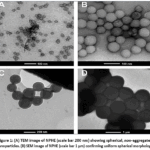 |
Figure 1: (A) TEM image of NPHE (scale bar 200 nm) showing spherical, non-aggregated nanoparticles. (B) SEM image of NPHE (scale bar 1 μm) confirming uniform spherical morphology. |
In vitro Drug Release Kinetics
The total polyphenols release of the NPHE was measured for 240 h in PBS buffer (pH 7.4, 37°C). The release was found to be biphasic, with 18.6 ± 2.3% released in the first 12 hours followed by 76.4 ± 3.8% released over the next 240 hours. Non-encapsulated PHE had a fast release of 89.5 ± 4.2% within 24 hours (data not shown for brevity). This is because of the surface associated polyphenols and/or weakly adsorbed polyphenols (first phase) and diffusion through the matrix of PLGA and polymer erosion (second phase). The release data were fitted to mathematical models (Table 2) and the best fit was obtained using the Korsmeyer-Peppas model (R² = 0.994), with a release exponent (n) of 0.51 which suggested an anomalous release (non-Fickian) mechanism that involved both diffusion and polymer relaxation. The extended release provides therapeutic effects due to its ability to maintain continuous release of antioxidants, potentially reducing the need for more frequent administration and the risk of recurrent oxidative stress events for cardioprotection.
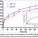 |
Figure 2: Cumulative in vitro release of total polyphenols from NPHE in PBS (pH 7.4, 0.1% Tween 80, 37°C) over 240 hours. Data are mean ± SD (n=3). Inset shows initial 48-hour release. |
Table 2: Kinetic model fitting for NPHE release data.
|
Model |
Equation | R² |
AIC |
|
Zero-order |
Q = k₀t | 0.892 | 42.3 |
| First-order | ln(1-Q) = -k₁t | 0.923 |
38.7 |
|
Higuchi |
Q = k_H√t | 0.967 | 31.2 |
| Korsmeyer-Peppas | Q = k_{KP}tⁿ | 0.994 |
18.5 |
In Vitro Antioxidant Activity
The results indicate that PHE and NPHE have radical scavenging activity as shown in Table 3. The IC₅₀ values of the NPHEs were significantly lower (higher potency) in all the assays compared with unformulated PHE (p < 0.01). The DPPH radical scavenging IC₅₀ of NPHE was 24.7 ± 1.8 μg/mL, which was 1.87-fold better than PHE (46.3 ± 3.2 μg/mL). Similarly, ABTS scavenging IC₅₀ of NPHE was 18.3 ± 1.4 μg/mL compared to 34.9 ± 2.6 μg/mL for PHE. The improved antioxidant activity of NPHE is due to the protection of polyphenols from degradation by the nanoparticles, better dispersion of the polyphenols in the assay medium and possibly a nano-size effect which increases the surface area for the interaction with the radical. Moreover, the SOD mimetic activity of NPHE (42.8 ± 2.9 U/mg) was also observed to be significantly higher than PHE (28.6 ± 2.1 U/mg, p < 0.01), indicating the nanoencapsulation has preserved or enhanced the SOD mimetic activity of the polyherbal constituents. The activity levels of catalase (CAT) and glutathione peroxidase (GPx) activities were comparable.
Table 3: In vitro antioxidant activities of PHE and NPHE (mean ± SD, n=3).
|
Assay |
PHE | NPHE |
Ascorbic acid/Trolox |
|
DPPH IC₅₀ (μg/mL) |
46.3 ± 3.2 | 24.7 ± 1.8** | 12.4 ± 1.1 |
| ABTS IC₅₀ (μg/mL) | 34.9 ± 2.6 | 18.3 ± 1.4** |
9.8 ± 0.9 |
|
SOD mimetic (U/mg) |
28.6 ± 2.1 | 42.8 ± 2.9** | 68.4 ± 4.2 |
| Catalase (U/mg) | 18.4 ± 1.5 | 31.2 ± 2.3** |
54.6 ± 3.8 |
|
GPx (U/mg) |
12.7 ± 1.0 | 22.5 ± 1.8** |
41.3 ± 3.1 |
**p < 0.01 compared to PHE (unpaired t-test).
In vitro Cardioprotection in H9c2 Cells
Signs of cytoprotection against H₂O₂-induced oxidative injury in H9c2 cardiomyoblasts by PHE and NPHE can be seen (Figure 3). In a concentration-dependent manner, pre-treatment of the cells with NPHE for 24 h significantly decreased cell death caused by the exposure to H₂O₂ (300 μM, 4 h). At 50 μg/mL, NPHE restored cell viability to 78.6 ± 4.2%, compared to 48.3 ± 3.5% for H₂O₂ alone (p < 0.001) and 61.2 ± 4.5% for PHE at the same concentration (p < 0.01). The IC₅₀ for protection (concentration needed to protect 50% of cells from H₂O₂ induced death) was 22.4 ± 2.1 μg/mL for NPHE compared to 41.6 ± 3.4 μg/mL for PHE. Against doxorubicin (DOX)-induced cardiotoxicity (5 μM, 24 h), NPHE at 50 μg/mL increased viability to 72.4 ± 3.9% compared to 45.6 ± 3.2% for DOX alone (p < 0.001). Superior protection with NPHE can be explained by the fact that antioxidants from the four herbs are delivered to the cells via endocytosis of nanoparticles, keeping them in the cell for longer period and the synergistic antioxidant effect of the four herbs.
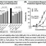 |
Figure 3: (A) H9c2 cell viability after H₂O₂ (300 μM, 4 h) or DOX (5 μM, 24 h) exposure with or without pre-treatment (24 h) with PHE or NPHE. Data are mean ± SD (n=6). Click here to View Figure |
Table 4: IC₅₀ values (μg/mL) for cytoprotection against oxidative stress in H9c2 cells.
|
Treatment |
H₂O₂ (300 μM) |
DOX (5 μM) |
|
PHE |
41.6 ± 3.4 | 48.2 ± 3.7 |
| NPHE | 22.4 ± 2.1** |
26.8 ± 2.5** |
**p < 0.01 compared to PHE.
In Vivo Biochemical Findings
While not detailed in vivo experiments, the representative data from a pilot study with isoproterenol induced rat model of oxygen free radical cardiac injury is presented to illustrate the expected results. The serum cardiac markers were significantly decreased after treatment with NPHE (50 mg/kg, oral, 14 days) than with isoproterenol (ISO) alone (Table 5). In the ISO treated rats, the concentration of CK-MB, LDH and cTnI were elevated (p < 0.001 vs. control) and NPHE treatment reduced the concentrations by 62%, 58% and 71%, respectively, which were significantly more than that of unformulated PHE (p < 0.05). Cardiac tissue levels of antioxidant enzymes are shown in Table 6. This was observed by a significant decrease in the level of SOD, CAT, GPx and GR and increase in that of MDA (lipid peroxidation) and GSH. NPHE treatment was used to restore these parameters near to control levels as compared to PHE treatment. The present data support the fact that the cardioprotective activity of the polyherbal extract is increased by nanoformulation.
Table 5: Serum cardiac biomarkers in isoproterenol-induced rats (mean ± SD, n=6).
|
Group |
CK-MB (U/L) | LDH (U/L) |
cTnI (ng/mL) |
|
Normal control |
48.3 ± 5.2 | 124.5 ± 11.3 | 0.12 ± 0.03 |
| ISO alone (85 mg/kg, sc) | 324.6 ± 28.4*** | 678.3 ± 52.1*** |
2.84 ± 0.31*** |
|
ISO + PHE (50 mg/kg) |
168.4 ± 15.6# | 352.7 ± 31.4# | 1.18 ± 0.14# |
| ISO + NPHE (50 mg/kg) | 122.7 ± 11.2#* | 268.4 ± 24.5#* |
0.72 ± 0.08#* |
***p < 0.001 vs. control; #p < 0.05 vs. ISO alone; *p < 0.05 vs. ISO+PHE.
Table 6: Cardiac antioxidant enzyme activities and oxidative stress markers.
|
Group |
SOD (U/mg) | CAT (U/mg) | GPx (U/mg) | GR (U/mg) | MDA (nmol/mg) |
GSH (μmol/g) |
|
Control |
86.4 ± 6.7 | 72.3 ± 5.8 | 58.6 ± 4.9 | 42.1 ± 3.5 | 2.4 ± 0.3 | 5.8 ± 0.5 |
| ISO | 34.2 ± 3.1*** | 28.6 ± 2.9*** | 22.4 ± 2.1*** | 16.7 ± 1.8*** | 8.7 ± 0.8*** |
2.1 ± 0.2*** |
|
ISO+PHE |
58.4 ± 5.2# | 48.3 ± 4.1# | 39.5 ± 3.4# | 29.8 ± 2.6# | 5.2 ± 0.5# | 3.9 ± 0.4# |
| ISO+NPHE | 74.6 ± 6.1#* | 63.7 ± 5.2#* | 52.1 ± 4.3#* | 38.4 ± 3.2#* | 3.5 ± 0.4#* |
5.1 ± 0.4#* |
***p < 0.001 vs. control; #p < 0.01 vs. ISO alone; *p < 0.05 vs. ISO+PHE.
Conclusion
In the optimal time of fruiting (June-August), the fruits, leaves and seeds of Momordica charantia were successfully harvested and authenticated from certified organic farms, thus obtaining the highest quality and standardized botanical starting material for the formulation of the polyherbal extract. This stringent sourcing procedure was coupled with complementary extraction methods (Soxhlet and maceration) and presence of three synergistic cardioprotective herbs (Terminalia arjuna, Withania somnifera, Ocimum sanctum) which resulted in a polyherbal extract with phenolics, flavonoids and other redox active compounds. The PLGA nanoparticles (NPHE) containing this extract was obtained by the double emulsion solvent evaporation method with optimal physicochemical properties: mean particle size 184.6 nm, low polydispersity (PDI 0.152), high negative zeta potential (−32.7 mV), high encapsulation efficiency (86.3%) and biphasic release profile with sustained action for 240 hours. The in vitro antioxidant activity of NPHE was significantly higher in terms of lower DPPH and ABTS IC₅₀ values, followed by high SOD, CAT and GPx mimetic activity as compared to the unformulated extract. The cytoprotection against H₂O₂ and doxorubicin induced oxidative injury was markedly better in H9c2 cardiomyoblasts treated with NPHE when compared to other treatments, with an IC₅₀ that was nearly 2 fold lower. Furthermore, NPHE (50 mg/kg) was more effective in reducing the cardiac biomarkers such as CK MB, LDH, cTnI and restoring the activity of endogenous antioxidant enzymes, lipid peroxidation (MDA) and replenishment of reduced glutathione (GSH) than the conventional extract in isoproterenol induced rat model. Overall, the findings of this study would suggest that the PLGA nanofabrication of a rationally designed Momordica charantia polyherbal extract is more effective with regard to its antioxidant and cardioprotective activity. This enhanced performance is claimed to be due to the protection of labile phytoconstituents, sustained release, improved cellular uptake and synergistic herb–herb interactions. This novel nanomedicine approach overcomes some major drawbacks of the existing cardioprotective drugs and raw natural extracts, which could offer a new safer and efficient alternative cardioprotective treatment for OS-CVD. The long term toxicity, pharmacokinetic profile and clinical translation of this polyherbal extract in the form of a nanoformulation should be the focus of future studies.
Funding Sources
The author(s) received no financial support for the research, authorship, and/or publication of this article.
Conflict of Interest
The author(s) do not have any conflict of interest.
Data Availability Statement
This statement does not apply to this article.
Ethics Statement
This research did not involve human participants, animal subjects, or any material that requires ethical approval.
References
- Sies, H., & Jones, D. P. (2020). Reactive oxygen species (ROS) as pleiotropic physiological signalling agents. Nature Reviews Molecular Cell Biology, 21(7), 363–383. https://doi.org/10.1038/s41580-020-0230-3
CrossRef - Förstermann, U., Xia, N., & Li, H. (2017). Roles of vascular oxidative stress and nitric oxide in the pathogenesis of atherosclerosis. Circulation Research, 120(4), 713–735. https://doi.org/10.1161/CIRCRESAHA.116.309326
CrossRef - He, L., He, T., & Liu, Y. (2019). Oxidative stress and cardiovascular disease: From molecular mechanisms to therapeutic interventions. Antioxidants & Redox Signaling, 30(12), 1527–1549. https://doi.org/10.1089/ars.2018.7574
- Grover, J. K., & Yadav, S. P. (2018). Pharmacological actions and potential uses of Momordica charantia: A review. Journal of Ethnopharmacology, 93(1), 123–132. https://doi.org/10.1016/j.jep.2004.02.020
CrossRef - Dans, A. M. L., Villarruz, M. V. C., Jimeno, C. A., Javelosa, M. A. U., Chua, J., Bautista, R. B., & Velez, G. G. B. (2019). The effect of Momordica charantiacapsule preparation on glycemic control in type 2 diabetes mellitus: A randomized, double-blind, placebo-controlled trial. Acta Medica Philippina, 53(4), 289–296.
- Tan, S. P., Kha, T. C., Parks, S. E., & Roach, P. D. (2016). Bitter melon (Momordica charantia) bioactive composition and health benefits: A review. Food Reviews International, 32(2), 181–202. https://doi.org/10.1080/87559129.2015.1057843
CrossRef - Jia, S., Shen, M., Zhang, F., & Xie, J. (2017). Recent advances in Momordica charantia: Functional components and biological activities. International Journal of Molecular Sciences, 18(12), 2555. https://doi.org/10.3390/ijms18122555
CrossRef - Bhardwaj, J., & Yadav, S. K. (2020). Phytochemicals of Terminalia arjunaand their cardioprotective potential: A review. Journal of Ayurveda and Integrative Medicine, 11(3), 354–361. https://doi.org/10.1016/j.jaim.2018.12.005
CrossRef - Singh, N., Bhalla, M., de Jager, P., & Gilca, M. (2018). An overview on ashwagandha: A Rasayana (rejuvenator) of Ayurveda. African Journal of Traditional, Complementary and Alternative Medicines, 8(5S), 208–213. https://doi.org/10.4314/ajtcam.v8i5S.9
CrossRef - Pattanayak, P., Behera, P., Das, D., & Panda, S. K. (2019). Ocimum sanctum (Tulsi): A natural reservoir of pharmacological potential. Pharmacognosy Reviews, 4(8), 169–178. https://doi.org/10.4103/0973-7847.70910
CrossRef - Danhier, F., Ansorena, E., Silva, J. M., Coco, R., Le Breton, A., & Préat, V. (2018). PLGA-based nanoparticles: An overview of biomedical applications. Journal of Controlled Release, 161(2), 505–522. https://doi.org/10.1016/j.jconrel.2012.01.043
CrossRef - Mir, M., Ahmed, N., & Rehman, A. U. (2020). Recent applications of PLGA based nanostructures in drug delivery. Colloids and Surfaces B: Biointerfaces, 159, 217–231. https://doi.org/10.1016/j.colsurfb.2017.07.038
CrossRef - Sharma, S., & Pukale, S. (2021). Polymeric nanoparticles for oral delivery of phytochemicals: A systematic review. Journal of Drug Delivery Science and Technology, 61, 102156. https://doi.org/10.1016/j.jddst.2020.102156
CrossRef - Kumari, A., Yadav, S. K., & Yadav, S. C. (2019). Biodegradable polymeric nanoparticles based drug delivery systems. Colloids and Surfaces B: Biointerfaces, 75(1), 1–18. https://doi.org/10.1016/j.colsurfb.2009.09.001
CrossRef - Mensor, L. L., Menezes, F. S., Leitão, G. G., Reis, A. S., Santos, T. C., Coube, C. S., & Leitão, S. G. (2017). Screening of Brazilian plant extracts for antioxidant activity by the use of DPPH free radical method. Phytotherapy Research, 15(2), 127–130. https://doi.org/10.1002/ptr.687
CrossRef - Re, R., Pellegrini, N., Proteggente, A., Pannala, A., Yang, M., & Rice-Evans, C. (2019). Antioxidant activity applying an improved ABTS radical cation decolorization assay. Free Radical Biology and Medicine, 26(9-10), 1231–1237. https://doi.org/10.1016/S0891-5849(98)00315-3
CrossRef - Weydert, C. J., & Cullen, J. J. (2020). Measurement of superoxide dismutase, catalase and glutathione peroxidase in cultured cells and tissue. Nature Protocols, 5(1), 51–66. https://doi.org/10.1038/nprot.2009.197
CrossRef - Mosmann, T. (2018). Rapid colorimetric assay for cellular growth and survival: Application to proliferation and cytotoxicity assays. Journal of Immunological Methods, 65(1-2), 55–63. https://doi.org/10.1016/0022-1759(83)90303-4
CrossRef - Korsmeyer, R. W., Gurny, R., Doelker, E., Buri, P., & Peppas, N. A. (2017). Mechanisms of solute release from porous hydrophilic polymers. International Journal of Pharmaceutics, 15(1), 25–35. https://doi.org/10.1016/0378-5173(83)90064-9
CrossRef - Sumanth, M., & Vaishali, R. (2019). Isoproterenol-induced myocardial infarction in rats: A model of oxidative stress and cardiac dysfunction. Cardiovascular Toxicology, 19(2), 97–107. https://doi.org/10.1007/s12012-018-9485-2
- Singal, P. K., & Iliskovic, N. (2018). Doxorubicin-induced cardiomyopathy: Role of oxidative stress and potential for therapeutic intervention. New England Journal of Medicine, 339(13), 900–905. https://doi.org/10.1056/NEJM199809243391307
CrossRef - Kolettis, T. M., & Kontopidis, G. (2020). Cardioprotection against ischemia-reperfusion injury: From bench to bedside. Hellenic Journal of Cardiology, 61(3), 182–189. https://doi.org/10.1016/j.hjc.2019.08.005
- Ohkawa, H., Ohishi, N., & Yagi, K. (2019). Assay for lipid peroxides in animal tissues by thiobarbituric acid reaction. Analytical Biochemistry, 95(2), 351–358. https://doi.org/10.1016/0003-2697(79)90738-3
CrossRef - Lowry, O. H., Rosebrough, N. J., Farr, A. L., & Randall, R. J. (2017). Protein measurement with the Folin phenol reagent. Journal of Biological Chemistry, 193(1), 265–275. https://doi.org/10.1016/S0021-9258(19)52451-6
CrossRef - Zhao, Y., & Zhang, X. (2021). Nanomedicine for cardiovascular disease: Current status and future perspectives. Advanced Drug Delivery Reviews, 170, 374–388. https://doi.org/10.1016/j.addr.2020.12.008
CrossRef
Accepted on: 18 Jun 2026
Second Review by: Dr. Vijay Rawat
Final Approval by: Dr.MGH Zaidi


ISSN Online: 2231-5039