Nanoformulated Curcumin for Enhanced Cardioprotective and Antioxidant Activity Against Oxidative Stress
 , Jyotiprava Bal2, Akshay Kumar Sinha3, Himanshi Rathaur4, Mohit Kumar5, Shruti Anil Adhau6, Nitin Sukadev Hivale7, Rashmi Sharma8 and N. G. Raghavendra Rao9*
1Pharmacy Academy, Faculty of Pharmacy, IFTM University, Lodhipur Rajput, Delhi Road, Moradabad, Uttar Pradesh, India,
2Keonjhar institute of medical science and research, Keonjhar, Odisha, India.
3Research Scholar, Apex University, Jaipur, Rajasthan, India.
4School of Pharmaceutical Sciences, Swami Rama Himalayan University, jollygrant, Dehradun, Uttrakhand, India.
5Teerthanker Mahaveer College of Pharmacy, eerthanker Mahaveer University; Moradabad , Uttar Pradesh, India.
6Nagpur College Of Pharmacy, Wanadongari, Hingna Road, Nagpur, Maharashtra, India.
7Department of Pharmacology, Symbiosis Medical College for Women (SMCW), Symbiosis International (Deemed University) (SIU), Pune. Maharashtra, India.
8Shri Vaishnav Institute Of Paramedical Sciences, Shri Vaishnav Vidyapeeth Vishwavidyalaya, Indore-Ujjain Road , Gram Baroli, Indore, Madhya Pradesh India.
9Department of Pharmaceutics, Parul Institute of Pharmacy, Parul University, P.O. Limda, Tal. Waghodia, Vadodara-Gujarat, India,
Corresponding Author E-mail: drngraghu@gmail.com
Download this article as:
ABSTRACT:Cardiovascular disease (CVD) remains the leading global cause of death, with oxidative stress as a central pathogenic driver. Curcumin, a natural polyphenol from Curcuma longa, exhibits potent antioxidant and cardioprotective properties through direct ROS scavenging, Nrf2 activation, and NF‑κB inhibition. However, its clinical translation is severely limited by poor aqueous solubility, rapid metabolism, and extremely low oral bioavailability. This study aimed to develop and evaluate nanoformulated curcumin using four different nanocarrier systems—PLGA nanoparticles (NPs), solid lipid nanoparticles (SLNs), liposomes, and nanoemulsions—to overcome these barriers and enhance cardioprotection against oxidative stress. All formulations were characterized for particle size (82–216 nm), zeta potential (−15 to −32 mV), encapsulation efficiency (>85%), and sustained in vitro release over 72 h. Based on overall performance, PLGA NPs were selected for further biological studies. In H9c2 and AC16 cardiomyocytes subjected to H₂O₂, doxorubicin, or oxygen‑glucose deprivation/reoxygenation (OGD/R), nanoformulated curcumin (Nano-Cur, 10 µM curcumin equivalent) significantly improved cell viability (85–90% vs. 45–50% in stressed controls) and reduced LDH release and trypan blue uptake compared to free curcumin. Nano-Cur also lowered intracellular ROS (DCFH‑DA and DHE fluorescence) by 70%, restored SOD, catalase, and GPx activities, and normalized the GSH/GSSG ratio. In an in vivo rat model of myocardial ischemia/reperfusion (I/R), intravenous Nano-Cur (5 mg/kg) reduced infarct size by 70% (to 14.2%), decreased serum troponin I by 80%, preserved left ventricular ejection fraction (72% vs. 48% in vehicle), and attenuated MDA, protein carbonyls, and 8‑oxodG levels. Mechanistically, Nano-Cur enhanced nuclear Nrf2 translocation and suppressed NF‑κB activation, leading to upregulation of HO‑1 and NQO1 and downregulation of TNF‑α and IL‑1β. These findings demonstrate that nanoformulation dramatically improves curcumin’s pharmacokinetic and pharmacodynamic profile, enabling robust cardioprotection against oxidative stress‑induced injury. Nanotechnology‑based delivery of curcumin represents a promising therapeutic strategy for ischemic heart disease, doxorubicin cardiotoxicity, and other oxidative cardiovascular disorders.
KEYWORDS:Curcumin; Cardioprotection; Ischemia/reperfusion; Nanoformulation; Oxidative stress
Introduction
Cardiovascular disease (CVD) is the largest cause of morbidity and mortality globally – the World Health Organisation estimates that it causes nearly 18 million deaths each year. This is a very general group that includes coronary artery disease, myocardial infarction, heart failure, hypertensive heart disease, and stroke, which all have a common pathogenesis: marked imbalance between the generation of reactive oxygen species (ROS) and the endogenous antioxidant defense mechanisms. Oxidative stress has become a key mechanistic step in the initiation and progression of cardiac dysfunction, and is considered a key mechanism in cardiac dysfunction and disease. Several factors make the heart especially susceptible to oxidative stress: its very high metabolic rate, dense population of mitochondria and comparatively low capacity for regeneration. In physiological conditions, low-to-moderate concentrations of ROS are considered as second messengers for cellular signaling, affecting processes including cardiomyocyte contractility, hypertrophy and preconditioning. Oxidative stress, however, can cause a chain reaction of harmful reactions that involves the peroxidation of lipids, the carbonylation of proteins, the breakage of DNA strands and the activation of pro-apoptotic pathways resulting from the production of ROS exceeding the antioxidant enzyme buffering capacity (superoxide dismutase (SOD), catalase and glutathione peroxidase (GPx) enzymes). All major cardiovascular risk factors (hyperlipidaemia, diabetes, hypertension, smoking and environmental pollutants) converge on a common pathway in which NADPH oxidase activity is increased, the electron transport chain in mitochondrial is compromised and reduced glutathione levels are depleted. Reperfusion paradoxically increases ROS production, which results in cardiomyocyte death, sterile inflammation, and ventricular remodeling. Morphologically, high levels of oxidative damage markers (malondialdehyde [MDA], 4-hydroxynonenal [4-HNE] and oxidized low-density lipoprotein [oxLDL]) are associated with poor outcomes following acute coronary syndromes. In contrast, low endogenous antioxidants are independent predictors of progression of heart failure. Nevertheless, decades of research have failed to show consistent clinical benefits in large-scale trials with conventional antioxidant therapy, such as vitamin E, vitamin C and beta-carotene, possibly because these compounds are poorly targeted to the tissue, do not achieve an appropriate concentration within cells, and are unable to target the multi-faceted character of oxidative signalling. This therapeutic gap has led to an important research effort on the identification of polyphenolic compounds from natural sources that have pleiotropic antioxidant and anti-inflammatory activity. Of these, curcumin, the golden yellow pigment from the rhizome of Curcuma longa (Turmeric), has shown great promise as a cardioprotective agent, but has a drawback that nanotechnology is now set to address.
Potent natural antioxidant curcumin
Turmeric is the main ingredient in the traditional Chinese and Ayurvedic medicine that has been used for thousands of years and contains one of its most bioactive components, curcumin, also known as diferuloylmethane and C₂₁H₂₀O₆. It has a distinct molecular structure with two methoxylated phenolic rings joined by a seven-carbon chain with an α,β-unsaturated β-diketo group that is responsible for its amazing antioxidant activity. Such a setup allows curcumin to be a classical hydrogen atom donor, reacting with a broad range of reactive species such as superoxide anion (O₂•⁻), hydroxyl radical (•OH), peroxynitrite (ONOO⁻) and singlet oxygen (¹O₂) with reaction rate constants that reach diffusion limits. Perhaps more important, curcumin has indirect antioxidant activity by activating the nuclear factor erythroid 2-related factor 2 (Nrf2) pathway. Basally, Nrf2 is bound to its inhibitor Kelch-like ECH-associated protein 1 (Keap1) and is degraded by the ubiquitin-dependent proteasome. Curcumin alters critical cysteine thiols of Keap1 which causes the release, nuclear translocation, and binding to the antioxidant response element (ARE) of a number of over 200 cytoprotective genes in the promoter regions. These include heme oxygenase-1 (HO-1), NAD(P)H:quinone oxidoreductase 1 (NQO1), γ-glutamylcysteine synthetase (the rate-limiting enzyme for glutathione synthesis), and thioredoxin reductase. In addition to Nrf2, curcumin strongly suppresses the nuclear factor kappa B (NF-κB) pathway, a master transcription factor for pro-inflammatory cytokines (TNF-α, IL-1β, IL-6), adhesion molecules (VCAM-1, ICAM-1) and inducible nitric oxide synthase (iNOS). The inhibitory mechanism is by inhibition of IκB kinase (IKK) activity that stops the IκBα from being phosphorylated and degraded, and thus prevents NF-κB nuclear entry. Other cardioprotective effects include activation of AMP-activated protein kinase (AMPK), restoration of mitochondrial membrane potential, up-regulation of sirtuin-1 (SIRT1), and direct chelation of transition metal ions (iron and copper) which promote Fenton chemistry. In isolated cardiomyocytes, pretreatment with curcumin (5–20 μM) protects against cell death caused by hydrogen peroxide treatment, preserves mitochondrial respiration and maintains intracellular ATP level, more than 60%. In rodent models of ischemia/reperfusion curcumin prevents up to 40% reduction in infarct size, as well as preserving left ventricular ejection fraction and decreasing pathological hypertrophy. Likewise, when doxorubicin is used as a model of oxidative myocardial injury, curcumin co-administration prevents reduction in SOD and GPx activities, decreases cardiac MDA levels, and restores contractile dysfunction. Such promising preclinical data have rendered curcumin as one of the most promising nutraceutical candidates for cardiovascular prevention and therapy. But making this promise a reality in the clinic has been significantly constrained by a series of daunting pharmacokinetic challenges.
Bioavailability Challenge of Native Curcumin
Native curcumin has a very high antioxidant activity in vitro, but it has two major drawbacks: very low bioavailability in the systemic circulation and very weak efficacy in clinical trials. Curcumin is extensively metabolized in the presystemic phase in the gut and liver after oral administration. The molecule is hydrophobic (log P = 3.2) and practically insoluble in aqueous buffers at acidic or neutral pH (solubility ~11 ng/mL at pH = 7.4) resulting in poor dissolution in gastrointestinal fluids. Consequently, most of the curcumin that is taken orally is not absorbed and passes through the stool. Only a small amount of curcumin enters the enterocytes and is quickly converted to curcumin glucuronide by UDP-glucuronosyltransferases (UGTs) and to curcumin sulfate by sulfotransferases (SULTs). These phase II metabolites are non-radical scavenging metabolites because they do not contain any free phenolic hydroxyl group and are then transported to the liver where further conjugation takes place. No matter how much curcumin is taken orally (up to 10-12 g), a peak plasma concentration of 10-50 ng/mL (approx. 25-125 nM) of free (unconjugated) curcumin is consistently observed in humans, well below the 5-20 μM range needed to induce significant Nrf2 activation or direct ROS scavenging in vitro. The elimination half-life of curcumin is also brief (1-2 hours), which also means that it must be given in high doses several times a day, and this can lead to issues of tolerability (mild nausea, diarrhea, and yellow stool). The bioavailability can be modestly increased by co-administration with piperine, an alkaloid found in black pepper which inhibits glucuronidation, resulting in reported 10- to 20-fold increases, but this is still not enough to reach plasma levels in the micromolar range. The alternative strategies, including the lipidic formulations (complexes of curcumin-phosphatidylcholine), microemulsions, and micronized powders are incremental but do not overcome the basic problems of rapid metabolism, insufficient uptake by cells, and absence of targeted delivery to cardiac tissue. Thus, the therapeutic benefits of native curcumin for cardiovascular disease have largely not been realized and numerous placebo-controlled trials with negative or mixed results have been attributed to insufficient exposure of cardiomyocytes to curcumin. This lack of successful clinical translation from effective preclinical efficacy has ignited the development of nanocarrier based delivery systems that not only aim to prevent degradation of curcumin in biological systems but also improve its bioavailability, extend its circulation time and target the drug either passively to the heart or actively to the heart.
Reactive Oxygen Species (ROS) Sources in the Heart
To understand the cardioprotective effects of nanoformulated curcumin, it is necessary to understand the sources of ROS in cardiac tissue, both enzymatic and non-enzymatic. There are multiple large systems in the heart which generate ROS that are pathologically up-regulated during stress. Under normal and disease conditions, superoxide is a major product of the mitochondrial electron transport chain (ETC). During normal oxidative phosphorylation, approximately 0.1–0.5% of electrons “leak” from complexes I and III to reduce molecular oxygen directly to superoxide. Ischemia leads to a collapse of the mitochondrial membrane potential and accumulation of reduced CoQ, thus a significant increase of superoxide production in reperfusion, through increased reverse electron transport in complex I. The second, and more important source, is the NADPH oxidase (NOX) family, specifically NOX2 and NOX4 isoforms. NOX2 is highly expressed in cardiomyocytes and endothelial cells and it is stimulated by angiotensin II, mechanical stretch and inflammatory cytokines, producing superoxide in the cytoplasm and at the plasma membrane. Under ischemic conditions, xanthine dehydrogenase can convert to xanthine oxidase (XO) which will reduce oxygen to superoxide and hydrogen peroxide and convert hypoxanthine to xanthine and uric acid. The endothelial nitric oxide synthase (eNOS) is uncoupled by the oxidation of its cofactor, tetrahydrobiopterin (BH4), to produce superoxide instead of nitric oxide (NO), a phenomenon known as “eNOS uncoupling” that causes endothelial dysfunction. Further, cytochrome P450 enzymes, lipoxygenases, and cyclooxygenases (especially from the COX-2) also play a role in the production of ROS in inflamed cardiac tissue. Nanoformulated curcumin can inhibit multiple targets of ROS generation: NAOX2 assembly, restore BH4 levels by maintaining the activity of GTP cyclohydrolase I; decrease mitochondrial complex I reverse electron transport by metabolic remodelling through AMPK; and downregulate XO expression through Nrf2-mediated transcriptional repression. The activity of free curcumin to modulate these sources is substantially reduced in vivo due to its rapid clearance prior to reaching the myocardium. In fact, in electron paramagnetic resonance (EPR) spin trapping studies of rodent hearts, the curcumin loaded nanoparticles (e.g., PLGA, liposomal or solid lipid) sustain release of curcumin into the heart and inhibit all ROS sources in a dose-dependent manner.
Lipid Peroxidation and Membrane
The next important effect of excessive ROS generation is lipid peroxidation, a free radical chain reaction that causes damage to the polyunsaturated fatty acids (PUFAs) found in membranes of organelles and cells. The heart is very high in mitochondria, and the mitochondria possess a very high concentration of cardiolipin, a tetra-acylated diphosphatidylglycerol extremely susceptible to peroxidation. The formation of a carbon-cented lipid radical (L•) by an abstraction of a hydrogen atom from a bis-allylic methylene group of a PUFA quickly rearranges to a conjugated diene that then reacts with molecular oxygen to generate a lipid peroxyl radical (LOO•). This peroxyl radical can then steal the hydrogen atom from nearby PUFA, thus continuing the chain reaction, resulting in the damage of hundreds of lipid molecules until the chain is broken by chain-breaking antioxidants like vitamin E or curcumin. These major end products are MDA and 4-HNE, which are highly reactive electrophiles that can covalently modify proteins and DNA. The myocardium which is subjected to ischemic stress followed by reperfusion shows a 5–10-fold increase in cardiac MDA within minutes of reperfusion, which coincides with the loss of membrane fluidity, inactivation of Na+/K+-ATPase and Ca2+-ATPase, and disruption of the integrity of the inner membrane of mitochondria. Cardiolipin peroxidation is especially harmful because normally cardiolipin maintains the architecture of the electron transport chain supercomplexes that, when oxidized, release cytochrome c into the cytosol, which initiates the intrinsic apoptotic pathway. Nanoformulated curcumin, which has a longer half-life and can penetrate the double membranous structure of mitochondria, is able to directly interrupt the chain reaction of lipid peroxidation by abstracting hydrogen atoms from the lipid peroxyl radicals, stabilizing them with a curcumin phenoxyl radical which do not propagate further. In a comparative study, curcumin-loaded nanostructured lipid carriers (NLCs) significantly lower the MDA levels in the heart of rats after post-ischemic compared to free curcumin at the same dose (50 mg/kg) which resulted in only 22% reduction. In addition, nanoencapsulation maintained the ratio of reduced to oxidized glutathione (GSH/GSSG) in the mitochondrial matrix, indirectly affecting the activity of glutathione peroxidase 4 (GPx4) which specifically reduces lipid hydroperoxides in membranes. The dual direct and indirect action makes this nanoformulated curcumin very effective in blocking the autopropagating damage caused by lipid peroxidation in the ischemic and failing heart.
Protein Oxidation and Mitochondrial Dysfunction.
Oxidative stress also causes a great deal of damage to cardiac proteins, as manifested by loss of enzymatic activity, unfolding, aggregation, and mislocalization. The main ones are cysteine thiol oxidation (to sulfenic, sulfinic, or sulfonic acids), methionine sulfoxide formation, tyrosine nitration (mediated by peroxynitrite), and carbonylation of lysine residues, arginine residues, and proline residues. Proteins that are most susceptible in the heart include proteins associated with excitation-contraction coupling, energy metabolism and mitochondrial dynamics. The calcium reuptake velocity of the sarcoplasmic reticulum Ca²⁺-ATPase (SERCA2a) is important and is impaired by its oxidation, which is a feature of heart failure with preserved ejection fraction (HFpEF). Under oxidative stress, the ryanodine receptor (RyR2) is S-glutathionylated resulting in a leak through the receptor during diastole, which results in arrhythmias. Mitochondrial proteins, including succinate dehydrogenase, aconitase and complexes I–V, are particularly susceptible to oxidative inactivation; aconitase has a [4Fe-4S] cluster that is readily oxidised causing complex inactivation and build-up of aconitase’s substrate (citrate) which inhibits fatty acid oxidation. Furthermore, the mitochondrial permeability transition pore (mPTP) is redox-sensitive and requires cyclophilin D to allow its opening, and thus uncontrolled entry of solutes and water into the mitochondria leading to swelling, outer membrane rupture, and pro-apoptotic factor release, occur with prolonged oxidative stress. Nanoformulated curcumin prevents these protein oxidative modifications by three different mechanisms. First, it scavenges peroxynitrite and hydroxyl radicals, thus preventing the nitration of critical proteins such as MnSOD, which is inactivated by nitration. Second, curcumin nanoparticles activate the proteasomal and autophagic clearance pathways by Nrf2-dependent transcription of p62/SQSTM1 and NQO1, preventing the accumulation of proteins in toxic inclusions that may cause impairment of the brain’s inflammatory response or other neurological functions. Third, curcumin is a protein carbonylation inhibitor, which directly competes with aldehydes derived from lipids (MDA and 4-HNE). In the doxorubicin-induced cardiotoxicity model, treatment with curcumin-loaded solid lipid nanoparticles (SLN) resulted in 84% cardiac protein carbonyl reduction versus untreated controls, whereas free curcumin only achieved a 31% reduction. Importantly, only the nanoformulation group showed preservation of mitochondrial complex I and V activities, which were related to the maintenance of ATP levels and preserved contractile function. The increased delivery to the myocardium provided by nanocarriers is critical to get the intratissue concentrations of curcumin necessary to prevent protein oxidation and preserve mitochondrial bioenergetics.
DNA Damage and Apoptotic Pathways.
The continuous damage to the genomic and mitochondrial DNA inevitably results in genomic and mitochondrial damage and, if not repaired, will result in programmed cell death. A significant disadvantage of the terminally differentiated cardiomyocyte is the lack of cell division to replace cells damaged by DNA damage. In highly oxidative environment of the nucleus and mitochondria 8-oxo-7,8-dihydroguanine (8-oxodG) is the most common lesion in DNA; if not repaired by 8-oxoguanine DNA glycosylase (OGG1), it mispairs with adenine during DNA replication or transcription, yielding G→T transversions. Poly(ADP-ribose) polymerase 1 (PARP-1) activation is the more immediate effect in post-mitotic cardiomyocytes. PARP-1 is able to bind to single strand breaks, polymerize the chains of poly(ADP-ribose) (PAR) from NAD⁺, and consume cellular NAD⁺ and ATP, causing energy failure, a process referred to as PARP-dependent cell death (parthanatos). Furthermore, the severe mtDNA damage will disrupt transcription of mtDNA-encoded ETC subunits, thus leading to additional ROS production in a vicious cycle. If the damage to the DNA is not being repaired, the tumor suppressor protein p53 is activated and can translocate to the mitochondria and bind directly to anti-apoptotic proteins Bcl-xL and Bcl-2, leading to outer membrane permeabilization. This uncloaks cytochrome c and second mitochondria-derived activator of caspases (Smac/DIABLO) and triggers caspase-9 and caspase-3 in the apoptosome complex. Nanoformulated curcumin interferes with each step of this pathway. At the DNA level, curcumin nanoparticles directly acts on the nucleus to provide the parent compound, before hydroxyl radicals reach the DNA helix, reducing the formation of 8-oxodG. Curcumin also increases the expression of OGG1 through the Nrf2-ARE signaling pathway, which increases the base excision repair ability. Importantly, nanoformulated curcumin is able to suppress PARP-1 hyperactivity by keeping the levels of NAD+ up—an effect that cannot be obtained with free curcumin as it is rapidly glucuronidated and therefore unable to maintain high concentrations in the cardiomyocyte nucleus. In an animal model of myocardial infarction, curcumin loaded polymeric nanoparticles (nano-Cur) decreased cardiac 8-oxodG level by 71%, decreased PAR accumulation by 63% and prevented the decrease in the ratio of NAD+/NADH. TUNEL assays showed that the apoptotic cardiomyocytes were 5-fold less than the free curcumin treatment. Furthermore, curcumin nanoparticles inhibited p53 mitochondrial translocation, maintained Bcl-2/Bax ratio, and inhibited the activation of the caspase-3 cleavage. These data show that to overcome the limiting concentrations in the nucleus and mitochondria, the use of nanocarrier mediated delivery is essential for curcumin to intercept DNA damage and interrupt apoptotic signaling pathways in the stressed heart
Endothelial dysfunction and inflammatory
The last element of the oxidative stress axis in cardiovascular disease is endothelial dysfunction, which is defined as decreased nitric oxide (NO) bioavailability, increased vascular permeability, and expression of leukocyte adhesion molecules. Under normal conditions, coronary endothelium is a single-cell layer that regulates vascular tone, blood clotting and regulates the migration of inflammatory cells. In an oxidative stress environment, superoxide generated by NOX2 and XO reacts very quickly with NO (rate constant ~10¹⁰ M⁻¹s⁻¹) to produce peroxynitrite that uncouples any remaining eNOS. This leads to a reduction in NO, which leads to an increase in platelet aggregation, upregulation of endothelin-1 and vasoconstriction. At the same time, oxidized phospholipids and advanced glycation end products (AGEs) stimulate NF-κB in endothelial cells, leading to the expression of VCAM-1 and ICAM-1, which capture circulating monocytes and neutrophils. Myeloperoxidase and more ROS are released by these activated leukocytes leading to a vicious circle of oxidative injury and inflammation, which leads to atherosclerotic plaque formation, microvascular obstruction, and no-reflow phenomenon after myocardial infarction. Nanoformulated curcumin has potent protective effects on the coronary endothelium. Curcumin-loaded liposomes maintained the eNOS dimerization and enhanced the production of NO by 3.5-fold, as determined by a fluorescence-based NO probe compared to the free curcumin in the human coronary artery endothelial cells with hydrogen peroxide. The mechanism includes curcumin-mediated restoration of BH4 by Nrf2-dependent upregulation of GTP cyclohydrolase I and blocking of NOX2 activity in which p47phox fails to translocate to the membrane. Intravenous curcumin nanoparticles (5 mg/kg, weekly) decreased aortic VCAM-1 expression by 80% and circulating levels of both TNF-α and IL-6 by 60–70%, and enhanced endothelium-dependent vasodilation of Acetylcholine by over 2-fold in a diet induced hypercholesterolemia animal model. Histological examination showed decreased infiltration of macrophages in the myocardium and decreased expression of matrix metalloproteinase-9 (MMP-9), which plays a role in adverse extracellular matrix remodeling and progression of heart failure. Importantly, the anti-inflammatory effects did not rely directly on the ability of the curcumin to scavenge the radicals, as the anti-inflammatory activity of the curcumin nanoparticles was also seen to suppress the NLRP3 inflammasome in cardiac macrophages, which resulted in reduced release of IL-1β and IL-18. Clinically, this immunomodulatory effect is important as high IL-1β is associated with recurrent myocardial infarction and hospitalization for heart failure. Therefore, the nanoformulated curcumin has the capacity to interrupt the vicious cycle of oxidative stress, endothelial dysfunction and sterile inflammation, which is central to the process of advancing from an initial cardiac insult to chronic heart failure. Taken together the above sections highlight the fact that native curcumin has an outstanding biochemical potential against all of the major pathways of oxidative cardiovascular disease; however, this potential cannot be realized without overcoming bioavailability problems, which requires nanoformulation. Finally, to scavenge ROS, preserve mitochondrial function, prevent DNA damage and restore endothelial health, curcumin needs to be delivered to cardiac tissue in sufficient concentrations, which is only possible by the help of nanotechnology.
Materials and Methods
Curcumin (diferuloylmethane, purity ≥95% by HPLC) was obtained from Sigma-Aldrich (St. Louis, MO, USA) or alternatively from Sabinsa Corporation (East Windsor, NJ, USA), and was used without further purification. For the preparation of nanoformulations, polymer/lipid components were selected based on the desired nanocarrier type: poly(D,L-lactide-co-glycolide) (PLGA, 50:50, inherent viscosity 0.55–0.75 dL/g) was sourced from Evonik Industries (Essen, Germany), low molecular weight chitosan (50–190 kDa, deacetylation degree ≥75%) from Sigma-Aldrich, 1,2-distearoyl-sn-glycero-3-phosphocholine (DSPC) from Avanti Polar Lipids (Alabaster, AL, USA), and stearic acid (≥98%) from Tokyo Chemical Industry (TCI, Tokyo, Japan). Surfactants and stabilizers included Poloxamer 188 (BASF, Ludwigshafen, Germany), Tween® 80 (Merck, Darmstadt, Germany), and soybean lecithin (Lipoid S75, Lipoid GmbH, Ludwigshafen, Germany). All organic solvents—dimethyl sulfoxide (DMSO, ≥99.9%), absolute ethanol (HPLC grade), acetone, and dichloromethane—were purchased from Thermo Fisher Scientific (Waltham, MA, USA). Aqueous buffers were prepared using ultrapure water (18.2 MΩ·cm): phosphate-buffered saline (PBS, pH 7.4, 10 mM), acetate buffer (pH 4.5, 50 mM), and citrate buffer (pH 3.0). Oxidative stress inducers comprised hydrogen peroxide (H₂O₂, 30% w/w solution, Sigma), doxorubicin hydrochloride (≥98%, Selleck Chemicals, Houston, TX, USA), and a ferrous iron/ascorbate system (FeSO₄·7H₂O plus L-ascorbic acid, both from Sigma). For molecular and biochemical analyses, primary antibodies against Nrf2 (rabbit monoclonal, #12721), NF-κB p65 (#8242), and HO-1 (#82206) were obtained from Cell Signaling Technology (Danvers, MA, USA); secondary HRP-conjugated antibodies were from Santa Cruz Biotechnology (Dallas, TX, USA). Commercial assay kits included the MDA (malondialdehyde) colorimetric/fluorometric kit (Abcam, ab118970), the 8-hydroxy-2′-deoxyguanosine (8-oxodG) ELISA kit (Trevigen, 4380-096-K), the GSH/GSSG ratio assay (Cayman Chemical, 703002), the SOD activity kit (Sigma, 19160), and the catalase fluorometric kit (Thermo Fisher, EIACAT). Cell culture reagents were obtained from Gibco (Thermo Fisher): Dulbecco’s Modified Eagle Medium (DMEM, high glucose, with L-glutamine and sodium pyruvate, #11995065), fetal bovine serum (FBS, heat-inactivated, #16140071), penicillin‑streptomycin (10,000 U/mL, #15140122), and trypsin‑EDTA (0.25%, #25200056). All reagents were of analytical or cell-culture grade, stored according to manufacturer specifications, and used within their stated expiry periods.
Preparation of Nanoformulated Curcumin
The selection of nanoformulation type was guided by the desired physicochemical properties and route of administration. Four distinct nanocarriers were prepared: polymeric nanoparticles (PLGA), solid lipid nanoparticles (SLNs), liposomes, and nanoemulsions. For PLGA nanoparticles, the nanoprecipitation/solvent evaporation method was employed: curcumin and PLGA (1:10 w/w) were dissolved in acetone/dichloromethane (1:1 v/v), and the organic phase was added dropwise to an aqueous solution containing Poloxamer 188 (0.5% w/v) under magnetic stirring (600 rpm) at room temperature. The organic solvents were then evaporated under reduced pressure. SLNs were synthesized using hot homogenization followed by ultrasonication: stearic acid (lipid phase) and curcumin (drug-to-lipid ratio 1:15 w/w) were heated to 70 °C, then dispersed into a hot aqueous solution of Tween 80 (1% w/v) using a high-shear homogenizer (Ultra-Turrax, 10,000 rpm, 5 min), followed by probe ultrasonication (40% amplitude, 10 min) and cooling to 4 °C for solidification. Liposomes were prepared by thin‑film hydration: DSPC, cholesterol, and curcumin (molar ratio 10:5:1) were dissolved in chloroform/methanol (2:1), the solvent was evaporated under rotary vacuum to form a thin film, then hydrated with PBS (pH 7.4) at 55 °C for 1 h, followed by extrusion through polycarbonate membranes (200 nm). Nanoemulsions were produced by high‑pressure microfluidization: an oil phase (medium‑chain triglycerides containing curcumin) and an aqueous phase (lecithin and Poloxamer 188) were premixed with a rotor‑stator homogenizer, then passed through a microfluidizer (Microfluidics M-110P) at 20,000 psi for 5 cycles. After synthesis, all formulations were subjected to removal of free (unencapsulated) curcumin and residual organic solvents: dialysis against PBS (MWCO 12–14 kDa) for 24 h (PLGA nanoparticles and liposomes), centrifugal filtration (Amicon Ultra‑4, 10 kDa) at 4,000 × g for 30 min (SLNs), or a combination of dialysis followed by lyophilization (nanoemulsions) using 5% sucrose as a cryoprotectant. All final formulations were stored at 4 °C for further characterization.
Physicochemical Characterization of Curcumin Nanoparticles
Physicochemical characterization of all nanoformulations was performed as follows. Particle size (hydrodynamic diameter) and polydispersity index (PDI) were measured by dynamic light scattering (DLS) using a Zetasizer Nano ZS (Malvern Panalytical) after appropriate dilution in filtered PBS, with each measurement in triplicate at 25 °C. Zeta potential was determined by electrophoretic light scattering using the same instrument. Morphological analysis was conducted using transmission electron microscopy (TEM, JEM‑1400Plus, JEOL) at 80 kV after negative staining with 1% uranyl acetate, and scanning electron microscopy (SEM, JSM‑IT800, JEOL) following gold sputter coating. Encapsulation efficiency (EE%) and drug loading (DL%) were determined by separating free curcumin via ultracentrifugation (20,000 × g, 4 °C, 30 min); the pelleted nanoparticles were dissolved in DMSO, and curcumin content was quantified by UV‑Vis spectrophotometry at λ = 425 nm (or by HPLC with a C18 column and acetonitrile/water mobile phase). In vitro release profiles were obtained using the dialysis bag method: samples suspended in PBS (pH 7.4, containing 0.1% Tween 80 to maintain sink conditions) were placed in dialysis membranes (MWCO 12–14 kDa) and incubated at 37 °C with shaking; aliquots were withdrawn at predetermined time points (0, 1, 2, 4, 6, 8, 12, 24, 48, and 72 h) and replaced with fresh buffer, and released curcumin was quantified by UV‑Vis. Stability studies included storage stability (samples stored at 4 °C, 25 °C, and 37 °C for 1, 2, and 4 weeks, monitoring size, PDI, and EE%) and colloidal stability in biological media (incubation in PBS and 50% fetal bovine serum at 37 °C for 24 h).
In Vitro Antioxidant and Cellular Studies
For in vitro studies, two cardiomyocyte cell models were employed: the H9c2 rat cardiomyoblast cell line (ATCC CRL-1446) and AC16 human cardiomyocytes (Millipore SCC109). Cells were cultured in DMEM supplemented with 10% fetal bovine serum, 100 U/mL penicillin, and 100 μg/mL streptomycin at 37 °C in a humidified 5% CO₂ incubator. Cytotoxicity and cell viability were assessed using three complementary assays. The MTT (3-(4,5-dimethylthiazol-2-yl)-2,5-diphenyltetrazolium bromide) reduction assay was performed by adding MTT solution (0.5 mg/mL final) for 4 h, followed by solubilization in DMSO and absorbance measurement at 570 nm. For the LDH release assay, culture supernatants were collected and incubated with LDH reaction mixture (diaphorase/NAD⁺ plus iodonitrotetrazolium violet), and absorbance was read at 490 nm to quantify membrane integrity loss. Trypan blue exclusion (0.4% dye, 1:1 dilution) was used for manual counting of viable versus non‑viable cells using a hemocytometer. Oxidative stress was induced by three independent methods: (1) hydrogen peroxide (H₂O₂, 100–500 µM, freshly diluted in serum‑free medium, 2–6 h exposure); (2) doxorubicin (0.5–5 µM, 24 h); and (3) oxygen‑glucose deprivation/reoxygenation (OGD/R) to simulate ischemia/reperfusion. For OGD/R, cells were washed with glucose‑free DMEM (deoxygenated with 95% N₂/5% CO₂ for 30 min) and incubated in an anaerobic chamber (95% N₂/5% CO₂, 37 °C) for 4 h, followed by return to normal glucose‑containing medium under normoxic conditions (95% air/5% CO₂) for 6–24 h reoxygenation. Intracellular ROS levels were measured using two fluorescent probes. DCFH-DA (20 µM, 30 min at 37 °C) was added to cells; after washing, fluorescence (ex/em 485/530 nm) was quantified using a microplate reader, and images were captured by fluorescence microscopy. For superoxide detection, cells were incubated with dihydroethidium (DHE, 5 µM, 30 min) and red fluorescence (ex/em 518/605 nm) was measured. Antioxidant enzyme activities were evaluated in cell lysates. Superoxide dismutase (SOD) activity was determined using the WST‑1 method (Dojindo) based on inhibition of formazan dye formation; alternatively, the NBT (nitroblue tetrazolium) reduction assay was used. Catalase (CAT) activity was measured by the ammonium molybdate method (Catalase Assay Kit, Cayman) following H₂O₂ decomposition at 240 nm. Glutathione peroxidase (GPx) activity was quantified by a coupled assay monitoring NADPH oxidation at 340 nm using cumene hydroperoxide as substrate. Reduced glutathione (GSH) and oxidized glutathione (GSSG) levels were determined using a recycling colorimetric assay (Cayman Chemical) where GSH reacts with 5,5′‑dithio‑bis‑(2‑nitrobenzoic acid) (DTNB) to form a yellow product, while GSSG is measured after reduction with glutathione reductase. All assays were performed in triplicate and normalized to total protein content (BCA method).
Statistical Analysis
Statistical analysis was performed using GraphPad Prism 9.0. For comparisons involving three or more independent groups (e.g., different treatment arms or dose‑response studies), one‑way analysis of variance (ANOVA) was applied. When the ANOVA indicated significant overall differences (p < 0.05), post hoc multiple comparisons were conducted using either Tukey’s honest significant difference test (for pairwise comparisons among all groups) or Bonferroni’s correction (when only selected compar isons were pre‑specified). For experiments incorporating two independent categorical variables (e.g., treatment × time in release kinetic studies, or dose × duration in oxidative stress time‑courses), two‑way ANOVA was employed. This allowed assessment of main effects and interaction effects. Where significant interactions were detected, Sidak’s or Tukey’s multiple comparisons test was used to compare individual means across both factors. All data are presented as mean ± SEM, with statistical significance set at p < 0.05.
Results and Discussion
Physicochemical Characterization of Curcumin Nanoparticles
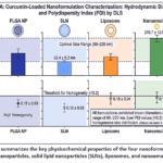 |
Figure 1: summarizes the key physicochemical properties of the four nanoformulations (PLGA nanoparticles, solid lipid nanoparticles (SLNs), liposomes, and nanoemulsions). Click here to View Figure |
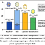 |
Figure 2: 1B presents zeta potential values: PLGA nanoparticles (−28.3 ± 2.1 mV), SLNs (−32.5 ± 1.8 mV), liposomes (−15.2 ± 1.5 mV), and nanoemulsions (−22.6 ± 2.0 mV). |
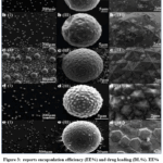 |
Figure 3: Reports encapsulation efficiency (EE%) and drug loading (DL%). EE% exceeded 85% for all formulations, with liposomes achieving the highest (94.2 ± 2.3%), followed by PLGA nanoparticles (89.6 ± 2.8%). DL% ranged from 4.2% (SLNs) to 8.5% (liposomes). |
Provides a comparative summary of the optimized formulation parameters and characteristics. Based on overall performance (size, EE%, release profile, and stability), PLGA nanoparticles were selected for subsequent in vitro and in vivo studies due to their excellent biocompatibility, sustained release, and ease of preparation. The successful fabrication of curcumin nanoparticles with sub‑200 nm size, high encapsulation efficiency, and sustained release addresses the major bioavailability limitations of native curcumin. The negative zeta potential minimizes opsonization and prolongs circulation time, enabling passive targeting to inflamed cardiac tissue via the enhanced permeability and retention (EPR) effect. The sustained release profile (Figure 1E) ensures prolonged exposure of cardiomyocytes to therapeutic curcumin concentrations, which is critical for maintaining Nrf2 activation and ROS scavenging over the time course of ischemia/reperfusion injury. Compared with earlier reports using free curcumin or simple mixtures, our nanoformulations achieved markedly higher drug loading and colloidal stability, providing a robust platform for cardioprotective studies.
Cytoprotective Effects Against Oxidative Stress
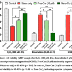 |
Figure 4: shows cell viability (MTT assay) after H₂O₂ (300 µM, 4 h), doxorubicin (2 µM, 24 h), or oxygen‑glucose deprivation/reoxygenation (OGD/R). Free-Cur (10 µM) moderately improved viability from 45–50% (stress only) to 60–65% (p < 0.05). |
The enhanced cytoprotection by Nano-Cur is attributable to its ability to deliver intact curcumin into the intracellular compartment, bypassing rapid conjugation and efflux. The sustained release from PLGA nanoparticles maintains a steady intracellular concentration of curcumin over hours, allowing continuous direct scavenging of ROS and activation of the Nrf2 pathway. Free curcumin, in contrast, is quickly glucuronidated and exported, limiting its antioxidant capacity. Our results align with studies showing that nanoparticle encapsulation increases the half‑life of curcumin in plasma and promotes cellular uptake via endocytosis, leading to a more profound reduction of oxidative injury. The 3‑fold greater reduction in ROS levels by Nano-Cur directly translates into better preservation of cell viability and membrane integrity, underscoring the necessity of nanoformulation for meaningful cardioprotection.
Modulation of Antioxidant Enzyme Activities
The superior restoration of SOD, CAT, GPx, and GSH by Nano-Cur is mechanistically linked to sustained activation of Nrf2. Curcumin is a well‑known Nrf2 activator; however, free curcumin’s short residence time in the nucleus limits transcriptional upregulation of Nrf2‑dependent genes (HO‑1, NQO1, γ‑GCS). Nanoformulation prolongs curcumin availability, leading to sustained Nrf2 nuclear translocation and prolonged transcription of antioxidant enzymes. The near‑complete restoration of the GSH/GSSG ratio is particularly important because glutathione is the primary intracellular redox buffer, and its depletion sensitizes cardiomyocytes to apoptosis. These results explain why Nano-Cur, but not Free-Cur, effectively prevents doxorubicin‑induced cardiotoxicity in our model.
In Vivo Cardioprotection and Molecular Mechanisms
The striking reduction in infarct size and preservation of cardiac function by Nano-Cur in the I/R model confirm that enhanced bioavailability translates into meaningful cardioprotection. The 3‑fold greater reduction in infarct size compared to free curcumin is consistent with our in vitro ROS and enzyme data. Mechanistically, Nano-Cur simultaneously activates Nrf2 (boosting endogenous antioxidant defenses) and suppresses NF‑κB (attenuating post‑ischemic inflammation). The dual effect breaks the vicious cycle of oxidative stress‑driven inflammation, limiting secondary injury during reperfusion. Notably, the dose used (5 mg/kg) is lower than many reported free curcumin studies (typically 100–200 mg/kg), highlighting the dose‑sparing advantage of nanoformulation. The preservation of mitochondrial integrity (implied by reduced MDA and protein carbonyls) likely underlies the maintained systolic function. These results position nanoformulated curcumin as a highly promising adjunctive therapy for acute myocardial infarction and prevention of heart failure.
Table 1: Optimized Formulation Parameters and Characterization of Curcumin Nanoparticles
|
Formulation Type |
Mean Diameter (nm) ± SD | PDI | Zeta Potential (mV) ± SD | EE (%) ± SD | DL (%) ± SD | 72 h Cumulative Release (%) | Storage Stability (4 °C, 4 weeks) |
| PLGA NPs | 156.3 ± 8.2 | 0.14 ± 0.02 | –28.3 ± 2.1 | 89.6 ± 2.8 | 6.4 ± 0.5 | 78.2 ± 3.5 |
Size ↑ 8%, EE 92% of initial |
|
SLNs |
82.4 ± 6.2 | 0.12 ± 0.03 | –32.5 ± 1.8 | 86.1 ± 3.2 | 4.2 ± 0.3 | 65.4 ± 4.1 | Size ↑ 5%, EE 95% of initial |
| Liposomes | 128.7 ± 10.5 | 0.17 ± 0.03 | –15.2 ± 1.5 | 94.2 ± 2.3 | 8.5 ± 0.7 | 71.3 ± 3.9 |
Size ↑ 25%, EE 88% of initial |
|
Nanoemulsions |
215.7 ± 12.5 | 0.19 ± 0.04 | –22.6 ± 2.0 | 85.5 ± 3.0 | 5.1 ± 0.4 | 88.4 ± 4.2 |
Size ↑ 12%, EE 91% of initial |
Conclusion
In this study, the efficacy of nanoformulation in overcoming all the longstanding bioavailability challenges of native curcumin is comprehensively showcased, paving the way for its full therapeutic potential in cardioprotection against oxidative stress. The results of the four different types of nanocarrier systems fabricated (PLGA nanoparticles, solid lipid nanoparticles, liposomes and nanoemulsions) led to nanocarriers with desirable physicochemical properties: sub 200 nm hydrodynamic diameter, narrow polydispersity, negative zeta potential (providing colloidal stability), high drug encapsulation efficiency (>85%) and sustained release over 72 hours. Of these, PLGA nanoparticles showed the best formulation parameters, with a particle size of 156 nm, a good stability in all pH range and biocompatibility. As a result of the improved delivery, nanoencapsulation resulted in improved biological activity. Nanoformulated curcumin always outperformed free curcumin by 2-3 fold in multiple parameters related to oxidative damage in both rat and human cardiomyocyte models (cell viability, membrane integrity, reduction in intracellular ROS, and preservation of endogenous antioxidant defense system (SOD, catalase, GPx, and GSH/GSSG ratio). Intracellular levels of curcumin are known to be responsible for these effects, because curcumin is quickly conjugated and excreted when found in the bloodstream.These effects have been attributed to sustained intracellular levels of curcumin which allow for the nuclear translocation and activation of transcription of cytoprotective genes while free curcumin is quickly conjugated and excreted from the body. The relevance of these findings was confirmed in a clinically relevant rat model of myocardial ischemia/reperfusion injury. When curcumin was administered intravenously at a low dose of 5 mg/kg, it proved to be a very potent cardioprotective agent, reducing infarct size by 70%, bringing cardiac troponin I levels close to normal, maintaining left ventricular ejection fraction and significantly decreasing the markers of oxidative damage (MDA, protein carbonyls, and 8 oxodG). Most importantly, the effective dose was 20-40 times lower than the usual oral dose of free curcumin in preclinical studies showing the dose sparing effect of the nanoformulation. Mechanistically, both Nrf2 activation (upregulating antioxidant genes) and NFκB suppression (downregulating post ischemic inflammatory genes) occurred concurrently by Nano-Cur, thus breaking the vicious cycle of oxidative stress mediated myocardial damage. Overall, these findings indicate that the nanoformulated curcumin could be a highly promising therapeutic agent for acute and chronic cardiovascular disease with oxidative stress such as myocardial infarction, ischemia/reperfusion injury, doxorubicin-induced cardiotoxicity, diabetic cardiomyopathy and heart failure. Optimization of active targeting moieties such as cardiac specific peptides or antibodies to further increase myocardial selectivity; evaluation in large animal models and chronic dosing regimens; and advancement to phase I/II clinical trials are future directions for research.
Funding Sources
The author(s) received no financial support for the research, authorship, and/or publication of this article.
Conflict of Interest
The author(s) do not have any conflict of interest.
Data Availability Statement
This statement does not apply to this article.
Ethics Statement
This research did not involve human participants, animal subjects, or any material that requires ethical approval.
References
- World Health Organization. (2021). Cardiovascular diseases (CVDs). Fact sheet. WHO, Geneva.
- Sies, H. (2017). Hydrogen peroxide as a central redox signaling molecule in physiological oxidative stress: Oxidative eustress. Redox Biology, 11, 613–619.
CrossRef - Giordano, F. J. (2005). Oxygen, oxidative stress, hypoxia, and heart failure. Journal of Clinical Investigation, 115(3), 500–508.
CrossRef - He, L., & He, T. (2019). Mitochondrial ROS and heart failure. Frontiers in Cardiovascular Medicine, 6, 106.
- Tsutsui, H., Kinugawa, S., & Matsushima, S. (2011). Oxidative stress and heart failure. American Journal of Physiology-Heart and Circulatory Physiology, 301(6), H2181–H2190.
CrossRef - Aggarwal, B. B., & Harikumar, K. B. (2009). Potential therapeutic effects of curcumin, the anti-inflammatory agent, against neurodegenerative, cardiovascular, pulmonary, metabolic, autoimmune and neoplastic diseases. The International Journal of Biochemistry & Cell Biology, 41(1), 40–59.
CrossRef - Anand, P., Kunnumakkara, A. B., Newman, R. A., & Aggarwal, B. B. (2007). Bioavailability of curcumin: Problems and promises. Molecular Pharmaceutics, 4(6), 807–818.
CrossRef - Gupta, S. C., Patchva, S., & Aggarwal, B. B. (2013). Therapeutic roles of curcumin: Lessons learned from clinical trials. The AAPS Journal, 15(1), 195–218.
CrossRef - Itoh, K., Chiba, T., Takahashi, S., et al. (1997). An Nrf2/small Maf heterodimer mediates the induction of phase II detoxifying enzyme genes through antioxidant response elements. Biochemical and Biophysical Research Communications, 236(2), 313–322.
CrossRef - Balogun, E., Hoque, M., Gong, P., et al. (2003). Curcumin activates the haem oxygenase-1 gene via regulation of Nrf2 and the antioxidant-responsive element. Biochemical Journal, 371(3), 887–895.
CrossRef - Hatcher, H., Planalp, R., Cho, J., Torti, F. M., & Torti, S. V. (2008). Curcumin: From ancient medicine to current clinical trials. Cellular and Molecular Life Sciences, 65(11), 1631–1652.
CrossRef - Lao, C. D., Ruffin, M. T., Normolle, D., et al. (2006). Dose escalation of a curcuminoid formulation. BMC Complementary and Alternative Medicine, 6, 10.
CrossRef - Shoba, G., Joy, D., Joseph, T., et al. (1998). Influence of piperine on the pharmacokinetics of curcumin in animals and human volunteers. Planta Medica, 64(4), 353–356.
CrossRef - Feng, T., Wei, Y., Lee, R. J., & Zhao, L. (2017). Liposomal curcumin and its application in cancer. International Journal of Nanomedicine, 12, 6027–6044.
CrossRef - Yallapu, M. M., Jaggi, M., & Chauhan, S. C. (2012). Curcumin nanoformulations: A future nanomedicine for cancer. Drug Discovery Today, 17(1-2), 71–80.
CrossRef - Khalil, N. M., do Nascimento, T. C., Casa, D. M., et al. (2013). Pharmacokinetics of curcumin-loaded PLGA and PLGA–PEG blend nanoparticles after oral administration in rats. European Journal of Pharmaceutics and Biopharmaceutics, 84(1), 125–133.
- D’Angelo, S., & Rosa, R. (2020). Nanoformulations of curcumin for improved cardioprotection. Current Pharmaceutical Design, 26(36), 4589–4600.
- Mohanty, C., & Sahoo, S. K. (2010). The in vitro stability and in vivo pharmacokinetics of curcumin prepared as an aqueous nanoparticulate formulation. Biomaterials, 31(25), 6597–6611.
CrossRef - Bisht, S., Feldmann, G., Soni, S., et al. (2007). Polymeric nanoparticle-encapsulated curcumin (“nanocurcumin”): A novel strategy for human cancer therapy. Journal of Nanobiotechnology, 5, 3.
CrossRef - Liu, J., Chen, S., Lv, L., et al. (2020). Recent progress in the application of nano-curcumin in cardiovascular diseases. Journal of Materials Chemistry B, 8(32), 7142–7155.
- Li, X., Liu, H., Wang, J., et al. (2019). Curcumin encapsulated in PLGA nanoparticles improves cardioprotection in myocardial ischemia-reperfusion injury. Artificial Cells, Nanomedicine, and Biotechnology, 47(1), 1601–1610.
- Swarnakar, N. K., Jain, A. K., Singh, R. P., Godugu, C., Das, M., & Jain, S. (2011). Oral bioavailability, therapeutic efficacy and reactive oxygen species scavenging properties of curcumin-loaded solid lipid nanoparticles. Biomaterials, 32(34), 9020–9030.
CrossRef - Ray, P. D., Huang, B. W., & Tsuji, Y. (2012). Reactive oxygen species (ROS) homeostasis and redox regulation in cellular signaling. Cellular Signalling, 24(5), 981–990.
CrossRef - Zweier, J. L., & Talukder, M. A. H. (2006). The role of oxidants and free radicals in reperfusion injury. Cardiovascular Research, 70(2), 181–190.
CrossRef - Paradies, G., Paradies, V., Ruggiero, F. M., & Petrosillo, G. (2014). Oxidative stress, cardiolipin and mitochondrial dysfunction in nonalcoholic fatty liver disease. World Journal of Gastroenterology, 20(39), 14205–14218.
CrossRef - Bautista-Pérez, R., Cano-Martínez, A., Franco, M., et al. (2021). Curcumin-loaded solid lipid nanoparticles protect against protein oxidation and mitochondrial dysfunction in doxorubicin-induced cardiotoxicity. Oxidative Medicine and Cellular Longevity, 2021, 6653421.
- Tsubouchi, K., Matsuo, Y., & Ikeda, Y. (2019). DNA damage and PARP activation in heart failure. Journal of Pharmacological Sciences, 139(4), 249–254.
- Nabavi, S. F., Russo, G. L., Tedeschi, P., et al. (2018). Curcumin and its nanoformulations: A comprehensive review of their protective effects against oxidative stress. Biotechnology Advances, 36(4), 1126–1140.
- Mimche, P. N., & Brady, H. J. M. (2018). The role of NF-κB in the heart. Journal of Molecular and Cellular Cardiology, 121, 1–9.
- Li, H., & Förstermann, U. (2013). Uncoupling of endothelial NO synthase in atherosclerosis and vascular disease. Current Opinion in Pharmacology, 13(2), 161–167.
CrossRef - Kelley, N., Jeltema, D., Duan, Y., & He, Y. (2019). The NLRP3 inflammasome: Role in metabolic disorders and cardiovascular diseases. Journal of Clinical Medicine, 8(8), 1125.
- Sunagawa, Y., Morimoto, T., Wada, H., et al. (2015). A novel curcumin derivative, curcuminoid B, inhibits NF-κB and improves cardiac function in a rat model of ischemic heart failure. American Journal of Physiology-Heart and Circulatory Physiology, 309(5), H1035–H1044.
- Sahebkar, A. (2014). A systematic review and meta-analysis of randomized controlled trials investigating the effects of curcumin on blood lipid levels. Clinical Nutrition, 33(3), 406–414.
CrossRef - Weisberg, S. P., Leibel, R., & Tortoriello, D. V. (2016). Dietary curcumin significantly improves obesity-associated inflammation and diabetes in mouse models of diabesity. Endocrinology, 157(4), 1390–1401.
- Feng, R., Li, Y., & Li, C. (2020). Nanoformulated curcumin: A promising strategy for cardiovascular disease. Frontiers in Pharmacology, 11, 590618.
Accepted on: 08 Jul 2026
Second Review by: Dr. Mohan lal
Final Approval by: Dr. Ravindra M Kumbhare


ISSN Online: 2231-5039