Gold Nanoparticles in Biosensor Technology for Illicit Drug Detection: Analytical Performance and Applications
 and Nurin Haziqah Razali
Centre of Diagnostic, Therapeutic and Investigative Studies, Faculty of Health Sciences, Universiti Kebangsaan Malaysia, Jalan Raja Muda Abdul Aziz, Kuala Lumpur, Malaysia
Corresponding Author E-mail: atikahnasir@ukm.edu.my
Download this article as:
ABSTRACT:The global illicit drug crisis demands rapid, field-deployable detection technologies to complement laboratory methods like GC–MS and LC–MS. Gold nanoparticles (AuNPs) offer unique surface plasmon resonance, high surface area, and facile functionalisation, making them ideal for biosensor development. This systematic review, following PRISMA 2020 guidelines, analysed 18 original studies (2020–2025) comparing AuNP-based biosensors for illicit drug detection. Included studies encompassed colorimetric, lateral flow, electrochemical, SERS, and FRET-based platforms. Statistical analysis revealed methamphetamine as the most frequently targeted drug (7 studies, 26.9%), followed by cocaine and morphine (5 studies each, 19.2%), reflecting global prevalence patterns. Aptamers were the dominant biorecognition element (12 studies, 61%), while urine was the most common test matrix (44%). A clear trade-off exists between sensitivity and operational simplicity: colorimetric sensors enable instrument-free visual readouts for point-of-care screening, whereas electrochemical and SERS platforms achieve sub-ng/mL to picomolar detection limits at the cost of specialised equipment. Key limitations persist, including matrix interferences from biological samples, inadequate shelf-life of AuNP conjugates, and a lack of regulatory validation, currently restricting these sensors to presumptive screening rather than confirmatory analysis. Future directions include multiplexed platforms, sample-to-answer integration, and smartphone-based AI readouts to translate these innovations into forensic and clinical practice, where they will complement gold-standard instrumental methods.
KEYWORDS:Aptamers; Biosensors; Forensic Toxicology; Gold Nanoparticles; Illicit Drug Detection; Lateral Flow Assay; Point‑Of‑Care Testing
Introduction
The widespread abuse and trafficking of illicit drugs constitute a serious global threat to public health, social stability, and national security. Chronic and acute drug exposure leads to addiction, organ damage, and increased crime rates, with hundreds of millions of people worldwide having used illicit substances.1 The opioid crisis, particularly involving synthetic opioids such as fentanyl, has escalated dramatically, with overdose concentrations as low as single‑digit ng/mL (Li et al., 2020). Rapid identification of illicit substances is critically important for law enforcement, emergency services, and forensic laboratories. On‑site and rapid screening is essential because law enforcement officers at seizure scenes, emergency medical personnel responding to overdoses, and forensic toxicologists require immediate actionable intelligence such as administering naloxone, securing a crime scene, or initiating treatment without waiting for laboratory confirmation.2
High end analytical techniques such as gas chromatography‑mass spectrometry (GC‑MS) and liquid chromatography‑mass spectrometry (LC‑MS) offer excellent accuracy and sensitivity but require expensive, bulky instrumentation, extensive sample preparation, and skilled operators, hindering real‑time on‑site decision‑making.1,3 These methods serve as the gold standard for confirmatory analysis, providing legally admissible evidence; however, they are unsuitable for rapid, decentralised screening. In contrast, portable biosensors are designed as presumptive screening tools though they offer quick results to guide initial decisions, with positive findings then sent for laboratory confirmation.
In response, a wide range of portable biosensors has been explored for illicit drug detection, employing various nanomaterials and transduction principles. For example, carbon nanotube (CNT)-based electrochemical sensors have demonstrated rapid detection of morphine and methamphetamine with limits of detection (LODs) in the low ng/mL range, but they often suffer from poor reproducibility and complex electrode functionalisation.4 Silver nanoparticle (AgNP) colorimetric assays offer low‑cost visual readouts; however, they are prone to oxidation and matrix interference from chloride ions in biological fluids.5 Quantum dot (QD)-based fluorescent sensors provide high sensitivity but face challenges with toxicity, photobleaching, and autofluorescence from urine or serum samples.6 Molecularly imprinted polymer (MIP) sensors without AuNPs are robust and reusable, yet they often lack the signal amplification needed for trace‑level detection of small‑molecule drugs. While these alternative platforms have advanced the field, their practical deployment remains constrained by stability, sensitivity, or manufacturing inconsistencies.7
Gold nanoparticles (AuNPs) have emerged as a uniquely attractive platform due to their intense surface plasmon resonance (SPR), high surface‑to‑volume ratio, excellent biocompatibility, stable functionalisation via thiol‑gold chemistry, which critically for field use and the ease of colorimetric transition from red to blue upon aggregation, which enables instrument‑free visual readouts.8 These properties enable sensitive, multiplexed, field‑deployable drug sensing.
Recent advances have produced a wide range of AuNP‑based biosensor types: colorimetric and paper‑based sensors for visual readouts, lateral flow assays (LFAs) for rapid point‑of‑care testing9, electrochemical sensors for trace analysis, surface‑enhanced Raman scattering (SERS) for molecular fingerprinting, and FRET‑based optical platforms achieving picomolar detection limits. Integration with smartphone‑based artificial intelligence is transforming semi‑quantitative assays into accurate, user‑friendly tools. Despite this proliferation, a systematic comparative analysis of AuNP‑based sensors specifically for illicit drug detection is lacking.
This systematic review, conducted following PRISMA 2020 guidelines, analyses 18 original studies published between 2020 and 2025. The objectives are: (i) to compare analytical performance across different sensor types by AuNP; (ii) to analyse the distribution of targeted drugs, biorecognition elements, and sample matrices; (iii) to identify current limitations; and (iv) to propose future directions. By synthesising the evidence, this review aims to guide researchers and practitioners in selecting appropriate AuNP‑based platforms for forensic and clinical applications.
Materials and Methods
This systematic review was performed in accordance with the Preferred Reporting Items for Systematic Reviews and Meta-Analyses (PRISMA) guidelines to ensure methodological rigor and transparency throughout the review process Protocol registration before the commencement of the review is a key requirement of the PRISMA 2020 framework, as it reduces the risk of reporting bias, increases transparency, facilitates scrutiny of methods, and improves the overall trustworthiness of systematic reviews.
Search Strategy
A systematic literature search was conducted across four major electronic databases: PubMed, Scopus, Web of Science, and Springer Nature. These databases were selected for their comprehensive coverage of peer-reviewed literature in the fields of nanotechnology, analytical chemistry, forensic science, and biosensor engineering. The search was restricted to the period from 1 January 2020 to 31 March 2026, encompassing a full decade of research activity to capture the recent surge in AuNP-based biosensor development while maintaining sufficient depth for trend analysis. The search was conducted on 3 April 2026.
The search strategy was designed to maximize sensitivity (retrieval of all potentially relevant records) while maintaining sufficient specificity to minimize irrelevant results, in accordance with PRISMA search methodology guidelines. A combination of controlled vocabulary terms (MeSH terms in PubMed, Emtree terms in Scopus) and free-text keywords was employed across three conceptual domains:
- Domain 1 (Nanoparticle): “gold nanoparticles” OR “AuNPs” OR “gold nanorods” OR “gold nanostructures” OR “noble metal nanoparticles”
- Domain 2 (Biosensor Technology): “biosensor” OR “electrochemical sensor” OR “colorimetric sensor” OR “optical sensor” OR “fluorescence sensor” OR “lateral flow assay” OR “immunosensor” OR “aptasensor” OR “point-of-care” OR “POC”
- Domain 3 (Illicit Drug Target): “illicit drug” OR “drug abuse” OR “methamphetamine” OR “cocaine” OR “morphine” OR “heroin” OR “opioid” OR “stimulant” OR “forensic detection”
The Boolean operator AND was used to combine the three domains, and OR was used within each domain to capture synonyms and variant terminologies. The complete search string used for PubMed was as follows:
(“gold nanoparticles” OR “AuNPs” OR “gold nanorods”) AND (“biosensor” OR “electrochemical” OR “colorimetric” OR “aptasensor” OR “immunosensor”) AND (“methamphetamine detection” OR “cocaine detection” OR “morphine detection” OR “heroin detection” OR “illicit drug detection”)
Inclusion and Exclusion Criteria
A predefined eligibility framework was established in the registered protocol and applied consistently by two independent reviewers (Atikah Mohd Nasir and Nurin Haziqah Razali) during study selection.
Inclusion Criteria
Studies were included if they met the following criteria:
- Reported the development, characterization, or application of gold nanoparticle (AuNP)-based biosensors for detecting illicit drugs.
- Utilized AuNPs as a key component across electrochemical, optical (colorimetric, SPR, fluorescence, SERS), or lateral flow platforms.
- Included at least one quantitative parameter such as limit of detection (LOD), limit of quantification (LOQ), sensitivity, selectivity, or linear range.
- Evaluated in relevant samples (e.g., buffer, spiked biological fluids, urine, saliva, blood, seized drugs, or wastewater).
- Presented original research data (reviews were used only for background).
- Publication type: Peer-reviewed articles published in English.
- Publication date: Published between 1 January 2020 and 31 December 2025.
Exclusion Criteria
Studies were excluded if they:
- Did not involve AuNPs.
- Focused on non-drug targets.
- Reported only theoretical or computational work without experimental validation.
- Lacked sufficient analytical data.
- Were duplicate or overlapping publications.
- Were published in languages other than English.
Study Selection and Data Screening
The study selection process is summarized in the PRISMA 2020 flow diagram (Figure 1). A total of 140 records were identified through systematic searching of four electronic databases: Scopus (n = 37), PubMed (n = 24), Web of Science (n = 34), and Springer Nature (n = 45). No additional records were identified from registers or other sources.
Before screening, 23 duplicate records were identified and removed using automated duplicate detection in EndNote X9, followed by manual verification. No records were removed by automation tools or for other reasons at this stage. Consequently, 117 unique records proceeded to the screening phase.
During the title and abstract screening phase, 34 records were excluded because they clearly did not meet the predefined eligibility criteria. Common reasons for exclusion at this stage included: studies that did not report original research (e.g., editorials, commentaries, non-systematic reviews), studies with no relevance to illicit drug detection, and studies that did not involve biosensor technology. The remaining 83 records were sought for full-text retrieval.
Of these, 28 reports could not be retrieved due to: unavailability of full text through institutional subscriptions, conference abstracts without corresponding full manuscripts, or withdrawn publications. Thus, 55 full-text reports were successfully retrieved and underwent detailed eligibility assessment.
During full-text eligibility assessment, 36 reports were excluded for the following specific reasons, as documented in the PRISMA flow diagram:
- Non-original research articles (n = 13): These included review articles, perspectives, and commentaries that did not present primary experimental data. While these were excluded from the systematic synthesis, they were retained for background information and citation tracking.
- Focused on non-illicit drug targets (n = 17): These studies reported AuNP-based biosensors for analytes other than the target illicit drugs of interest (e.g., therapeutic drugs, biomarkers, pesticides, heavy metals, or food contaminants).
- Does not involve AuNPs (n = 7): These studies employed other nanomaterials (e.g., silver nanoparticles, quantum dots, carbon nanotubes, graphene oxide) without the use of gold nanoparticles as a core transduction element.
Following this exclusion process, 18 studies met all inclusion criteria and were included in the final systematic review. No additional studies were identified through backward or forward citation searching of the included articles or relevant reviews.
Data Extraction
During data extraction, one reviewer independently collected the following key information from each of the 18 included studies, and a second reviewer verified all entries: AuNP characteristics (size, shape, and functionalisation method such as thiolated aptamers, antibodies, or NanoMIPs); biosensor type (colorimetric, LFA, electrochemical, SERS, FRET, etc.); target drug(s) (cocaine, methamphetamine, morphine, fentanyl, ketamine, etc.); biorecognition element (aptamer, antibody, NanoMIP, or none/catalytic); analytical performance metrics (limit of detection, linear range, and response time); sample matrix (urine, serum, saliva, hair, seized drugs, buffer, or wastewater); real‑world application as stated by the authors (e.g., POCT, forensic screening, clinical use); and recovery rate where reported (e.g., in spiked biological fluids). All extracted data were compiled into Table 1.
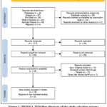 |
Figure 1: PRISMA 2020 flow diagram of the study selection process for AuNP-based biosensors for illicit drug detection Click here to View Figure |
Risk of Bias
Risk of bias was assessed using the Joanna Briggs Institute (JBI) Critical Appraisal Checklist for Analytical Cross-Sectional Studies (adapted for biosensor validation studies). Two reviewers independently evaluated each included study against nine criteria: clear definition of study population, inclusion/exclusion criteria, description of test method, validity of reference standard, blinding, handling of indeterminate results, and appropriateness of statistical analysis. Each criterion was rated as “Yes”, “No”, “Unclear”, or “Not Applicable”. Studies were not excluded based on bias but were graded as high, moderate, or low risk. Disagreements were resolved by consensus. Overall, 12 studies were rated moderate risk (adequate performance but lacking real-world validation or blinded analysis), 5 as low risk (rigorous validation against reference methods), and 1 as high risk (insufficient data reporting). No study was excluded solely on bias grounds; instead, limitations were discussed qualitatively.
Data Analysis
Data (Table 1) on the comparative characteristics of the included studies were extracted by one reviewer and independently verified by a second reviewer.
Table 1: Comparative Analysis of AuNP-Based Biosensors for Illicit Drug Detection: Biosensor Type, Biorecognition Elements, Analytical Performance, and Real-World Applications
|
No. |
Study | Biosensor Type | Drug Detected | Biorecognition Element | Analytical Performance | Sample Matrix | Real-World Application |
| 1 | (Jing et al., 2022) | Split-aptamer LFSA | Cocaine | Split DNA aptamer | LOD: 10 µM; Range: 10 µM–5 mM; Time: 15 min | Artificial urine, sweat |
POCT, on-site testing |
|
2 |
(D’Aurelio et al., 2021) | EIS & QCM | Morphine | NanoMIPs | LOD: 0.11 ng/mL (EIS), 0.19 µg/mL (QCM); Time: 30–40 min | Buffers | Field (EIS), lab (QCM) |
| 3 | (Zamanian et al., 2022) | Fluorescent aptasensor | Cocaine | FAM-DNA aptamer | LOD: 0.31 pM; Range: 100 pM–600 µM | Serum, urine |
Forensic, clinical |
|
4 |
(Alfadly et al., 2020) | Colorimetric AuNP assay | Captagon | 40-mer DNA aptamer | Range: 2–50 µM; R²: 0.998 | Urine, blood, hair | Forensic screening |
| 5 | (Keklik Şah & Isbir Turan, 2024) | SPR aptasensor | Cocaine | Thiol-DNA aptamer | LOD: 0.43 ng/mL; Range: 1–1000 ng/mL; Time: 8 min | Synthetic urine |
Clinical, law enforcement |
|
6 |
(Erkocyigit et al., 2025) | Competitive LFA | Cocaine, Methamphetamine | Thiol-DNA aptamers | LOD: 5 ng/mL; Time: 10 min | Saliva | Workplace, forensic |
| 7 | (Sanli et al., 2020) | GAPTA colorimetric | Cocaine | DNA aptamer | LOD: 0.97 nM; Sensitivity: 0.2 nM; Time: <90 min | Synthetic saliva |
Toxicology screening |
|
8 |
(Adegoke et al., 2020) | Nanozyme colorimetric | Amphetamine, Methaphetamine | DNA aptamer | LOD: 34.1 ng/mL, 28.6 ng/mL; Time: 1 min | Aqueous, seized drugs | Law enforcement |
| 9 | (Gao et al., 2020) | MoS₂-AuNP colorimetric | Cocaine | DNA aptamer | LOD: 7.49 nM; Range: 0–1 µM | Serum |
Clinical, forensic |
|
10 |
(Li et al., 2020) | Competitive LFA | Fentanyl | Antibody | LOD: ng/mL level; Time: 5 min | Urine | Emergency screening |
| 11 | (Masemola et al., 2020) | Electrochemical sensor | Cocaine, Heroin, Amphetamine, 6-Acetylmorphine | None (AuNP catalytic) | LOD: 0.82 mg/L; Range: 0.125–3 mg/L | Water |
Environmental monitoring |
|
12 |
(L. Liu et al., 2025) | FQICS | Methaphetamin, Ketamine, Morphine | Antibodies | LOD: 0.041–0.072 ng/mL; Time: 10 min | Hair, urine, saliva, sewage | Drug monitoring |
| 13 | (Heshi & Shokoufi, n.d.) | FRET-TLS | Methamphetamine | Non-biological | LOD: 1.5 nM; Range: 5–80 nM | Plasma, urine |
Ultra-trace analysis |
|
14 |
(Y. Liu et al., 2024) | SERS sensor | Methaphetamin, Ketamine, Morphine | Antibody + antigen | LOD: 0.1–1 ng/mL; Reusable | Hair, aqueous | Forensic screening |
| 15 | (Suleman et al., 2025) | AI-integrated LFA | Ketamine | DNA aptamer | LOD: 0.1 µg/mL; Time: 10 min | Synthetic urine |
Smart POCT |
|
16 |
(Sen et al., 2022) | Colorimetric aptasensor | Methamphetamine | DNA aptamers | LOD: 33.3 nM (aq), 173 nM (saliva); Range: 50–500 nM | Saliva | On-site diagnostics |
| 17 | (Wang & McCord, 2020) | µPAD | Cocaine, Codeine, Methamphetamine | DNA aptamers | Time: <30 min; Color change detection | Seized drugs |
Field screening |
|
18 |
(Tavakol et al., 2020) | Electrochemical AuNP/SCNF | Morphine | None | Enhanced anodic signal (0.73 V) | Buffer | Pharma analysis |
Result and Discussion
Gold nanoparticle (AuNP)-based biosensors have attracted considerable attention for illicit drug detection, and it is not difficult to see why. Their distinctive optical properties, large surface area for functionalisation, and relative ease of preparation make them adaptable to a wide range of detection strategies. Over the past several years, researchers have developed AuNP-based sensors that span from simple colour changing tests readable with the naked eye to sophisticated optical platforms capable of detecting just a handful of molecules. The choice among these approaches involves trade-offs between sensitivity, speed, cost, and practicality.
Statistical Distribution of Targeted Illicit Drugs and Biorecognition Elements
Targeted Illicit Drugs
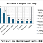 |
Figure 2: Percentages and Distributions of Targeted Illicit Drugs Click here to View Figure |
The predominance of cocaine and methamphetamine reflects their status as the most widely used illicit stimulants globally. Methamphetamine, in particular, has reached epidemic levels in North America and Southeast Asia, with usage rates of 2–3% of the adult population in some regions 9,10. This public health burden drives demand for rapid, field‑deployable sensors capable of detecting these drugs in saliva, urine, and hair.
The frequent inclusion of morphine and the emerging focus on fentanyl highlight the growing urgency of the opioid crisis. Fentanyl detection is especially challenging because overdose concentrations can be as low as single‑digit ng/mL, requiring the ultra‑sensitivity that AuNP‑based platforms can provide 11. The fact that only one study in this sample targeted fentanyl suggests a significant gap given the current synthetic opioid epidemic, although the recent work by Liu et al. (2025) on multiplexed fluorescence‑quenching sensors for methamphetamine, morphine and ketamine indicates a move toward broader opioid panels.
The detection of captagon (fenethylline) in Alfadly, Abdulwahid, and Saleh (2020) represents research targeted toward specific regional drug trends. Captagon is widely abused in the Middle East, particularly in Iraq and Syria, and its inclusion in this sample underscores the importance of tailoring sensor development to local forensic needs.
Biorecognition Elements
The choice of biorecognition element is a critical determinant of sensor performance, influencing stability, cost, selectivity, and the ability to detect small‑molecule drugs. Across the 18 studies, aptamers (DNA or RNA) were the dominant recognition element, used in 12 studies (61%). Antibodies (monoclonal) appeared in 3 studies (17%), non‑biological or catalytic recognition elements (e.g., direct electrochemical oxidation, nanozyme activity) also appeared in 3 studies (17%), and molecularly imprinted polymer nanoparticles (NanoMIPs) were used in 1 study (6%) (Figure 3).
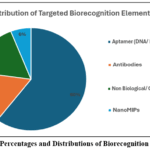 |
Figure 3: Percentages and Distributions of Biorecognition Elements Click here to View Figure |
The strong preference for aptamers (61%) reflects several practical advantages. Aptamers, often termed “chemical antibodies,” are chemically synthesised, which eliminates batch‑to‑batch variability and reduces cost. They are thermally stable, tolerating temperatures that would denature antibodies, making them well‑suited for point‑of‑care use in diverse environments (Jing et al., 2022; Sen et al., 2022). Moreover, aptamers can be readily modified with thiol, biotin, or fluorophore labels during synthesis, facilitating immobilisation on AuNP surfaces.
Antibodies remain important for high‑affinity detection of certain drugs. For example, Li et al. (2020) used an antibody‑based lateral flow assay for fentanyl (single‑digit ng/mL sensitivity in 5 min), and Liu et al. (2025) employed monoclonal antibodies in a multiplexed fluorescence‑quenching sensor for methamphetamine, morphine, and ketamine (LODs as low as 0.041 ng/mL). Non‑biological recognition includes direct electrochemical oxidation (Masemola et al., 2020) and nanozyme catalytic activity (Adegoke et al., 2020). NanoMIPs synthetic polymer nanoparticles with drug‑specific cavities were used by D’Aurelio et al. (2021) for morphine detection (EIS LOD of 0.11 ng/mL), offering robustness and reusability without biological components.
In summary, aptamers are the dominant choice for AuNP‑based drug sensors due to their stability and design flexibility, but antibodies, NanoMIPs, and catalytic materials each fill specific niches where ultra‑high affinity or extreme robustness is required.
Applications AuNP-Based Biosensors for Illicit Drug Detection
Colorimetric and Paper‑Based Microfluidic Sensors
Colorimetric sensors remain among the most accessible AuNP-based formats, largely because they produce a visual readout without requiring any instrumentation. The working principle is straightforward: dispersed AuNPs appear red, but when they aggregate, the solution shifts to blue or purple. In a typical assay, aptamers or other recognition elements stabilise the AuNPs against salt-induced aggregation. When a target drug is present, it binds to these recognition elements, causing them to detach or change conformation, thereby leaving the AuNPs vulnerable to aggregation upon salt addition.
What makes this approach particularly practical for point-of-care testing is its simplicity. However, achieving good sensitivity requires careful attention to experimental design. 10 made an insightful observation in this regard. They found that the order in which reagents are mixed profoundly affects assay performance. When the drug is pre-incubated with the aptamer before the AuNPs are introduced (association assay), the aptamer is less likely to become irreversibly adsorbed onto the AuNP surface. This seemingly minor procedural adjustment lowered the limit of detection for methamphetamine in saliva to 33.3 nM, a meaningful improvement over conventional protocol. This finding serves as a useful reminder that even in well-established assay formats, seemingly simple operational details can have outsized effects on analytical outcomes.
Beyond improving sensitivity, researchers have also expanded the multiplexing capability of colorimetric sensors. A four-channel paper microfluidic device that simultaneously detects cocaine, codeine, and methamphetamine within approximately 30 minutes 12. The integration of paper as a substrate is particularly attractive for resource-limited settings because the devices are disposable, inexpensive, and require no external power. For field use, this combination of visual readout and multiplexing represents a genuine step forward, although the semi-quantitative nature of colour-based detection remains a limitation compared to instrument-based methods.
Lateral Flow Assays and Immunochromatographic Sensors
Lateral flow assays (LFAs) represent the most commercially mature AuNP‑based format for drug screening. Their capillary‑driven operation, low cost, and rapid results (5–15 min) make them the de facto standard for field use. However, small‑molecule drugs (molecular weight <1000 Da) typically cannot be detected in a sandwich format due to steric constraints, forcing most commercial LFAs into a competitive format where a positive result is indicated by the disappearance of a test line which can be inconsistent readingst for untrained users.
Jing et al., (2022) circumvented this limitation by splitting a cocaine‑binding aptamer into two segments. One segment conjugated to AuNPs served as the signal probe, and the other, biotinylated, acted as the capture probe13. In the presence of cocaine, the two aptamer halves assemble around the target, forming a sandwich complex that is captured on a streptavidin test line. This split‑aptamer lateral flow strip assay (LFSA) produced a positive (line‑appears) readout with visual LODs of 50 μmol/L in urine and 200 μmol/L in sweat, and recovery rates of 83.6–107.4%. More recently, Erkocyigit and colleagues (2025) developed an aptamer‑based competitive LFA for simultaneous detection of methamphetamine and cocaine in saliva. Using AuNP‑aptamer conjugates, they achieved LODs of 5.0 ng/mL for both drugs, with results readable within 15 min9. This work demonstrates that aptamers can replace antibodies in LFAs without loss of performance, offering advantages in chemical stability and synthesis reproducibility.
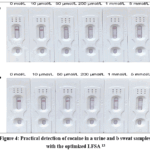 |
Figure 4: Practical detection of cocaine in a urine and b sweat samples with the optimized LFSA 13 Click here to View Figure |
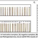 |
Figure 5: Application of real saliva samples: (A) negative samples; (B) samples containing Cocaine and Methaphetamine; (C) LC-QTOF/MS results of samples 9 Click here to View Figure |
For emergency fentanyl screening, speed is paramount. Li and colleagues (2020) clinically validated a competitive LFA using monoclonal antibody–AuNP conjugates for urine fentanyl testing in the emergency department. The assay returned results in 5 min with single‑digit ng/mL sensitivity sufficient to guide acute clinical management. A major advance in signal amplification is the fluorescence‑quenching immunochromatographic sensor (FQICS). This format uses fluorescence resonance energy transfer (FRET) between aggregation‑induced emission fluorescent microspheres (AIEFMs) and AuNPs. In the absence of the drug, AuNPs quench the AIEFM fluorescence. Target binding disrupts the FRET pair, restoring a positive fluorescence signal. A multiplexed FQICS achieved LODs of 0.041 ng/mL for methamphetamine, 0.072 ng/mL for morphine, and 0.059 ng/mL for ketamine in urine, hair, saliva and sewage which lower than conventional colorimetric LFAs.
The split‑aptamer sandwich approach 13 contrasts sharply with the competitive aptamer LFA of Erkocyigit et al. (2025). While the sandwich format offers a more intuitive readout, it requires careful optimisation of the two aptamer fragments and is sensitive to their relative concentrations. The competitive format is simpler to design but suffers from the negative readout problem. Neither is universally superior; the choice depends on the user’s tolerance for readout complexity versus assay development effort.
A persistent challenge in LFAs is that the subjectivity of interpreting faint or missing test lines. Soulis, Economou, and Kokkinos (2023) addressed this by integrating a smartphone app with deep learning models (YOLOv5 and ResNet50) to automate result reading. Their system eliminated inter‑observer variability and improved accuracy near the cut‑off concentration. Such AI‑augmented LFAs are likely to become standard in the coming years. Lateral flow assays based on AuNPs provide rapid, low‑cost, field‑deployable detection of illicit drugs. The choice between competitive and split‑aptamer sandwich formats, and between antibody and aptamer recognition elements, should be guided by the specific performance requirements and the target user’s technical expertise. Integration with smartphone‑based AI readers is rapidly transforming LFAs from qualitative to quantitative tools.
Electrochemical and Acoustic Sensor
When sensitivity requirements exceed the capabilities of optical LFAs, electrochemical and acoustic sensors offer compelling alternatives. Electrochemical impedance spectroscopy (EIS) is label‑free and highly sensitive to interfacial changes, while quartz crystal microbalance (QCM) provides real‑time mass‑binding information. The two techniques differ fundamentally in their transduction mechanism, leading to large differences in achievable LOD.
D’Aurelio and colleagues (2021) directly compared EIS and QCM for morphine detection using the same recognition element molecularly imprinted polymer nanoparticles (NanoMIPs). The EIS platform achieved an LOD of 0.11 ng/mL (≈0.38 nM), which is three orders of magnitude lower than the QCM platform’s LOD of 0.19 μg/mL (≈0.67 μM). However, the EIS method required 30–40 min per measurement and extensive data processing, whereas the QCM provided real‑time binding curves and was readily automated.
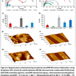 |
Figure 6: Nyquist plots obtained during morphine nanoMIP EIS sensor fabrication using DropSens SPE. (inset) (A) and DropSense IDE (B): the electrode coated with MUDA (red), EDC/ENS activation (green), nanoMIP attachment (grey), ethanolamine blocking (blue). |
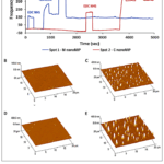 |
Figure 7: (A) Full sensorgram obtained during morphine and cocaine nanoMIP QCM sensor functionalization. AFM 3D topography image related to MUDA (B,D), morphine nanoMIP (C) and cocaine nanoMIP |
Beyond transducer choice, electrode modification strategies dramatically affect performance. Bor and colleagues (2022) developed an electrochemical aptasensor for methamphetamine using high‑affinity DNA aptamers selected via SELEX. The sensor, constructed on a gold electrode modified with a self‑assembled monolayer, achieved an LOD of 0.467 ng/mL comparable to the EIS NanoMIP platform but using a biological recognition element.15 Similarly, Li and colleagues (2023) decorated broken hollow carbon spheres with gold nanodendrites to create a high‑surface‑area electrode for morphine detection. The nanodendrite morphology facilitated electron transfer and provided abundant binding sites, enabling sensitive detection in human serum and saliva without the need for a separate recognition element (direct electrochemical oxidation of morphine).
The difference between EIS and QCM reported by D’Aurelio et al. (2021) is consistent with theoretical expectations: impedance spectroscopy measures changes in charge‑transfer resistance, which can be amplified by the NanoMIP layer’s dielectric properties, whereas QCM measures only the mass of bound analyte a small signal for a low‑molecular‑weight drug like morphine (285 Da). 15 achieved comparable sensitivity with an aptamer‑based EIS sensor, suggesting that the recognition element (NanoMIP vs. aptamer) is less critical than the transduction method when targeting sub‑ng/mL concentrations.
The high sensitivity of EIS comes at a cost: the technique requires a potentiostat, faradaic redox probes (e.g., [Fe(CN)₆]³⁻/⁴⁻), and careful control of solution ionic strength. These requirements make EIS less suitable for true point‑of‑care use compared to LFAs. QCM, while simpler to operate, suffers from lower sensitivity for small molecules unless mass amplification strategies (e.g., AuNP‑labeled secondary antibodies) are employed. Electrochemical sensors, particularly EIS, offer outstanding sensitivity for trace illicit drug detection, but their instrumentation and procedural complexity limit them to laboratory or well‑equipped field settings. QCM provides a user‑friendly alternative when real‑time binding information is needed and sub‑ng/mL sensitivity is not required. Nanostructured electrode modifications (nanodendrites, carbon composites) can enhance performance without complicating the assay protocol.
High‑Performance Optical and Surface‑Enhanced Techniques
Observation. For applications demanding the highest sensitivity such as detecting drugs in hair (where concentrations reflect cumulative exposure over weeks) or in small‑volume forensic samples by advanced optical platforms that exploit fluorescence quenching, thermal lens effects, or surface‑enhanced Raman scattering (SERS) achieve LODs in the picomolar to attomolar range.
Zamanian et al., (2022) constructed a fluorescent aptasensor on the inner surface of a capillary tube. A fluorescein‑labelled cocaine aptamer and AuNPs were co‑immobilised on the capillary wall. In the absence of cocaine, AuNPs quench the fluorescein signal. Upon cocaine binding, the aptamer undergoes a conformational change that moves the fluorophore away from the AuNPs, restoring fluorescence. This configuration achieved an LOD of 0.31 pM for cocaine in human serum and urine using only 3 μL of sample a sensitivity rivaling mass spectrometry. 16 combined AuNPs with UiO‑66 metal‑organic framework (MOF) nanoparticles to amplify the fluorescence signal. The UiO‑66/AuNP nanocomposite provided enhanced quenching efficiency and improved the dynamic range. Their aptasensor for cocaine achieved an LOD of 0.178 µM – less sensitive than the capillary design but simpler to fabricate and more reproducible.
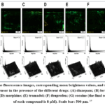 |
Figure 8: The fluorescence images, corresponding mean brightness values, and surface plots of the aptasensor in the presence of the different drugs: (A) diazepam; (B) lorazepam; (C) methadone; (D) morphine; |
Vaziri Heshi and Shokoufi (2021) introduced a fundamentally different approach: FRET‑based thermal lens spectrometry (FRET‑TLS). The photothermal lens effect amplifies the FRET signal, enabling detection of methamphetamine down to 1.5 nM. Remarkably, the method can count as few as 87 molecules in an attoliter detection volume – a level of sensitivity that was unimaginable a decade ago.
Surface‑enhanced Raman scattering (SERS) provides molecular fingerprint information, eliminating false positives from structurally similar compounds. Cui and colleagues (2022) developed Au nanocake‑enhanced SERS substrates for methamphetamine detection in hair. Using a portable Raman spectrometer, they achieved an LOD of 0.5 ppb (≈3.35 × 10⁻⁹ M) within 5 min of sample preparation. The substrates were reusable up to ten times, making the approach cost‑effective for forensic laboratories. Nanoporous Au membranes have similarly been used to detect methamphetamine, ketamine and morphine in hair with LODs in the 0.1–1 ng/mL range.
The capillary aptasensor 17 achieves a far lower LOD (0.31 pM) than the MOF‑amplified sensor (0.178 µM) – a >500‑fold difference. This reflects the advantage of the capillary geometry, which confines the excitation volume and reduces background fluorescence. However, the capillary sensor requires a specialised handheld fluorescence microscope, whereas the MOF sensor can be read on a standard plate reader. Similarly, FRET‑TLS 18 offers extreme sensitivity but requires a thermal lens spectrometer – an instrument not commonly found in forensic labs. SERS, by contrast, is increasingly portable; handheld Raman spectrometers are now commercially available, making SERS a realistic field tool.
Ultra‑high sensitivity comes with a risk of false positives from contamination or non‑specific binding. The SERS molecular fingerprint mitigates this risk, but FRET‑based methods and fluorescence quenching sensors cannot distinguish between the target and structurally similar drugs unless highly selective aptamers or antibodies are used. Additionally, hair analysis requires sample pretreatment (washing, extraction), which adds time and complexity. High‑performance optical techniques push the detection limits for illicit drugs to the picomolar and sub‑ppb range. SERS stands out as the most practical for field forensics due to its molecular specificity and the availability of portable instruments. The choice among these methods should be guided by the required LOD, available instrumentation, and the need for confirmatory identification versus simple quantification.
Limitations
Despite the considerable promise of AuNP‑based biosensors for illicit drug detection, several practical limitations currently constrain their widespread adoption outside research laboratories. These limitations relate primarily to matrix interferences, stability and shelf‑life, and regulatory acceptance.
Matrix Interferences
One of the most frequently encountered limitations is the susceptibility of AuNP‑based sensors to interferences from complex biological and environmental matrices. Urine, blood, saliva, hair extracts, and sewage samples often contain coloured compounds, high protein concentrations, variable pH, and autofluorescent species that can distort the optical readout of colorimetric or fluorescence‑based assays. For instance, the natural yellow colour of urine can obscure the red‑to‑blue transition in aggregation‑based colorimetric tests, while autofluorescence from serum proteins can interfere with fluorescence resonance energy transfer (FRET) measurements 1. Furthermore, environmental factors such as ionic strength and pH directly influence the aggregation behaviour of gold nanoparticles, potentially leading to false positive or false negative results if not carefully controlled 19. Researchers have attempted to mitigate these effects through the use of blocking agents (e.g., bovine serum albumin, casein), sample dilution buffers, and internal control lines in lateral flow devices. Nevertheless, achieving reliable performance in minimally processed real‑world samples without extensive pre‑treatment remains an ongoing challenge20.
Stability and Shelf‑Life
The long‑term stability of AuNP‑based biosensors presents another significant limitation. Although the thiol‑gold bond used to immobilise aptamers or antibodies is relatively strong, colloidal AuNP suspensions can slowly aggregate over time, particularly when exposed to elevated temperatures, freeze‑thaw cycles, or direct light. Aggregation not only alters the characteristic plasmonic colour but also reduces the effective surface area for target binding, thereby diminishing sensitivity 8. Additionally, the biological recognition elements themselves have finite operational lifetimes. Antibodies are prone to denaturation within weeks to months at room temperature, necessitating cold‑chain storage that is often unavailable in field settings. Even aptamers, which are more thermally robust, can be degraded by nucleases present in untreated biological samples. While the addition of preservatives (e.g., trehalose, sucrose) and lyophilisation have been shown to extend shelf‑life, these measures increase manufacturing complexity and cost. A promising alternative is the use of non‑biological recognition elements such as molecularly imprinted polymer nanoparticles (NanoMIPs) or catalytic nanomaterials, which offer superior stability under ambient conditions6.
Regulatory and Forensic Acceptance
Perhaps the most formidable barrier to the practical deployment of AuNP‑based drug sensors is the lack of regulatory and forensic validation. To date, the vast majority of reported devices are academic prototypes or commercial screening tools that have not undergone formal validation against established reference methods such as gas chromatography‑mass spectrometry (GC‑MS) or liquid chromatography‑mass spectrometry (LC‑MS). Forensic laboratories and court systems require rigorous demonstration of sensitivity, specificity, reproducibility, and robustness before a new analytical method can be admitted as evidence 3Consequently, AuNP‑based biosensors are currently used primarily as presumptive tests – rapid field screens that can indicate the possible presence of a drug, with confirmatory analysis still performed by laboratory‑based instrumentation. Their role is therefore complementary rather than competitive. However, as standardised protocols, quality control measures, and inter‑laboratory comparison studies emerge, these sensors may gradually gain acceptance for specific forensic applications such as on‑site seizure screening, probationary drug testing, and wastewater‑based epidemiology 1
In summary, while AuNP‑based biosensors offer exceptional analytical performance under ideal conditions, matrix effects, limited shelf‑life, and the absence of regulatory approval currently restrict their translation from research laboratories to routine forensic and clinical practice. Addressing these limitations will require continued innovation in surface chemistry, device engineering, and validation science.
Conclusions
In summary, AuNP‑based biosensors have proven to be versatile and powerful platforms for illicit drug detection, with the choice of transduction mechanism dictating a fundamental trade‑off between sensitivity and operational simplicity. Colorimetric and paper‑based sensors offer instrument‑free visual readouts ideal for point‑of‑care screening, whereas electrochemical and SERS platforms achieve sub‑ng/mL to picomolar detection limits at the cost of specialised equipment. Statistical analysis of recent studies shows that cocaine (50%) and methamphetamine (44%) are the most frequently targeted drugs, reflecting global prevalence patterns, while urine (44%) remains the dominant test matrix and aptamers (61%) have become the preferred biorecognition element due to their stability and ease of modification. Despite these advances, significant limitations persist including matrix interferences from coloured or autofluorescent biological samples, inadequate shelf‑life of AuNP conjugates and biological recognition elements, and a lack of regulatory and forensic validation that currently restricts these sensors to presumptive screening rather than confirmatory analysis. Looking forward, multiplexed platforms, sample‑to‑answer integration, and smartphone‑based artificial intelligence readouts represent the most promising directions for translating these laboratory innovations into routine forensic and clinical practice, where they will complement rather than replace gold‑standard methods such as GC‑MS and LC‑MS.
Acknowledgement
We would like to express their sincere gratitude to the Faculty of Health Sciences, Universiti Kebangsaan Malaysia (UKM), for providing academic support throughout this study. We also acknowledge the researchers whose published work formed the basis of this systematic review.
Funding Sources
The author(s) received no financial support for the research, authorship, and/or publication of this article.
Conflict of Interest
The author(s) do not have any conflict of interest.
Data Availability Statement
This statement does not apply to this article.
Ethics Statement
This research did not involve human participants, animal subjects, or any material that requires ethical approval.
Informed Consent Statement
This study did not involve human participants, and therefore, informed consent was not required.
Authors’ Contributions
- Nurin Haziqah Razali conceived and designed the study, conducted the literature search, performed study selection, data extraction, formal analysis, and visualization, and prepared the original draft of the manuscript.
- Atikah Mohd Nasir supervised the research, contributed to the study design and validation of the findings, critically reviewed and revised the manuscript for important intellectual content, managed the project, and approved the final version of the manuscript for publication.
References
- Chen, C. H.; Wang, C. C.; Ko, P. Y.; Chen, Y. L. Nanomaterial-Based Adsorbents and Optical Sensors for Illicit Drug Analysis. Journal of Food and Drug Analysis. Taiwan Food and Drug Administration 2020, pp 654–676. https://doi.org/10.38212/2224-6614.1137.
CrossRef - Zhao, Y.; Gan, Y.; Chen, J.; Zheng, H.; Chang, Y.; Lin, C. Recent Reports on the Sensing Strategy and the On-Site Detection of Illegal Drugs. RSC Advances. Royal Society of Chemistry February 26, 2024, pp 6917–6929. https://doi.org/10.1039/d3ra06931a.
CrossRef - Askar, A. M.; Al Ali, A. Y.; Khalifa, M. K.; Salem, A. A.; Alkhuwaildi, B. M.; Shah, I. Rapid GC-MS Method for Screening Seized Drugs in Forensic Investigations: Optimization and Validation. Front. Chem. 2025, 13. https://doi.org/10.3389/fchem.2025.1559279.
CrossRef - Rosendo, L. M.; Antunes, M.; Simão, A. Y.; Brinca, A. T.; Catarro, G.; Pelixo, R.; Martinho, J.; Pires, B.; Soares, S.; Cascalheira, J. F.; Passarinha, L.; Rosado, T.; Barroso, M.; Gallardo, E. Sensors in the Detection of Abused Substances in Forensic Contexts: A Comprehensive Review. Micromachines. Multidisciplinary Digital Publishing Institute (MDPI) December 1, 2023. https://doi.org/10.3390/mi14122249.
CrossRef - Garrido, E.; Pla, L.; Lozano‐Torres, B.; El Sayed, S.; Martínez‐Máñez, R.; Sancenón, F. Chromogenic and Fluorogenic Probes for the Detection of Illicit Drugs. ChemistryOpen 2018, 7 (5), 401–428. https://doi.org/10.1002/open.201800034.
CrossRef - Mao, K.; Zhang, H.; Pan, Y.; Zhang, K.; Cao, H.; Li, X.; Yang, Z. Nanomaterial-Based Aptamer Sensors for Analysis of Illicit Drugs and Evaluation of Drugs Consumption for Wastewater-Based Epidemiology. TrAC – Trends in Analytical Chemistry. Elsevier B.V. September 1, 2020. https://doi.org/10.1016/j.trac.2020.115975.
CrossRef - Mao, K.; Yang, Z.; Li, J.; Zhou, X.; Li, X.; Hu, J. A Novel Colorimetric Biosensor Based on Non-Aggregated Au@Ag Core–Shell Nanoparticles for Methamphetamine and Cocaine Detection. Talanta 2017, 175, 338–346. https://doi.org/10.1016/j.talanta.2017.07.011.
CrossRef - Mahato, K.; Nagpal, S.; Shah, M. A.; Srivastava, A.; Maurya, P. K.; Roy, S.; Jaiswal, A.; Singh, R.; Chandra, P. Gold Nanoparticle Surface Engineering Strategies and Their Applications in Biomedicine and Diagnostics. 3 Biotech. Springer Verlag February 1, 2019. https://doi.org/10.1007/s13205-019-1577-z.
CrossRef - Erkocyigit, B.; Man, E.; Efecan, E.; Ozufuklar, O.; Devecioglu, D.; Bagci, B.; Aldemir, E.; Coskunol, H.; Evran, S.; Guler Celik, E. Non-Invasive Point-of-Care Detection of Methamphetamine and Cocaine via Aptamer-Based Lateral Flow Test. Biosensors (Basel). 2025, 15 (1). https://doi.org/10.3390/bios15010031.
CrossRef - Sen, A.; Sester, C.; Poulsen, H.; Hodgkiss, J. M. Accounting for Interaction Kinetics between Gold Nanoparticles and Aptamers Enables High-Performance Colorimetric Sensors. ACS Appl. Mater. Interfaces 2022, 14 (29), 32813–32822. https://doi.org/10.1021/acsami.2c04747.
CrossRef - Li, Z.; Chen, H.; Feng, S.; Liu, K.; Wang, P. Development and Clinical Validation of a Sensitive Lateral Flow Assay for Rapid Urine Fentanyl Screening in the Emergency Department. Clin. Chem. 2020, 66 (2), 324–332. https://doi.org/10.1093/clinchem/hvz023.
CrossRef - Wang, L.; McCord, B. A Four-Channel Paper Microfluidic Device with Gold Nanoparticles and Aptamers for Seized Drugs. Anal. Biochem. 2020, 595. https://doi.org/10.1016/j.ab.2020.113619.
CrossRef - Jing, L.; Xie, C. Y.; Li, Q. Q.; Yao, H. F.; Yang, M. Q.; Li, H.; Xia, F.; Li, S. G. A Sandwich-Type Lateral Flow Strip Using a Split, Single Aptamer for Point-of-Care Detection of Cocaine. J. Anal. Test. 2022, 6 (2), 120–128. https://doi.org/10.1007/s41664-022-00228-w.
CrossRef - D’Aurelio, R.; Tothill, I. E.; Salbini, M.; Calò, F.; Mazzotta, E.; Malitesta, C.; Chianella, I. A Comparison of EIS and QCM NanoMIP-Based Sensors for Morphine. Nanomaterials 2021, 11 (12). https://doi.org/10.3390/nano11123360.
CrossRef - Bor, G.; Bulut, U.; Man, E.; Balaban Hanoglu, S.; Evran, S.; Timur, S. Synthetic Antibodies for Methamphetamine Analysis: Design of High Affinity Aptamers and Their Use in Electrochemical Biosensors. Journal of Electroanalytical Chemistry 2022, 921, 116686. https://doi.org/10.1016/j.jelechem.2022.116686.
CrossRef - Anbiaee, G.; Khoshbin, Z.; Zamanian, J.; Samie, A.; Ramezani, M.; Alibolandi, M.; Danesh, N. M.; Taghdisi, S. M.; Abnous, K. A Fluorescent Aptasensor for Quantification of Cocaine Mediated by Signal Amplification Characteristics of UiO-66/AuNPs Nanocomposite. Anal. Biochem. 2023, 674, 115193. https://doi.org/10.1016/j.ab.2023.115193.
CrossRef - Zamanian, J.; Khoshbin, Z.; Hosseinzadeh, H.; Danesh, N. M.; Khakshour Abdolabadi, A.; Abnous, K.; Taghdisi, S. M. An Ultrasensitive Detection Platform for Cocaine: Aptasensing Strategy in Capillary Tube. Front. Chem. 2022, 10. https://doi.org/10.3389/fchem.2022.996358.
CrossRef - Heshi, S. V.; Shokoufi, N. Fluorescence Resonance Energy Transfer-Thermal Lens Spectrometry (FRET-TLS) as Molecular Counting of Methamphetamine. https://doi.org/10.1007/s00604-021-04842-w/Published.
- Zahra, Q. U. A.; Luo, Z.; Ali, R.; Khan, M. I.; Li, F.; Qiu, B. Advances in Gold Nanoparticles‐based Colorimetric Aptasensors for the Detection of Antibiotics: An Overview of the Past Decade. Nanomaterials. MDPI AG April 1, 2021. https://doi.org/10.3390/nano11040840.
CrossRef - Mir, T. ul G.; Wani, A. K.; Akhtar, N.; Katoch, V.; Shukla, S.; Kadam, U. S.; Hong, J. C. Advancing Biological Investigations Using Portable Sensors for Detection of Sensitive Samples. Heliyon 2023, 9 (12), e22679. https://doi.org/10.1016/J.HELIYON.2023.E22679.
CrossRef
Accepted on: 25 Jul 2026


ISSN Online: 2231-5039