In silico Design of N-Substituted Quinoline-Isatin Hybrids for Multiple Targeted enzymes for Anti-Microbial Activity
 , Revathi Gnanavelou, Sowmiya Perinbaraj, Hemachandran Vinayagam, Aruvisal Hananth Arun Dayanand and Konda Reddy Girija*
Department of Pharmaceutical Chemistry, College of Pharmacy, Mother Theresa Post Graduate, and Research Institute of Health Science. (A. Govt. Puducherry Institution) Puducherry, India.
Corresponding Author E-mail:girijanarasimhan66@gmail.com
DOI : http://dx.doi.org/10.13005/ojc/420410
Download this article as:

The World Health Organization reports that approximately 700,000 people are affected by Antimicrobial Resistance (AMR) each year. This growing concern has accelerated the search for new antibacterial agents. Fluoroquinolones are key broad-spectrum antibiotics and recent interest in Isatin derivatives has revealed promising antimicrobial activity. Combining quinoline and Isatin pharmacophores offers a strategy to develop novel agents with unique mechanisms to overcome resistance. Based on SAR and literature data, a series of N-substituted quinoline-isatin conjugates were designed to target bacterial enzymes such as Enoyl-ACP reductase, BioA, and DNA gyrase. Docking studies identified QI7f as a lead compound with superior binding scores (-10.35, -12.38, -8.93 kcal/mol) compared to Standards. In silico ADMET analysis predicted favorable pharmacokinetic and toxicity profiles. The compounds were synthesized and structurally confirmed via FTIR, NMR, and mass spectrometry. Q17f showed notable in-vitro antimicrobial activity (2-10 mg/mL) comparable to gentamicin, likely due to electron-withdrawing groups enhancing target interactions.
KEYWORDS:Antimicrobial resistance; Isatin; Molecular hybridization; Quinoline
Introduction
Mycobacterium tuberculosis is the primary cause of tuberculosis (TB), a chronic infectious disease. Although it primarily affects the pulmonary system, it can spread to other organs such as the brain, spine, and kidneys. spread by airborne droplets released during a cough, sneeze, or speech by an infected individual. It is one of the most deadly infectious illnesses in the world, especially in low-income areas.Annually, over 10 million individuals are diagnosed with active TB, resulting in approximately 1.5 million fatalities.2 Although TB is treatable with antibiotics, the emergence of drug-resistant strains complicates its management. Drug-resistant tuberculosis (DR-TB) presents an escalating public health challenge, underscoring the necessity for novel therapeutic interventions3. In 2021, According to the World Health Organization (WHO), there were 1.6 million TB-related fatalities and 11 million new TB infections. Due to resistance to second-line medications, multidrug-resistant TB is a more severe form of the disease with greater fatality rates. 4 Isoniazid, Rifampicin, Pyrazinamide, and Ethambutol are among the first-line drugs for tuberculosis. While XDR-TB shows resistance to these and other important medications, MDR-TB is characterized by resistance to at least isoniazid and rifampicin. Despite the essential role of these first-line drugs in treatment, they can induce significant adverse effects, such as hepatotoxicity, neurotoxicity, and visual impairment, potentially affecting patient adherence and contributing to resistance. Over the past five decades, The USFDA, or US Food and Drug Administration, has authorized only a limited number of new anti-TB agents, highlighting the substantial challenges in developing novel treatments.5 Bedaquiline, a newer medication for MDR-TB and XDR-TB, disrupts bacterial energy production; however, resistance to bedaquiline has emerged, raising concerns about its efficacy. Bedaquiline should be administered in conjunction with other effective drugs and under close monitoring.6
To address the rise of drug-resistant TB, this study targeted on development of innovative Anti-TB medications, specifically N-substituted quinoline-isatin conjugate derivatives. Quinoline and isatin are recognized for their antibacterial properties.7 By combining these two compounds, there is a promising opportunity to develop new drugs with enhanced efficacy and safety. Molecular hybridization is a strategic approach in drug design, wherein pharmacophoric elements from various bioactive compounds are combined to form new hybrid entities. The objective of these hybrids is to improvise their affinity, efficacy, and selectivity while minimizing adverse drug effects compared to their original counterparts. This approach is particularly advantageous for creating multitarget drugs that can address complex diseases by simultaneously engaging with multiple targets.8, 9
Design of N-Substituted Quinoline-Isatin Hybrids
Quinoline moiety: The quinoline moiety, encompassing both natural and synthetic compounds (Figure 1), has significantly contributed to medicinal advancements over the past five decades. The introduction of various functional groups to the quinoline structure has profoundly influenced its biological activities.10 Numerous Quinoline and quinolone derivatives exhibit a broad spectrum of biological effect , including antituberculosis,11 anticancer,12 antimalarial, 13antimicrobial,14 anti-inflammatory, and antiviral properties.15
N-Substitution on Quinoline: According to literature and structure-activity relationship (SAR) studies, the Introducing various substituents (alkyl, aryl, or heterocyclic groups) at the nitrogen atom of the quinoline ring can:
Enhance lipophilicity or hydrophilicity.
Improve cell permeability.
Modulate interaction with the biological target.16
Isatin moiety: Isatin derivatives have shown potential as inhibitors of enoyl-ACP reductase (InhA), an enzyme associated with bacterial “fatty acid metabolic pathway”.22 By attaching isatin scaffold to the quinoline ring, the hybrid molecule could exhibit dual-target antibacterial activity. The isatin group may act as a lead compound for targeting InhA. Due to its structural similarity to the enzyme’s natural substrate, it is a potential target for drug development, anti-tuberculosis activity, and anti-bacterial activity.17
Linker: Schiff bases are versatile compounds widely used as linkers in hybrid molecules for enhancing biological activity. Their imine (-C=N-) functional group plays a crucial role in bioactivity, enabling Schiff base hybrids to exhibit diverse pharmacological actions such as antimicrobial, anticancer, antioxidant, and anti-inflammatory effects.18, 19
Design Strategy
Keep considering the biological importance of the two chemical moietie isatin and quinoline. Thus, the current work describes the production of new hybrids for anti-tubercular activity.
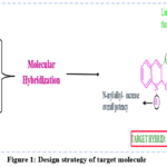 |
Figure 1: Design strategy of target molecule Click here to View Figure |
The development of molecular hybridization technique, combining quinoline’s membrane-disrupting properties with isatin’s enzymatic inhibition, could accelerate progress towards WHO’s 2035 TB elimination targets.20
Materials and Methods
All compounds were designed using ChemSketch software. Molecular docking analyses of the developed molecules were carried with Molinspiration and Swiss ADME employed to evaluate” drug-likeness ,drug absorption and distribution characteristics parameters.
Experimental Section
Design of Compounds
Considering the benefit offered by this hybridization technique in tackling tuberculosis, and based on a comprehensive literature review and assessment of synthetic feasibility, the scaffolds Quinoline and Isatin were selected and designed for further investigation on this research work. (scheme1 and table 1).
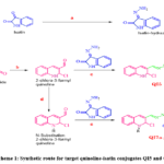 |
Scheme 1: Synthetic route for target quinoline-isatin conjugates QI5 and QI7a-j. Click here to View Scheme |
Reagents and conditions: a) Ethanol hydrazine hydrate (99%) was heated for 10 min. b) DMF, POCl₃, 80-90°C reflux 4-16 hrs. c) Ethanol, glacial acetic acid, reflux for 2-4 hrs. d) DMF, K₂CO₃, R-X (alkyl/aryl halide), reflux for 3-4 hrs.
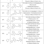 |
Table 1: Structure and scheme details of designed compounds Click here to View Table |
In silico Studies
Molecular docking is a method that investigates the compatibility of different molecular structures, including drugs, enzymes, or proteins, to gain insights into their interactions and how they fit together. Docking is a molecular modeling technique used to predict the interactions between proteins (enzymes) and small molecules (ligands). The main goal of this approach is to identify the precise locations of ligands within a protein’s binding pocket and evaluate the strength of the interactions between the ligand and the protein.21
Protein Preparation
This study employed AUTODOCK 1.5.7 to carry out Molecular Docking analysis. Crystallographic structures of the target molecules were saved in a standard 3D format after being taken from the Protein Data Bank (PDB). The Multiple Target of Mycobacterium tuberculosis-InhA protein complexed with NADH (PDB ID: 4DRE) was the main focus of this investigation. BIOA (DAPA synthase). (PDB-ID-6GE8), DNAgyrase. (PDB-ID: 3M4I). 22 The active sites of the target proteins were identified using the online Pdbsum online tool.23 Before initiating the docking procedure, the water molecules and heteroatoms were removed from the target protein.
Ligand Preparation
Marvin sketch/chem Sketch software was used to draw the structure of the Quinoline-isatin hybrids, and a 3D structure was generated and saved in PDB format.24
Grid Generation
Utilizing a technique called grid box generation, which functions in three dimensions (3D), molecules are systematically arranged within a cubic grid. This method evaluates interaction energies at each grid point using a predetermined probe. A 3D grid box of 60 × 60 × 60 in with a spacing of 0. A total of 375 points were established, specifying the (x, y, z) coordinates. The docking parameters for the x-, y-, and z-axes are defined as 4. 787, 64. 876, and 56. 214, respectively. The AutoGrid application played a crucial role in selecting macromolecules and ligands in the PDBQT format. Subsequently, a grid parameter file (GPF) was generated, and the associated grid log file (GLG) from AutoGrid was executed.25, 26
Docking
Auto Dock Tools is an established program for automated docking known for its effectiveness in conformation searches, especially for ligands with approximately 10 rotatable bonds. The protein and ligand were chosen from the selections provided by AutoGrid and subjected to a docking process. This involved conducting 25 runs of the Genetic Algorithm (GA), which produced a docking parameter file (DPF) and log file for docking (DLG). In order to record the final docked coordinates,AUTODOCK in a docking log file (DLG). 26
Docking analysis
This process is crucial in drug discovery as it helps predict the binding affinity and mode of interaction between a ligand (small molecule) and a protein receptor is analysed by Discovery Studio of the docked compounds.27
Evaluation of Lipinski’s rule of five
The Swiss ADME online tool was used to assess the developed compounds’ drug-likeness characteristics and Lipinski’s rule of five. Various properties, such as TPSA, number of rotatable bonds, log p, molecular weight, hydrogen bond acceptor, and donor were predicted. This rule is applied to assess whether a compound meets specific physicochemical criteria that favor oral absorption.28
in silico ADME Prediction
The Swiss ADME online program was used for ADME screening in order to forecast the physicochemical and pharmacokinetic, Lipophilicity and Water solubility of Quinoline-Isatin conjugate derivatives. Whether the compound inhibits cytochrome P450 could also be envisaged. Other attributes including skin penetration, blood-brain barrier penetration, gastrointestinal absorption, and Lipinski, Ghose, and Veber-like drug-likeness properties, can also be predicted.29
Bioactivity
The Molinspiration online platform was used to analyze ion channels, nuclear ligand receptors, proteases, tyrosine kinase-linked receptors, enzyme inhibitors, and G-protein-coupled receptors.30
Toxicity Assessment
The toxicity profile Mutagenicity, tumorigenicity, irritancy, and reproductive effects of the designed N-Substituted quinoline-isatin hybrids were predicted using Osiris property explorer.31
Antimicrobial Study
The antibacterial activities of the synthesized compounds against Gram-positive (Staphylococcus aureus and Enterococcus faecalis) and gram-negative (Escherichia coli) bacteria were investigated in vitro using the agar well diffusion method.
Media used
For antibacterial screening, nutrient media gelled with 2% agar (bacteriological grade) was utilized. The following components are present in the nutritional media:
Nutrient broth: 13g/l
Agar: 15.00 g/l
Final pH at 25°C: 7.4(±0.2)
Media Preparation and Sterilization
Heat was used to dissolve the components in distilled water, then diluted acid or alkali was used to bring the pH down to 7.4 (±0.2). 20–25 milliliters of nutritional After being moved to Petri plates, the medium was sealed. The medium was autoclaved for 15 psi (121°C) of pressure.
15 min. min (approximately 25 min), respectively.
Microorganisms used: Gram-negative Escherichia coli, gram-positive Staphylococcus aureus, and gram-positive Enterococcus faecalis.
Test drug: Synthesized compound used QI7f
Standard drug: Gentamicin 10 mg/ml
Concentration used: 2 mg/ml,4 mg/ml, 6 mg/ml and ,10 mg/ml
Control: DMSO
Solvent used: DMSO
Method: Agar well diffusion method
Gram negative (Escherichia coli) and gram positive (Stapylococcus aureus and Enterococcus faecalis) bacteria were tested for in vitro antibacterial activity using the synthesized compounds.
organisms using well diffusion method at The concentrations were 2 mg/ml, 4 mg/ml, 6 mg/ml, and 10 mg/ml. Zones of inhibition for compounds larger than 15 mm were
quantitatively studied.
Nutrient medium plates were prepared aseptically and dried at 37°C before inoculation. Microrganisms Gram-negative Escherichia coli, gram-positive Staphylococcus aureus, and gram-positive Enterococcus faecalis were inoculated utilising a aproach that involved removing excess inoculums, streaking the medium, and drying at room temperature. The compound QI7f were poured into wells, and plates were incubated at 37°C for 18 to 24 hours. The test compound’s zone of inhibition was measured and evaluated. with that of the standard drug gentamicin (10 mg/ml), as outlined in the experimental protocol.32
Result and Disscussion
Design of Compounds
A Series of quinolone-isatin conjugate derivatives were desgined successfully. (scheme1 and table1)
Molecular Docking Study
The docking studies were performed for all the designed compounds using AUTODOCK software version 1.5.7. to predict the interaction between the designed ligands and the targeted enzymes – Eonyl-ACP reductase (INHA), BIOA (DAPA synthase), DNA gyrase (table 2, table 3 and table 4.)
Eonyl-ACP reductase (INHA): The binding energy of all the The range of the intended compounds was -8.49 to -10.35 kcal/mol. With an interatomic distance of 1.923A, the molecule QI7f established a single hydrogen bond with the residue Ile194 and demonstrated the best binding affinity of -10.35 kcal/mol.
compared with standard Isoniazid (-5.66 kcal/mol) produced two hydrogen bonds Ile194 and Val65 with the distance 1.769, 2.104 respectively. The result showed that the Similar amino acid residues Ile194 appeared in the test and standard compound can be considered for validation.
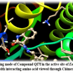 |
Figure 2: 3D Binding mode of Compound QI7f in the active site of Eonyl-ACP reductase (INHA), along with interacting amino acid viewed through Chimera 1.17 software. Click here to View Figure |
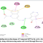 |
Figure 3: 2D Binding interaction image of Compound QI7f in the active site of Eonyl-ACP reductase (INHA), along with interacting amino acid viewed through Discovery Studio software. |
Binding Energy: -10.80;
H-Bond Interaction: Lysa:165, Ile A:194;
H-Bond Distance A0: 2.169, 2.072
BioA 7,8-diamino-pelargonic acid (DAPA) synthase: The binding energy of all the designed compounds ranged from -9.10 to -12.38 kcal/mol. The compound QI7i showed best binding affinity of -12.38 kcal/mol formed two hydrogen bonds with the residue Lys283, Leu292 with the interatomic distance of 993 A0 and 2.071 A0 compared with standard Isoniazid (-6.05 kcal/mol) which produced three hydrogen bonds- Lys283, Thr318(A) and Thr318(B) with the distance 2.492, 1.952, 1.827 respectively. The result showed that the Similar amino acid residues Lys283 appeared in the test and standard compound can be considered for validation.
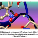 |
Figure 4: 3D Binding mode of Compound QI7i in the active site of BioA 7,8-diamino-pelargonic acid (DAPA) synthase along with interacting amino acid viewed through Chimera 1.17 software. |
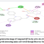 |
Figure 5: 2D Binding interaction image of Compound QI7i in the active site of Eonyl-ACP reductase (INHA), along with interacting amino acid viewed through Discovery Studio software. |
Binding Energy: -12.38
H-Bond Interaction: Lys283, Leu292
H-Bond Distance A0: 1.993, 2.071
DNA-Gyrase:The binding energy of all the designed compounds ranged from -7.51 to -8.93 kcal/mol. The compound QI7f showed best binding affinity of -8.93 kcal/mol generate a hydrogen bond with the Gln565 residue at an interatomic distance of 2.048 A0 compared with standard Moxifloxacin (-5.66 kcal/mol) produced two hydrogen bonds Gln565 and Ser541 with the distance 1.866, 1.853 respectively. The result showed that the Similar amino acid residues Gln565 appeared in the test and standard compound can be considered for validation.
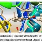 |
Figure 6: 3D Binding mode of Compound QI7f in the active site of DNA-Gyrase along with interacting amino acid viewed through Chimera 1.17 software. Click here to View Figure |
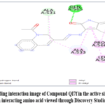 |
Figure 7: 2D Binding interaction image of Compound QI7f in the active site of DNA-Gyrase along with interacting amino acid viewed through Discovery Studio software. Click here to View Figure |
Binding Energy: -8.93
H-Bond Interaction: Gln565
H-Bond Distance A0: 2.048
Evaluation of Lipinski’s Rule of Five
The Designed compounds’ molecular properties revealed partition coefficient values ranging from 2.45 to 3.64, which suggested they were naturally lipophilic and therefore changed the bioavailability and pharmacokinetic. The molecules that are responsible for bioavailability range from strongly to moderately polar, according to the TPSA values, which range from 57.06 Ų to 102.88 Ų. The compounds’ molar weights range from 352.40 to 405.29 Da. which is smaller than 500 Da indicates that the molecules are more readily absorbed, distributed, and generally bioavailable, making them rapidly soluble and eliminated. The Lipinski Rule of Five states that compounds having flexible linkages, H-bond donors, and H-bond acceptors are within the permissible range for oral delivered medications. When the number of rotatable bonds is within a specific threshold, typically fewer than 10, it indicates that the molecule may still possess satisfactory bioavailability, as they were found to be within the acceptable range. The H bond donors and acceptors have a major impact on oral bioavailability, permeability, and solubility, which results in decreased effectiveness together result in good absorption therefore the compounds shows good bioavailability. The evaluation’s findings are listed in Table5.
Evaluation of drug-likeness
The assessment of drug-likeness, including the bioavailability and the Lipinski, Ghose, Veber, and Muegge rules score, indicates that all the designed compounds comply with these criteria and fall within the bioavailability score of 0.055 (Table 6).
In silico ADME Prediction
The ADME characteristics of the compounds that were evaluated using the SWISS ADME software. The compounds that were designed exhibited a high gastrointestinal absorption. All compounds developed are less likely to be transported by P-glycoprotein, which may result in improved absorption and distribution. For a drug to be both effective and safe, it should exhibit minimal interactions with cytochrome P450 (CYP) enzymes to prevent drug-drug interactions and potential toxicity. All developed quinolone-isatin conjugates are unlikely to inhibit CYP1A2 and CYP2D6, indicating that they are expected to have a negligible effect on the metabolism of drugs that are substrates for these enzymes. Table 7 presents the results of this assessment.
Lipophilicity and water solubility
The lipophilicity and solubility of a compound play a vital part in determining its absorption and bioavailability. The data revealed that each of the developed compounds could successfully penetrate biological membranes and demonstrated moderate solubility. (Table 8)
Bioactivity
The observed range of GPCR ligand values fell within the moderately active category (-0. 23 to 0. 49), suggesting minimal interactions. GPCRs may function as potent antagonists or weak agonists. Similarly, significant interactions were noted between enzyme and kinase inhibition. In contrast, ion channel receptors, nuclear receptor ligands, and protease modulators exhibited moderate to low bioactive effects, indicating that these compounds also demonstrated weak inhibitory properties. Table 9 presents the results of this assessment.
Toxicity Properties
The OSIRIS server serves as an online tool for forecasting toxicity parameters. The findings indicated that all the designed hybrids were falls on Low-risk in terms of Mutagenic, Tumorogenic, Irritant, and Reproductive effects, except for hybrid QI7d, which exhibited toxicity in these areas and falls on High-risk. This information is illustrated in the accompanying table 10, where the green circle denotes nontoxicity.
Table 2: Molecular interaction of designed ligand with Mycobacterium tuberculosis InhA (ENOYL-ACP REDUCTASE) in associates with NADH. PDB-ID: 4DRE
|
Compound Code |
No. of hydrogen
bond formed |
H-bond interaction | H-bond distance A0 | Binding Energy
(kcal/mol) |
|
QI5 |
1 | Val65 | 2.194 |
-8.49 |
| QI7a | 2 | Try158, Ile194 | 1.905, 1.786 |
-9.44 |
|
QI 7b |
2 | Try158, Ile194 | 1.802, 2.055 | 9.91 |
| QI7c | 2 | Gly14, Thr39 | 2.169, 2.072 |
-9.28 |
|
QI7d |
2 | Try158, Ile194 | 1.953,2.206 | -9.41 |
| QI7e | 2 | Try158, | 1.919, 1.938 |
-10.13 |
|
QI7f |
1 | Ile194 | 1.923 | -10.35 |
| QI7g | 1 | Ile194 | 1.772 |
-9.29 |
|
QI7h |
1 | Try158 | 1.921 | -9.15 |
| QI7i | 3 | Ile21, Try158
Ile194 |
1.731, 2.232
2.173 |
-9.71 |
|
QI7j |
1 | Ala22 | 2.076 | -9.32 |
| Isoniazid | 2 | Ile194, Val65 | 1.769, 2.104 |
-5.66 |
Table.3: Molecular interaction of synthesized substances in the active region of the protein mycobacterium tuberculosis BIOA (DAPA SYNTHASE). (6GE8)
|
Compound Code |
No. of hydrogen bond formed |
H-bond interaction |
H-bond distance A0 |
Binding Energy (kcal/mol) |
| QI5 | 1 | Lys283 | 2.067 | -9.10 |
| QI7a | 2 | Thr318,Phe319 | 2.248, 2.226 | -11.72 |
| QI7b | 1 | Phe319 | 2.139 | -10.85 |
| QI7c | 1 | Ser123 | 1.804 | -1.28 |
| QI7d | 1 | Phe319 | 2.187 | -11.74 |
| QI7e | 2 | Lys283, Thr318 | 2.21, 1.979 | -11.25 |
| QI7f | 1 | Lys283 | 2.174 | -10.92 |
| QI7g | 1 | Ser125 | 1.882 | -10.18 |
| QI7h | 3 | Thr318, Phe319,
Thr318 |
2.038, 1.961
1.9089 |
-11.82 |
| QI7i | 2 | Lys283, Leu292 | 1.993, 2.071 | -12.38 |
| QI7j | 3 | Thr318, Phe319, Thr318 | 2.015, 2.240
2.094 |
-10.98 |
| Isoniazid | 3 | Lys283, Thr318,
Thr318 |
1.952, 1.827, 2.492 | -6.05 |
Table 4: Molecular interaction of ligand compounds in active region of the protein mycobacterium tuberculosis DNA gyrase. (3M4I)
|
Compound Code |
No. of hydrogen bond formed | H-bond interaction | H-bond distance | Binding Energy
(kcal/mol) |
| QI5 | 2 | Ala531, Ser541 | 2.213, 1.962 | -8.11 |
| QI7a | 1 | Ser541 | 2.073 | -7.55 |
| QI7b | 1 | Ser541 | 2.126 | -8.10 |
| QI7c | 1 | Gln565 | 2.237 | -7.76 |
| QI7d | 1 | Gln565 | 2.093 | -8.68 |
| QI7e | 2 | Ala531, Ser541 | 1.779, 2.189 | -7.99 |
| QI7f | 1 | Gln565 | 2.048 | -8.93 |
| QI7g | 1 | Gln565 | 1.907 | -8.33 |
| QI7h | 1 | Ala531 | 1.972 | -7.59 |
| QI7i | 1 | Ser541 | 2.009 | -7.51 |
| QI7j | 1 | Ser541 | 2.0 | -7.61 |
| Moxifloxacin | 2 | Ser541, Gln565 | 1.866, 1.853 | -6.87 |
Table 5: Lipinski’s rule of five using Swiss ADME software
|
Compound code |
Molecular weight
(Gram) gram (g/ mol) |
Molecular
formula |
TPSA | HBA | HBD | RB |
LOGP |
|
QI5 |
316.36 | C19H16N4O | 65.85 Ų | 3 | 2 | 2 | 2.45 |
| QI7a | 414.89 | C24H17ClN4O | 57.06 Ų | 3 | 1 | 3 |
2.88 |
|
QI7b |
449.33 | C24H17Cl2N4O | 57.06 Ų | 3 | 1 | 3 | 3.30 |
| QI7c | 428.91 | C25H21ClN4O | 57.06 Ų | 3 | 1 | 3 |
3.64 |
|
QI7d |
429.90 | C24H20ClN5O | 83.08 Ų | 3 | 2 | 3 | 2.80 |
| QI7e | 426.90 | C25H19ClN4O | 57.06 Ų | 3 | 1 | 4 |
3.30 |
|
QI7f |
440.88 | C25H17ClN4O2 | 74.13 Ų | 4 | 1 | 4 | 2.84 |
| QI7g | 380.83 | C20H17ClN4O | 74.13 Ų | 4 | 1 | 3 |
2.60 |
|
QI7h |
415.87 | C23H18ClN5O | 69.95 Ų | 4 | 1 | 3 | 3.00 |
| QI7i | 457.87 | C24H16ClN5O3 | 102.88 Ų | 5 | 1 | 4 |
2.68 |
|
QI7j |
366.84 | C20H19ClN4O | 57.06 Ų | 3 | 1 | 3 |
2.89 |
Table 6: Drug likeness of designed compounds using Swiss ADME software
|
Compound code |
Lipinski | Ghose | Veber | Egan | Muegge | Bio-availability score |
| QI5 | yes | yes | yes | yes | yes |
0.55 |
|
QI7a |
yes | yes | yes | yes | yes | 0.55 |
| QI7b | yes | no | yes | yes | No |
0.55 |
|
QI7c |
yes | no | yes | yes | yes | 0.55 |
| QI7d | yes | no | yes | yes | yes |
0.55 |
|
QI7e |
yes | no | yes | yes | no | 0.55 |
| QI7f
|
yes
|
no
|
yes
|
yes
|
yes
|
0.55
|
|
QI7g |
yes | yes | yes | yes | yes | 0.55 |
| QI7h | yes | yes | yes | yes | yes |
0.55 |
|
QI7i |
yes | no | yes | yes | yes | 0.55 |
| QI7j | yes | yes | yes | yes | yes |
0.55 |
Table 7: Insilico drug pharmacokinetics of designed compounds using Swiss ADME software
|
Compound code |
GI | BBB | P-gp | CYP1A2 | CYP2D6 | CYP3A4 | Log Kp
(cm\s) |
| QI5 | High | Yes | No | Yes | No | Yes |
-6.04 |
|
QI7a |
High | Yes | No | Yes | No | Yes | -5.54 |
| QI7b | High | Yes | No | Yes | No | Yes |
-5.31 |
|
QI7c |
High | Yes | No | Yes | No | Yes | -5.37 |
| QI7d | High | Yes | No | Yes | No | Yes |
-6.12 |
|
QI7e |
High | Yes | No | No | No | Yes | -5.32 |
| QI7f | High | Yes | No | No | No | Yes |
-5.83 |
|
QI7g |
High | Yes | No | No | No | Yes | -6.98 |
| QI7h | High | Yes | No | Yes | No | Yes |
-6.31 |
|
QI7i |
High | No | No | No | No | Yes | -5.94 |
| QI7j
|
High
|
Yes
|
No
|
Yes
|
No
|
Yes
|
-6.10
|
Table 8: Lipophilicity and water solubility of designed compounds using swiss adme software
|
Compound Code |
|
Lipophilicity logpo/w |
Water solubility (logs)
|
||||||
| I
log p |
X logp3 | W log p | M
log p |
Silics -IT | Cons. Log p | E sol | Ali | Silicos- IT | |
| QI5 | 2.45 | 3.09 | 2.06 | 2.13 | 3.68 | 2.68 | -3.99 | -4.14 | -6.30 |
| QI7a | 3.47 | 4.63 | 4.63 | 3.29 | 4.44 | 3.98 | -5.43 | -5.55 | -6.91 |
| QI7b | 3.77 | 5.26 | 4.73 | 3.76 | 5.08 | 4.52 | -6.03 | -6.21 | -7.50 |
| QI7c | 3.64 | 4.99 | 4.39 | 3.49 | 4.96 | 4.29 | -5.73 | -5.93 | -7.29 |
| QI7d | 2.80 | 3.95 | 3.67 | 2.74 | 3.72 | 3.38 | -5.08 | -5.39 | -6.54 |
| QI7e | 3.30 | 5.05 | 3.87 | 3.42 | 5.17 | 4.16 | -5.83 | -5.99 | -8.65 |
| QI7f | 3.37 | 5.68 | 4.49 | 3.55 | 5.40 | 4.50 | -5.53 | -5.73 | -8.19 |
| QI7g | 2.60 | 2.31 | 2.42 | 1.90 | 3.33 | 2.51 | -3.62 | -3.51 | -4.76 |
| QI7h | 3.00 | 3.56 | 3.47 | 2.26 | 3.87 | 3.23 | -4.76 | -4.71 | -6.54 |
| QI7i | 2.68 | 4.94 | 4.58 | 2.50 | 3.00 | 3.54 | -5.93 | -6.84 | 8.08 |
| QI7j | 2.89 | 3.44 | 2.89 | 2.38 | 3.79 | 3.08 | -4.25 | -4.32 | -5.23 |
Table 9: Bioactivity of designed compounds using Molinspiration software
|
Compound code |
GPCR
Ligand |
Ion channel
Modulator |
Kinase
Inhibitor |
Nuclear
Receptor Ligand |
Protease
Inhibitor |
Enzyme |
|
QI5 |
-0.49 | -0.68 | -0.25 | -0.78 | -0.92 | -0.34 |
| QI7a | -0.33 | -0.51 | -0.13 | -0.56 | -0.75 |
-0.20 |
|
QI7b |
-0.33 | -0.50 | -0.13 | -0.57 | -0.78 | -0.24 |
| QI7c | -0.35 | -0.55 | -0.16 | -0.57 | -0.77 |
-0.24 |
|
QI7d |
-0.28 | -0.44 | -0.05 | -0.59 | -0.65 | -0.12 |
| QI7e | -0.31 | -0.49 | -0.19 | -0.55 | -0.68 |
-0.22 |
|
QI7f |
-0.33 | -0.52 | -0.18 | -0.60 | -0.68 | -0.26 |
| QI7g | -0.41 | -0.64 | -0.29 | -0.76 | -0.80 |
-0.32 |
|
QI7h |
-0.23 | -0.43 | 0.03 | -0.61 | -0.67 | -0.15 |
| QI7i | -0.41 | -0.50 | -0.23 | -0.58 | -0.79 |
-0.26 |
|
QI7j |
-0.39 | -0.62 | -0.26 | -0.68 | -0.85 |
-0.31 |
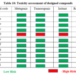 |
Table 10: Toxicity accessment of designed compouds Click here to View Table |
Low Risk – High-Risk
Evaluation of In Vitro Anti-Bacterial Activity
The binding energies led to the evaluation of the top compounds, the Synthesized Compound QI7f for their antibacterial properties in vitro against both gram-negative (Escherichia coli) and gram-positive (Staphylococcus aureus and Enterococcus faecalis) pathogens., respectively. This was performed using the well diffusion method at four concentrations: 2, 4, 6, and 10 mg/ml. Among the compounds tested, QI7f exhibited strong inhibitory effects at 10 mg/ml, with inhibition zones of 22 mm against Enterococcus faecalis, 15 mm against Escherichia coli and 19 mm against Staphylococcus aureus. (Table 11) are illustrated in the figure, with comparisons made with the standard gentamicin at 10 mg/ml.
Table 11: Evaluation of anti-microbial activity of the synthesized compound QI7f
|
Concentration of Compound QI7f |
Zone of inhibition (mm) | ||
| Escherichia Coli | Staphylococcus aureus | Enterococcus Faecalis | |
| 2mg/ml | 12mm | 16mm | 18mm |
| 4mg/ml | 13mm | 17mm | 19mm |
| 6mg/ml | 14mm | 18mm | 20mm |
| 10mg/ml | 15mm | 19mm | 22mm |
| Standard Gentamicin 10mg/ml | 28mm | 25mm | 27mm |
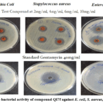 |
Figure 8: Anti-bacterial activity of compound QI7f against E. coli, S. aureus, and E. faecalis. Click here to View Figure |
Conclusion
In this study, Quinoline-isatin conjugate derivativeThey were created using the SAR analysis and literature survey as a guide, and they were then docked. All of the developed compounds showed good binding affinities when compared to the conventional medication, according to the molecular docking analysis.Among them, compound QI7f (-10.23kcal/mol) produced profound binding affinity against the targeted enzymes Enoyl acyl carrier protein reductase (InhA), Compound QI7i (-12.38kcal/mol) produced profound binding affinity against the targeted enzymes BioA 7,8-diamino-pelargonic acid (DAPA) synthase, compound QI7f (-8.38kcal/mol) produced profound binding affinity against the targeted enzymes DNA gyrase respectively. The results obtained revealed that these compounds showed promising binding energies and ligand efficiency in docking studies against the targeted enzymes. Also, compounds QI7f and QI7i were falls on Low-risk and expected to have good oral absorption and bioavailability, as evaluated by OSIRIS Property Explorer Software and Swiss ADME software, respectively. Hence, it is beneficial to carrying out further studies in the development of novel analogues because of their high biological potential in the treatment of tubercular infectious disease. Based on the binding energies, the best-hit compound QI7f were evaluated for in vitro antimicrobial properties. QI7f demonstrated significant antimicrobial activity, producing an inhibition zone of approximately 19mm against S. aureus, 22mm against E. faecalis and 15mm against E. coli at the concentration of 10mg/ml. These outcomes were contrasted with those of the conventional medication, Gentamicin, at a concentration of 10 mg/ml. Table 11 and Figure 8 present the results of this antimicrobial assessment. According to these results, compounds QI7f and QI7i have a great deal of promise as lead compounds in the development of anti-tubercular drugs.
Acknowledgement
We express our sincere thanks to the Dr. S. Govindarajan, Dean, Mother Theresa Post Graduate and Research Institute of Health Sciences, (Govt. of Puducherry Institution), Puducherry for his kind support. I would like to thank Dr. Joseph Selvin, head of the department of microbiology at Pondicherry University, for carrying out anti-microbial activity.
Funding Sources
The author(s) received no financial support for the research, authorship, and/or publication of this article.
Conflict of Interest
The author(s) do not have any conflict of interest.
Data Availability Statement
This statement does not apply to this article.
Ethics Statement
This research did not involve human participants, animal subjects, or any material that requires ethical approval.
References
- Moule, M. G., & Cirillo, J. D. (2020). Mycobacterium tuberculosis Dissemination Plays a Critical Role in Pathogenesis. Frontiers in Cellular and Infection Microbiology, 10.
CrossRef - Barter, D. M., Agboola, S. O., Murray, M. B., & Bärnighausen, T. (2012b). Tuberculosis and poverty: the contribution of patient costs in sub-Saharan Africa – a systematic review. BMC Public Health, 12(1).
CrossRef - Song, H., Tian, J., Song, H., Guo, S., Lin, Y., & Pan, J. (2024). Tracking multidrug resistant tuberculosis: a 30-year analysis of global, regional, and national trends. Frontiers in Public Health, 12.
CrossRef - Tuberculosis deaths and disease increase during the COVID-19 pandemic. (2022, October 27). PAHO/WHO | Pan American Health Organization. https://www.paho.org/en/news/27-10-2022-tuberculosis-deaths-and-disease-increase-during-covid-19-pandemic.
- Bakare, A. A., Moses, V. Y., Beckely, C. T., Oluyemi, T. I., Ogunfeitimi, G. O., Adelaja, A. A., Ayorinde, G. T., Gbadebo, A. M., Fagbenro, O. S., Ogunsuyi, O. I., Ogunsuyi, O. M., & Ige, O. M. (2022). The first-line antituberculosis drugs, and their fixed-dose combination induced abnormal sperm morphology and histological lesions in the testicular cells of male mice. Frontiers in Cell and Developmental Biology, 10.
CrossRef - Sarathy, J. P., Gruber, G., & Dick, T. (2019). Re-Understanding the mechanisms of action of the Anti-Mycobacterial drug bedaquiline. Antibiotics, 8(4), 261.
CrossRef - Abdelrahman, M. A., Almahli, H., Al-Warhi, T., Majrashi, T. A., Abdel-Aziz, M. M., Eldehna, W. M., & Said, M. A. (2022). Development of Novel Isatin-Tethered Quinolines as Anti-Tubercular Agents against Multi and Extensively Drug-Resistant Mycobacterium tuberculosis. Molecules, 27(24), 8807.
CrossRef - Zampieri, D., & Mamolo, M. G. (2020). Hybridization Approach to Drug Discovery Inhibiting Mycobacterium tuberculosis-An Overview. Current Topics in Medicinal Chemistry, 21(9), 777–788.
CrossRef - Singh, P., Jaiyeola, B., Kerru, N., Ebenezer, O., & Bissessur, A. (2017). A review of recent advancements in anti-tubercular molecular hybrids. Current Medicinal Chemistry, 24(37).
CrossRef - Panchal, N. B., & Vaghela, V. M. (2023). From molecules to medicine: the remarkable pharmacological odyssey of quinoline and it’s derivatives. Oriental Journal of Chemistry, 39(3), 546–567.
CrossRef - Tabassum, R., Ashfaq, M., & Oku, H. (2020). Current pharmaceutical aspects of synthetic quinoline derivatives. Mini-Reviews in Medicinal Chemistry, 21(10), 1152–1172.
CrossRef - Yadav, P., & Shah, K. (2021). Quinolines, a perpetual, multipurpose scaffold in medicinal chemistry. Bioorganic Chemistry, 109, 104639.
CrossRef - Ajani, O. O., Iyaye, K. T., & Ademosun, O. T. (2022). Recent advances in chemistry and therapeutic potential of functionalized quinoline motifs – a review. RSC Advances, 12(29), 18594–18614.
CrossRef - Elebiju, O. F., Ajani, O. O., Oduselu, G. O., Ogunnupebi, T. A., & Adebiyi, E. (2023). Recent advances in functionalized quinoline scaffolds and hybrids—Exceptional pharmacophore in therapeutic medicine. Frontiers in Chemistry, 10.
CrossRef - Kaur, R., & Kumar, K. (2021). Synthetic and medicinal perspective of quinolines as antiviral agents. European Journal of Medicinal Chemistry, 215, 113220. https://doi.org/10.1016/j.ejmech.2021.113220
CrossRef - Ökten, S., Aydın, A., Koçyiğit, Ü. M., Çakmak, O., Erkan, S., Andac, C. A., Taslimi, P., & Gülçin, İ. (n.d.). Quinoline‐based promising anticancer and antibacterial agents, and some metabolic enzyme inhibitors. Archiv Der Pharmazie, 353(9).
CrossRef - Suvaiv, N., Singh, K., Kumar, P., Hasan, S. M., Kushwaha, S. P., Kumar, A., Ismail, K. S., Mujeeb, S., Maurya, S. K., & Zaidi, S. M. H. (2023). Antibacterial potentiality of Isatin-Containing hybrid derivatives. Asian Journal of Chemistry, 35(4), 815–827.
CrossRef - Belay, Y., Muller, A., Mokoena, F.S. et al.1,2,3-triazole and chiral Schiff base hybrids as potential anticancer agents: DFT, molecular docking and ADME studies. Sci Rep 14, 6951 (2024).
CrossRef - El-Malah, A., Taher, E. S., Angeli, A., Elbaramawi, S. S., Mahmoud, Z., Moustafa, N., Supuran, C. T., & Ibrahim, T. S. (2022). Schiff bases as linker in the development of quinoline-sulfonamide hybrids as selective cancer-associated carbonic anhydrase isoforms IX/XII inhibitors: A new regioisomerism tactic. Bioorganic Chemistry, 131, 106309.
CrossRef - Singh, P., Jaiyeola, B., Kerru, N., Ebenezer, O., & Bissessur, A. (2017b). A review of recent advancements in anti-tubercular molecular hybrids. Current Medicinal Chemistry, 24(37).
CrossRef - Kitchen, D. B., Decornez, H., Furr, J. R., & Bajorath, J. (2004). Docking and scoring in virtual screening for drug discovery: methods and applications. Nature Reviews Drug Discovery, 3(11), 935–949.
CrossRef - Bank, R. P. D. (n.d.). Organization of 3D structures in the Protein Data Bank. https://www.rcsb.org/docs/general-help/organization-of-3d-structures-in-the-protein-data-bank.
- Team, E. W. (n.d.). PDBsum home page. https://www.ebi.ac.uk/thornton-srv/databases/pdbsum.
- Laxmi, K. L. K. (2022). Characterization of Ligand N’-[(1E)-1-phenylethylidene]-1, 3-benzothiazole-2-carbohydrazide by using Marvin Sketch 20.8 Software. Oriental Journal of Chemistry, 38(1), 77–84.
CrossRef - Wilantho, A., Tongsima, S., & Jenwitheesuk, E. (2008). Pre-docking filter for protein and ligand 3D structures. Bioinformation, 3(5), 189–193.
CrossRef - Wang, Y., Guo, Y., Qiang, S., Jin, R., Li, Z., Tang, Y., Leung, E. L. H., Guo, H., & Yao, X. (2021). 3D-QSAR, molecular docking, and MD simulations of anthraquinone derivatives as PGAM1 inhibitors. Frontiers in Pharmacology, 12.
CrossRef - Meng, X., Zhang, H., Mezei, M., & Cui, M. (2011). Molecular Docking: a powerful approach for Structure-Based drug discovery. Current Computer – Aided Drug Design, 7(2), 146–157.
CrossRef - Bayan Abdi, Mona Fekadu and Digafie Zeleke et al. Synthesis and Evaluation of the Antibacterial and Antioxidant Activities of Some Novel Chloroquinoline Analogs. J Chem. 2021. Vol. 2021:1-13.
CrossRef - Gandhi, P. V., Burande, S. R., Charde, M. S., & Chakole, R. D. (2021). Synthesis and Characterization of Isatin-Quinoline Hybrids as Antitubercular Agents. International Journal of Creative Research Thoughts (IJCRT), 9(10), d348
- Shilpa et al., World Journal of Pharmaceutical research (2018)
- Chodankar, R., & Mahajan, A. (2022). Characterization and in‐silico toxicity prediction of the oxidative degradation products of Pimozide. Separation Science Plus, 5(7), 275–284.
CrossRef - Elmongy, E. I., Ahmed, A. a. S., Sayed, I. E. T. E., Fathy, G., Awad, H. M., Salman, A. U., & Hamed, M. A. (2022). Synthesis, Biocidal and Antibiofilm Activities of New Isatin–Quinoline Conjugates against Multidrug-Resistant Bacterial Pathogens along with Their In Silico Screening. Antibiotics, 11(11), 1507.
CrossRef
 PlumX:
  Views: 606 Views
  PDF Downloads: 99
 Accepted on: 02 Mar 2026
Second Review by: Dr. K. Mujeeb
Final Approval by: Dr. Abdelwahab Omri


ISSN Online: 2231-5039