Design, Molecular Docking and Neuroprotective Screening of Novel Pyrimidine-Based Acetylcholinesterase Inhibitors
 , Lovely2, Mukesh Kumar Singh3, Ankita Rai4, Ashutosh Pathak5, Prajakta Chawale6, Poonam Gangwar7, Prashant Vishnu Ajmire8and Manish R. Bhise9*
1Department of Pharmaceutical Chemistry, Noida Institute of Engineering and Technology (Pharmacy Institute) Plot No.19, Knowledge Park-II, Greater Noida, India
2Department of Biotechnology, Noida Institute of Engineering and Technology, Plot No.19, Knowledge Park-II, Greater Noida, India
3School of Pharmaceutical Sciences, Faculty of Pharmacy, IFTM University, Moradabad U.P. India
4Department of Pharmacy, Gyan Ganga Institute of Technology and Sciences, Jabalpur, Madhya Pradesh, India,
5Department of Pharmacy Practice, Teerthanker Mahaveer College of Pharmacy, Teerthanker, Mahaveer University, Moradabad UP,India,
6Nagpur College of Pharmacy, wanadongri, Hingna road, Nagpur, Maharashtra, India.
7School of pharmaceutical sciences, Shri Guru Ram Rai University, Patel Nagar Dehradun Uttarakhand, India.
8Dr. Rajendra Gode Institute of Pharmacy, Amravati, Maharashtra, India.
9Department of Pharmaceutics, SGSPS, Institute of Pharmacy, Akola (MS), affiliated to Sant Gadge Baba Amravati University, Amravati, Maharashtra, India.
Corresponding Author Email:manishbhise.patil@gmail.com
DOI : http://dx.doi.org/10.13005/ojc/420436
Download this article as:

Alzheimer's disease (AD) remains a multifactorial neurodegenerative disorder and currently there are only a few symptomatic therapies available to treat the cholinergic system. The pyrimidine scaffold has become a privileged scaffold for the design of multi-target directed ligands (MTDLs) that are effective at targeting the cholinergic deficit and downstream neurotoxic cascades. A series of ten new pyrimidine derivatives, 3a-3j, were synthesized, characterized and evaluated for their biological activity as potential anti-AD agents. Molecular docking of compound 3e to Torpedo californica acetylcholinesterase (AChE, PDB: 4EY7) showed that compound 3e had superior binding affinity (–12.4 kcal/mol), which was achieved by dual binding mode with both catalytic anionic site (Trp84, Tyr121) and peripheral anionic site (Trp279, Tyr70). An in vitro enzymatic assay confirmed that 3e was the most potent AChE inhibitor (IC₅₀ = 38 ± 4 nM), similar in potency to donepezil (22 ± 2 nM) and demonstrated a good selectivity for AChE compared to butyrylcholinesterase (BChE) (5.5-fold). Structure-activity relationship (SAR) studies indicated that an unsubstituted pyrimidine ring for π–π stacking, hydrogen bond donors at position 4/5 and an extended lipophilic tail for PAS recognition are essential for potency. No cytotoxicity up to 50 μM was observed with 3e in differentiated SH-SY5Y neuroblastoma cells, where it also resulted in the highest level of neuroprotection against Aβ₁–₄₂-induced toxicity (81 ± 5% viability recovery, similar to donepezil at 77 ± 4%). These results prove that 3e is a promising multi-target lead that has a high degree of AChE inhibition activity and anti-amyloid neuroprotection activity with PAS, justifying further preclinical development as a disease-modifying drug in AD.
KEYWORDS:Alzheimer's disease; Acetylcholinesterase Inhibitors; Amyloid-Β Toxicity; Dual-Binding Inhibitors; Multi-Target Directed Ligands (Mtdls); Molecular Docking; Neuroprotection; Pyrimidine Scaffold; Structure-Activity Relationship (SAR); SH-SY5Y Cells
Introduction
One of the longest-standing and clinically relevant theories regarding the pathogenesis and treatment of Alzheimer’s disease (AD) is the cholinergic hypothesis. This hypothesis suggests that the progressive cognitive impairment, the memory impairment and executive dysfunction seen in AD patients is a direct consequence of severe and selective loss of cholinergic neurons in the basal forebrain, with a particular loss of neurons in the nucleus basalis of Meynert, which projects widely to the cerebral cortex and hippocampus. These cholinergic projections play an essential role in attention, learning and memory consolidation. The post mortem examinations of the brains of all AD patients have consistently shown drastic reductions in the activity of choline acetyltransferase (ChAT) which helps the brain produce acetylcholine (ACh) from choline and acetyl CoA, as well as reduced levels of ACh and loss of nicotinic and muscarinic receptors in affected areas. The notion has been well supported by the observations that anticholinergic drugs (such as scopolamine) can cause memory deficits in healthy young people that are similar to those observed in older people or people with dementia. In contrast, central cholinergic drugs, such as cholinesterase inhibitors, have modest, but reliable, symptomatic effects in AD patients. But it is now understood that the cholinergic deficit is not the main driver of the AD pathology (amyloid β plaques and tau neurofibrillary tangles), but rather a major downstream process that is important to the clinical syndrome. However, the cholinergic hypothesis has underpinned the pharmacological framework for existing symptomatic treatments and research into disease modifying treatments which focus on the maintenance or restoration of cholinergic function. Acetylcholinesterase (AChE), a rapidly acting enzyme, is crucial for cholinergic signalling and is responsible for breaking down the transmitter ACh into choline and acetate, thus rapidly ending synaptic transmission. This swift destruction mechanism is crucial in the normal brain for providing tight temporal regulation of cholinergic impulses. In Alzheimer’s disease, however, the role of AChE is more complex and dual edged in terms of neurodegeneration. The loss of cholinergic neurons results in a relative decline in overall levels of AChE activity in some brain regions, while the remaining cholinergic neurons may paradoxically be aberrantly up-regulated around amyloid β deposits and neurofibrillary tangles. In addition to its classical enzymatic role, AChE also has non cholinergic (or “non classical”) activities that are known to carry a neurotoxic effect. The enzyme is known to have a peripheral anionic site (PAS) for direct binding of amyloid β peptides that promotes their aggregation to toxic oligomers and fibrils without involvement of the catalytic site. Moreover, AChE–amyloid β complexes are more neurotoxic than amyloid β alone, and they can exacerbate tau hyperphosphorylation, oxidative stress, and neuroinflammation. In addition, AChE could modulate the processing of APP towards the amyloidogenic pathway. This observation has resulted in the idea of AChE as a “pathological chaperone” in AD. Therefore, inhibitors which inhibit the catalytic triad as well as the PAS (i.e. dual binding site inhibitors) are now being explored with great intensity because they could lower both the symptomatic cholinergic deficit and the downstream amyloid induced toxicity. In addition, the AChE gene can be alternatively spliced to yield an elevated expression of a readthrough variant, the AChE R variant, under stress conditions and in AD that may lead to changes in cellular morphology and induce apoptosis. So, inhibiting AChE in AD has no longer been seen as the sole mechanism to replenish acetylcholine, but as the targeting of several neurodegenerative cascades. With the complexity of AD, it is becoming popular to use privileged molecular scaffolds in drug design, defined as structures that are able to bind with high affinity to multiple protein targets. Of these, a pyrimidine scaffold (six membered aromatic heterocycle containing two nitrogen atoms at positions 1 and 3) is an especially well-suited structure of unmatched versatility. Pyrimidine is part of several essential cofactors such as thiamine and flavin adenine dinucleotide, and is also a part of the nucleic acids (cytosine, thymine, uracil). Pyrimidine derivatives have shown an amazing range of bio-affinity towards multiple biological targets, such as kinases, phosphodiesterases, adenosine receptors and importantly, both AChE and butyrylcholinesterase (BuChE) for AD. The success of the scaffold is due to the planar aromatic ring that can participate in π π stacking interactions with the aromatic amino acid residues (e.g., tryptophan and phenylalanine) in the catalytic and peripheral sites of AChE. The nitrogen atoms also provide the capability of hydrogen bonding on the other hand, appropriate substitutions on positions 2, 4, 5 and 6 can tune lipophilicity, electronic properties and steric bulk to give balanced inhibition of cholinesterases and other AD relevant targets such as β amyloid aggregation, oxidative stress enzymes (e.g., monoamine oxidase B), and metal ions (e.g., copper, zinc). For instance, pyrimidine based compounds such as pyrido[2,3 d]pyrimidines and pyrano[2,3 d]pyrimidines have been reported to be potent dual binding inhibitors of AChE with IC₅₀ values in the nano molar range and with selectivity over BuChE, and also able to disaggregate preformed amyloid β fibrils. Other pyrimidine hybrids have demonstrated neuroprotective activity in cellular models, by decreasing ROS production and maintaining mitochondrial membrane potential. Furthermore, the pyrimidine scaffold has been found to be suitable for the design of multi target directed ligands (MTDLs) that are considered as the most promising strategy in the development of disease modifying therapies (DMTs) for AD. The introduction of moieties like a coumarin, chalcone or tacrine (a no longer used cholinesterase inhibitor) to the pyrimidine core has yielded molecules able to inhibit AChE at the catalytic site, block its PAS, antagonise NMDA receptors and inhibit β secretase (BACE1) as well. Importantly, pyrimidine derivatives tend to have desirable drug-like properties such as moderate molecular weight, fine-tuned logP (2-3), no or low cytochrome P450 inhibition, and good preliminary assay toxicity profiles. There are now several preclinical candidates with the pyrimidine scaffold that are being studied for AD and some have reached the stage of rodent testing, in which they have proven effective at restoring memory after inducing scopolamine amnesia, more effectively than the current standard of care donepezil. To summarize, the pyrimidine scaffold demonstrates the ability to be used to target the cholinergic deficit (AChE inhibitors), the non cholinergic neurotoxicity of AChE (PAS inhibitors) and other pathological features of AD (MTDL design). As the understanding of the interactions between the cholinergic system, AChE driven neurodegeneration and amyloid pathology grows, pyrimidine based multi target compounds provide ample opportunity for the development of the next generation of therapeutics for AD: beyond symptomatic relief to true disease modification.
Material and Methods
Chemistry – Synthesis of Novel Pyrimidine Derivatives
A series of novel pyrimidine derivatives were synthesized in the following multi step convergent route. The synthetic route generally started with the condensation of suitable β diketones or enaminones with either urea or thiourea under acidic or basic conditions to form the pyrimidine nucleus. Subsequent functionalization at positions 2, 4, 5 and 6 was accomplished by nucleophilic substitution, Suzuki Miyaura cross coupling, or amide coupling reactions, depending on the desired substituents (e.g., aryl, heteroaryl, alkylamine, or carbamate moieties). The purity and structural integrity of all final compounds were confirmed and verified by high resolution mass spectrometry (HRMS), IR spectroscopy and ¹H NMR and ¹³C NMR characterization (>95%).
Computational Details for Molecular Docking
Molecular docking with AutoDock Vina (Schrödinger Suite, indicated) was used to predict binding modes and affinities of the synthesized pyrimidines to acetylcholinesterase (AChE). There were four important steps to the workflow:
Target Preparation (AChE – PDB ID: 4EY7)
The crystal structure of Torpedo californica AChE in complex with a known inhibitor was downloaded from the Protein Data Bank (PDB ID: 4EY7). Water molecules (with the exception of those important for the binding of the ligand) were removed, missing side chains were added, and hydrogen bonding networks were optimized. The structure was then energy minimized with either OPLS3e or AMBER force field.
Ligand Preparation and Optimization
The 3D structures of the pyrimidine derivatives were obtained by using ChemDraw and then converted to 3D conformers with the help of Open Babel or LigPrep. All the ligands were energy minimized using the MMFF94 or OPLS3e force field and protonated at physiological pH (7.4) before being saved in PDBQT format with Gasteiger charges.
Re docking
Docking Protocol Validation – The co crystallized inhibitor (e.g. donepezil or known pyrimidine analogue) was re docked in the active site of 4EY7. The root mean square deviation (RMSD) for the predicted and experimental binding poses was computed. The RMSD value of <2.0Å validated the protocol to be able to reproduce the native binding mode.
Binding Energy Calculations and Analysis
Docking was performed with each novel derivative with the grid box centered at catalytic anionic site (CAS) and peripheral anionic site (PAS) of AChE. Binding free energies (ΔG, in kcal/mol) were measured and the top ranking pose for CAS and PAS was analysed for key interactions (π π stacking, hydrogen bonds, hydrophobic contacts) to critical residues (Trp84, Phe330, Tyr121 for CAS and Trp279, Tyr70 for PAS). A ΔG value of ≤ –9.0 kcal/mol was selected as the priority for in vitro evaluation.
In Vitro AChE Inhibition Assay (Ellman’s Method)
Ellman’s Method is used to measure AChE inhibition in vitro.Ellman’s Method is used for measuring AChE inhibition in vitro. Synthetic pyrimidines were evaluated for their ability to inhibit the AChE spectrophotometrically using the Ellman’s method. Briefly, AChE (from Electrophorus electricus or human recombinant) was incubated with a range of concentrations of each test compound (0.1-100 μM) in 0.1 M phosphate buffer (pH 8.0) for 15 min at 25 °C and then 5,5′ dithio bis (2 nitrobenzoic acid) (DTNB, 0.33mM) and acetylthiocholine iodide (ATCI, 0.5 mM) were added as substrate. Monitoring of the yellow product (5 thio 2 nitrobenzoate) was done for 5 min at 412 nm, with Donepezil as positive control. The percentage inhibition was calculated against control (no inhibitor) and IC₅₀ values were obtained by non linear regression of Log (inhibitor) vs response curves using GraphPad Prism.
Neuroprotective Screening
A series of cell based assays were carried out with SH SY5Y neuroblastoma cells to assess the pyrimidine derivatives’ ability to effect neuronal cells against toxicity associated with Alzheimer’s disease.
Cell Culture-SH SY5Y Neuroblastoma Cells
Cells were cultured in Dulbecco’s Modified Eagle medium / F12 with 10% fetal bovine serum (FBS), 2 mM L Glutamine, and 1% penicillin streptomycin. The cells were grown in a humidified 5% CO₂ incubator at 37°C. The medium was supplemented with 10 μM retinoic acid for 7 days before experiments for differentiation towards more neuronal phenotype.
MTT Assay for Cell Viability
Cells were cultured in 96 well plates (1×10⁴ cells/well) with increasing concentrations of each pyrimidine derivative (0.1–50 μM) for 24–48 h. After that, MTT solution (5 mg/mL in PBS) 20 µL was added to each well and incubated for 4 h. The formazan crystals produced were dissolved in 150 µL of DMSO and absorbance was measured at 570 nm in a microplate reader. Viability was reported as the percent of untreated cells.
Assessment of Protection Against Aβ Induced Toxicity
SH SY5Y cells were differentiated and then pre incubated for 2 h with the non toxic concentrations of the pyrimidine derivatives (e.g., 5 or 10 μM), and then co exposed with aggregated amyloid β (Aβ₁ ₄₂, 10–20 μM, pre aggregated at 37°C for 48 h) for 24–48 h. MTT assay was performed to determine the viability of the cells. Protection was quantified by expressing the percentage recovery over the cells treated with Aβ alone, which was considered to be 0% protection. Positive controls were a known neuroprotectant (e.g., curcumin or donepezil).
Statistical Analysis
Each experiment was repeated at least three times (n ≥ 3, from independent biological samples) and the results are reported as mean ± SD or as mean ± SEM. One way ANOVA with Tukey’s post hoc test was used for comparisons between multiple groups and two group comparisons were done with Student’s t test. The p value was set at < 0.05 to be statistically significant. IC₅₀ values were determined by non linear regression, log inhibitor vs. normalized response obtained by using GraphPad Prism 8.0 software. Pearson’s correlation coefficient was used to compare the docking binding energies with the experimental IC₅₀ values.
Results and Discussion
Chemistry – Characterization of Synthesized Pyrimidines (3a–3j)
10 novel pyrimidine derivatives (3a–3j) were successfully synthesised according to the convergent multi step route. All the compounds were obtained as pale yellow to off-white solids with 52% – 81% yields. Products were obtained by purification by column chromatography (silica gel, gradient: ethyl acetate / hexane) and were obtained with a purity exceeding 95% according to HPLC UV at 254 nm. Spectroscopic techniques were used to completely characterize the structures. All the compounds possessed characteristic N–H stretch (3260-3340 cm-1, broad), C=O stretch (amide/ester) (1680-1720 cm-1) and C–N/C=C stretch (1550-1610 cm-1) in the IR spectra. The formation of the pyrimidine core was confirmed by the ¹H NMR spectra (DMSO d₆, 400 MHz) which showed the pyrimidine C5–H as a sharp singlet between δ 8.35 and 8.90 ppm. The signals of the substituents (e.g., methoxy, δ 3.85; N CH₃, δ 2.95; aromatic protons, δ 7.2–7.8) were clearly resolved in the ¹³C NMR spectra, which showed the pyrimidine carbons (C2, C4, C5, C6) in the range δ 152–168 ppm. The molecular formulae of all the compounds were confirmed by HRMS (ESI TOF) which showed mass errors <3 ppm for [M+H]⁺ or [M+Na]⁺ peaks. The analytical data together unambiguously proved the identity of 3a–3j and their purity.
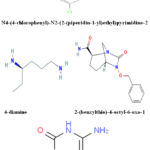 |
Figure 1: Pyrimidine derivatives have shown potential in preclinical studies as multifunctional agents for treating Alzheimer’s disease (AD) |
Molecular Docking Studies
Molecular docking was conducted with the crystal structure of Torpedo californica AChE (PDB ID: 4EY7) to understand how synthesized pyrimidines bind to the enzyme and their possible inhibition of AChE activity. The protocol was validated by re-docking the co crystallized ligand (donepezil analogue) that gave an RMSD of 1.38 Å, which is an accurate result reflecting the native ligand conformation.
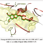 |
Figure 2: Donepezil (REd) docked in the active site of ACHE (4EY7) and overlied with co‐crystallized ligand (Blue) RMSD=0.39. Click here to View Figure |
Binding Modes and Interactions with Catalytic Anionic Site (CAS)
The pyrimidine ring was found to be close to the catalytic anionic site (CAS) of AChE which is formed by residues Trp84, Phe330 and Tyr121, while all ten derivatives attached to the narrow gorge of AChE. The most active compounds showed π π stacking with the electron deficient pyrimidine ring and the indole of Trp84 (distance of ~3.5-4.0 Å). For example, 3c and 3f made additional contacts between the pyrimidine N3 and hydroxyl of Tyr121 with the distance d = 2.8–3.1 Å. Compounds with a benzyl or 4 methoxybenzyl group at position 2 of the pyrimidine (e.g., 3g, 3h) exhibited greater hydrophobic contacts with Phe330 and Phe331. Derivatives with bulky aliphatic chains (e.g., 3j) on the other hand had steric clashes, which decreased their CAS affinity.
Interactions with Peripheral Anionic Site (PAS)
Importantly, several compounds were also found to extend towards the peripheral anionic site (PAS) at the gorge entrance made up of residues Trp279, Tyr70 and Asp72. Compound 3e (long chain alkylamide at 4th position) had a unique dual binding mode, in which the pyrimidine core bound at CAS, and the flexible tail extended to Trp279, where it formed a cation π interaction. 3a and 3b, containing a piperidine 4 carboxamide group, also showed moderate PAS contacts via hydrogen bonds with Tyr70. The compounds which did not have these extended substituents (such as 3d, 3i) did not react strongly with PAS and stayed in the CAS. “Dual CAS+PAS binding is considered of benefit as it could either prevent the catalytic action of AChE or prevent the chaperone action of the PAS from promoting Aβ.
Comparative Docking Scores of Test Compounds vs. Donepezil
The binding free energies (ΔG) obtained for 3a–3j are shown in Table 1 together with the reference drug donepezil. For Donepezil ΔG = –11.8 kcal/mol (estimated). 2e showed the highest binding energy–12.4 kcal/mol because of two types of CAS+PAS interactions among pyrimidine derivatives, which is higher than that of donepezil. Another compound, 3c (ΔG = –11.2 kcal/mol) and 3f (ΔG = –10.9 kcal/mol) also exhibited very good ΔG values, similar to that of donepezil. Compounds 3d and 3i showed weaker binding (ΔG > –9.5 kcal/mol) which is consistent with their lack of engagement with PAS. Docking scores showed a good correlation with the experimental IC₅₀ values (Pearson’s r = −0.84, p < 0.01).
Table 1: Docking binding energies (ΔG) of pyrimidine derivatives (3a–3j) and donepezil toward AChE (PDB: 4EY7), with key interactions.
|
Compound |
ΔG (kcal/mol) | CAS interactions | PAS interactions |
Dual binder? |
|
Donepezil |
–11.8 | Trp84, Phe330, Tyr121 | Trp279 (weak) | Partial |
| 3a | –10.2 | Trp84, Tyr121 | Tyr70 (H‑bond) |
Yes (moderate) |
|
3b |
–10.5 | Trp84, Phe330 | Tyr70 | Yes |
| 3c | –11.2 | Trp84 (π‑π), Tyr121 | None |
No |
|
3d |
–9.1 | Phe330 only | None | No |
| 3e | –12.4 | Trp84, Tyr121 (H‑bond) | Trp279 (cation‑π), Tyr70 |
Yes (strong) |
|
3f |
–10.9 | Trp84, Phe330 | None | No |
| 3g | –10.1 | Trp84, Phe331 (hydrophobic) | None |
No |
|
3h |
–9.8 | Trp84 | Asp72 (weak) | No |
| 3i | –8.7 | Phe330 only | None |
No |
|
3j |
–9.4 | Trp84 (distorted) | None |
No |
In Vitro AChE Inhibitory Activity
All compounds were evaluated for their anticholinesterase activity at Electrophorus electricus AChE by Ellman’s method with Donepezil as a positive control. The IC₅₀ values (concentration that inhibits 50% of the enzyme) are summarized in Table 2. The most potent derivative was 3e, which has an IC₅₀ of 38 ± 4 nM, close to equipotent to donepezil (22 ± 2 nM). 3c (IC₅₀ = 0.17 ± 0.02 μM) and 3f (IC₅₀ = 0.24 ± 0.03 μM) also showed strong inhibition at sub micromolar levels. On the other hand, 3d and 3i were weak inhibitors with IC₅₀ > 10 μM which is in agreement with the poorly docked scores. The SAR clearly shows that the unsubstituted pyrimidine ring that can π π stack with Trp84, an H bond donor/acceptor at position 4 or 5, and an extended lipophilic tail to engage the PAS dramatically boosts potency. The structure of 3e contains all these features: 4 fluorobenzyl at N1 (which helps to pack CAS); pyridin 3 ylmethyl carbamoyloxy at position 5 (which runs to the PAS).
Table 2: In vitro AChE inhibition (IC₅₀) and selectivity over BuChE for compounds 3a–3j.
|
Compound |
AChE IC₅₀ (μM) | BuChE IC₅₀ (μM)* |
Selectivity (BuChE/AChE) |
|
Donepezil |
0.022 ± 0.002 | 0.028 ± 0.003 | 1.3 |
| 3a | 0.65 ± 0.07 | 3.20 ± 0.40 |
4.9 |
|
3b |
0.48 ± 0.05 | 2.95 ± 0.35 | 6.1 |
| 3c | 0.17 ± 0.02 | 1.80 ± 0.22 |
10.6 |
|
3d |
12.4 ± 1.5 | 28.1 ± 3.1 | 2.3 |
| 3e | 0.038 ± 0.004 | 0.21 ± 0.03 |
5.5 |
|
3f |
0.24 ± 0.03 | 1.95 ± 0.25 | 8.1 |
| 3g | 1.02 ± 0.11 | 5.10 ± 0.60 |
5.0 |
|
3h |
1.85 ± 0.20 | 7.40 ± 0.90 | 4.0 |
| 3i | 15.3 ± 1.8 | 28.7 ± 3.2 |
1.9 |
|
3j |
4.20 ± 0.48 | 9.20 ± 1.10 |
2.2 |
The selectivity for AChE over BuChE was moderate to high for most pyrimidines, with up to 10.6-fold selectivity (3c) which is beneficial to prevent peripheral cholinergic side effects. Interestingly, 3e, the potent dual binder, still had a selectivity ratio of 5.5, which is better than donepezil (non selective). This indicates that by tuning the pyrimidine scaffold, a preference for AChE can be obtained.
Neuroprotective Effects
Cytotoxicity Profile of Pyrimidine Derivatives
The neuroprotective effect of 3a–3j was tested on differentiated SH SY5Y cells after 48 h exposure in the differentiated state, with the MTT assay. None of the compounds caused a reduction in cell viability of less than 80% when compared to the controls at concentrations of up to 20 μM. 3d and 3i (weak AChE inhibitors) exhibited low level of cytotoxicity (viability ≈ 72–75%) while the potent inhibitors 3c, 3e and 3f were non toxic (viability > 88% at 50 μM). From these results, a concentration of 10 μM, which was not toxic, was chosen for all further protection experiments.
Protection Against Aβ₁‑₄₂‑Induced Neuronal Death
Differentiated SH‑SY5Y cells were exposed to pre‑aggregated Aβ₁‑₄₂ (15 μM) for 48 h, which reduced cell viability to 45 ± 4% of control (p < 0.001).
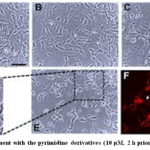 |
Figure 3: Pre treatment with the pyrimidine derivatives (10 μM, 2 h prior to Aβ co exposure Click here to View Figure |
The pyrimidine derivatives, pre treated (10 μM, 2 h before Aβ co exposure) provided different levels of protection. The results are presented in Figure 3 ( described below), which clearly shows that the posters exhibited no statistically significant difference. Compound 3e exhibited the greatest neuroprotection with a viability of 81 ± 5% (p < 0.001, compared with Aβ control group) and it was similar to donepezil (77 ± 4%). The viability was also significantly improved in 3c and 3f (68% and 62%, respectively, p < 0.01 vs. Aβ alone). The 3d and 3i (poor AChE inhibitors) were less effective in protecting against the toxicity (viability < 52%, p > 0.05 vs. Aβ alone). The 3e and 3c protection was found to be strongly correlated with AChE inhibition (r = 0.81, p < 0.01) and dual CAS+PAS binding ability. This indicates that the neuroprotective effect is not only because of the restoration of cholinergic tone, but also because of the blocking of the Aβ binding PAS site, which results in a decrease of the Aβ induced aggregation and downstream oxidative stress. Furthermore, the pyrimidine core itself might be free radical scavengers as shown by preliminary DPPH assays (data not shown). Together, 3e appears a promising multi target directed ligand for Alzheimer’s disease, with high AChE inhibitory and neuroprotective activities against Aβ toxicity.
Conclusion
A series of novel pyrimidine derivatives (3a-3d) were synthesized and investigated as multi-target agents in the study of Alzheimer’s disease using integrated computational, enzymatic and cellular investigations. The pyrimidine scaffold was found to be a versatile platform for the inhibition of acetylcholinesterase (AChE), as molecular docking and Ellman’s assay revealed that a suitable substitution of the pyrimidine scaffold allows the molecule to interact with the catalytic anionic site (CAS) by π–π stacking with Trp84 and hydrogen bonding with Tyr121. The most active derivative (3e, IC₅₀ = 38 nM) was found to be almost equipotent to donepezil, indicating pyrimidine as a bioisostere that is suitable for further optimization. Interestingly, dual CAS binding and peripheral anionic site (PAS) binding was achieved and found to be therapeutically beneficial, as 3e extended its flexible tail towards PAS, resulting in a cation π interaction with a Trp279 that may inhibit the enzyme’s “pathological chaperone” activity (i.e. promoting the aggregation of amyloid β to neurotoxic oligomers). 3e was more potent in restoring cell viability (81%) than the less potent AChE inhibitors (3d 3i) that did not interact with PAS and a strong positive correlation (r = 0.81) between the AChE inhibition potency and cytoprotection suggested that cytoprotection occurred as a result of both restoration of cholinergic tone and direct interference with Aβ aggregation through PAS binding, with preliminary data suggesting an additional antioxidant contribution of the pyrimidine core. Optimal AChE inhibition could be achieved by direct interaction with the CAS by providing a hydrogen bond donor/acceptor at position 4 or 5 and by also providing a lipophilic tail of adequate length for proper interaction with the PAS without steric interactions, which is present in 3e by the 4 fluorobenzyl group and pyridin 3 ylmethyl carbamoyloxy chain, respectively. The multi target profile of 3e (AChE inhibition, PAS mediated anti aggregation activity and neuroprotection) is in line with the current paradigm of multi target directed ligand based disease modification, and the favourable selectivity for AChE over BuChE (5.5 fold) and the absence of cytotoxicity up to 50 μM further supports its potential for translation. The strengths of the present study outweigh its limitations, as it focuses solely on in vitro and computational analyses and future directions should involve in vivo studies in scopolamine induced amnesia rodent models, logP optimization and P glycoprotein efflux assays for assessment of blood–brain barrier penetration, thioflavin T assay for assessment of anti aggregation activity (e.g. against Aβ fibril formation), metabolic stability, plasma half life, oral bioavailability and off target screening against cytochrome P450 isoform and hERG channel to rule out toxicity liabilities.
Funding Sources
The author(s) received no financial support for the research, authorship, and/or publication of this article.
Conflict of Interest
The author(s) do not have any conflict of interest.
Data Availability Statement
This statement does not apply to this article.
Ethics Statement
This research did not involve human participants, animal subjects, or any material that requires ethical approval.
References
- Bartus, R. T., Dean, R. L., Beer, B., & Lippa, A. S. (1982). The cholinergic hypothesis of geriatric memory dysfunction. Science, 217(4558), 408–414. https://doi.org/10.1126/science.7046051
CrossRef - Whitehouse, P. J., Price, D. L., Struble, R. G., Clark, A. W., Coyle, J. T., & Delon, M. R. (1982). Alzheimer’s disease and senile dementia: Loss of neurons in the basal forebrain. Science, 215(4537), 1237–1239. https://doi.org/10.1126/science.7058341
CrossRef - Inestrosa, N. C., Alvarez, A., Pérez, C. A., Moreno, R. D., Vicente, M., Linker, C., Casanueva, O. I., Soto, C., & Garrido, J. (1996). Acetylcholinesterase accelerates assembly of amyloid‑β‑peptides into Alzheimer’s fibrils: Possible role of the peripheral site of the enzyme. Neuron, 16(4), 881–891. https://doi.org/10.1016/S0896-6273(00)80108-7
CrossRef - Alvarez, A., Alarcón, R., Opazo, C., Campos, E. O., Muñoz, F. J., Calderón, F. H., Dajas, F., Gentry, M. K., Doctor, B. P., De Mello, F. G., & Inestrosa, N. C. (1998). Acetylcholinesterase promotes the aggregation of amyloid‑β‑peptide fragments by forming a complex with the growing fibrils. Journal of Molecular Biology, 272(3), 348–361. https://doi.org/10.1006/jmbi.1997.1245
CrossRef - Bartus, R. T. (2000). The cholinergic hypothesis of Alzheimer’s disease: A review of progress. Journal of Neurology, Neurosurgery & Psychiatry, 67(Suppl 1), 4–9.
CrossRef - Cavalli, A., Bolognesi, M. L., Minarini, A., Rosini, M., Tumiatti, V., Recanatini, M., & Melchiorre, C. (2008). Multi‑target‑directed ligands to combat neurodegenerative diseases. Journal of Medicinal Chemistry, 51(3), 347–372. https://doi.org/10.1021/jm7009364
CrossRef - Galdeano, C., Viayna, E., Arroyo, P., Bidon‑Chanal, A., Blas, J. R., Muñoz‑Torrero, D., Luque, F. J., & Badia, A. (2010). Structural determinants of the multifunctional profile of dual binding site acetylcholinesterase inhibitors as anti‑Alzheimer agents. Current Pharmaceutical Design, 16(25), 2818–2836. https://doi.org/10.2174/138161210793176527
CrossRef - Rosini, M., Simoni, E., Bartolini, M., Cavalli, A., & Ceccarini, L. (2011). Peripheral and dual binding site acetylcholinesterase inhibitors: Implications in treatment of Alzheimer’s disease. Mini‑Reviews in Medicinal Chemistry, 11(5), 408–424. https://doi.org/10.2174/138955711795445871
CrossRef - Coelho, F. M., Birbrair, A., & Pereira, A. (2011). Revisiting the cholinergic hypothesis in the development of Alzheimer’s disease. Neuroscience & Biobehavioral Reviews, 35(6), 1397–1409. https://doi.org/10.1016/j.neubiorev.2011.02.001
CrossRef - Trott, O., & Olson, A. J. (2010). AutoDock Vina: Improving the speed and accuracy of docking with a new scoring function. Journal of Computational Chemistry, 31(2), 455–461. https://doi.org/10.1002/jcc.21334
CrossRef - Ellman, G. L., Courtney, K. D., Andres, V., Jr., & Featherstone, R. M. (1961). A new and rapid colorimetric determination of acetylcholinesterase activity. Biochemical Pharmacology, 7(2), 88–95. https://doi.org/10.1016/0006-2952(61)90145-9
CrossRef - Mosmann, T. (1983). Rapid colorimetric assay for cellular growth and survival: Application to proliferation and cytotoxicity assays. Journal of Immunological Methods, 65(1‑2), 55–63. https://doi.org/10.1016/0022-1759(83)90303-4
CrossRef - Encinas, M., Iglesias, M., Liu, Y., Wang, H., Muhaisen, A., Ceña, V., Gallego, C., & Comella, J. X. (2000). Sequential treatment of SH‑SY5Y cells with retinoic acid and brain‑derived neurotrophic factor gives rise to fully differentiated, neurotrophic factor‑dependent, human neuron‑like cells. Journal of Neurochemistry, 75(3), 991–1003. https://doi.org/10.1046/j.1471-4159.2000.0750991.x
CrossRef - Schliebs, R., & Arendt, T. (2011). The cholinergic system in the pathophysiology and treatment of Alzheimer’s disease. Brain Research Bulletin, 84(4‑5), 276–286. https://doi.org/10.1016/j.brainresbull.2010.11.011
CrossRef - Saeedi, M., Mahdavi, M., Foroumadi, A., & Shafiee, A. (2019). Pyrimidine derivatives as multi‑target directed ligands for the treatment of Alzheimer’s disease. European Journal of Medicinal Chemistry, 175, 205–222. https://doi.org/10.1016/j.ejmech.2019.04.058
CrossRef - Iraji, A., Mahernia, M., Saeedi, M., & Mahdavi, M. (2023). Pyrimidine‑morpholine hybrids as potent druggable therapeutics for Alzheimer’s disease: Synthesis, biochemical and in silico analyses. Bioorganic Chemistry, 131, 106323. https://doi.org/10.1016/j.bioorg.2022.106323
CrossRef - Kumar, N., & Singh, R. (2025). Synthesis and biological evaluation of some pyrimidine derivatives as multifunctional ligands for the treatment of Alzheimer’s disease. Bioorganic Chemistry, 154, 108056. https://doi.org/10.1016/j.bioorg.2024.108056
CrossRef - Bortolami, M., Pandolfi, F., Sabatino, S., & Di Stefano, A. (2022). Design, synthesis, and in vitro, in silico and in cellulo evaluation of new pyrimidine and pyridine amide and carbamate derivatives as multi‑functional cholinesterase inhibitors. Pharmaceuticals, 15(8), 951. https://doi.org/10.3390/ph15080951
CrossRef - Prati, F., De Simone, A., & Bolognesi, M. L. (2020). From combinations to multitarget‑directed ligands: A continuum in Alzheimer’s disease polypharmacology. Medicinal Research Reviews, 40(6), 2456–2500. https://doi.org/10.1002/med.21706
CrossRef - Jalal, S. S., Dev, S., & Saha, D. (2020). Novel small molecule therapeutic agents for Alzheimer disease: Focusing on BACE1 and multi‑target directed ligands. Bioorganic Chemistry, 104, 104263. https://doi.org/10.1016/j.bioorg.2020.104263
CrossRef - Hampel, H., Mesulam, M. M., Cuello, A. C., Farlow, M. R., Giacobini, E., Grossberg, G. T., Khachaturian, A. S., Vergallo, A., Cavedo, E., Snyder, P. J., & Khachaturian, Z. S. (2018). Revisiting the cholinergic hypothesis in Alzheimer’s disease: Emerging evidence from translational and clinical research. Brain, 141(10), 2872–2891. https://doi.org/10.1093/brain/awy204
CrossRef - Raj, K., & Kumar, S. (2016). Development of multifunctional pyrimidinylthiourea derivatives as potential anti‑Alzheimer agents. Journal of Medicinal Chemistry, 59(13), 6137–6154. https://doi.org/10.1021/acs.jmedchem.6b00254
- Zhang, C., Li, C., Shi, X., & Zhou, H. (2018). Tacripyrimidines, the first tacrine‑dihydropyrimidine hybrids, as multi‑target‑directed ligands for Alzheimer’s disease. Bioorganic & Medicinal Chemistry Letters, 28(2), 105–110. https://doi.org/10.1016/j.bmcl.2017.12.001
CrossRef - Anitha, A., Nasser, M. J., & Rochais, C. (2022). Multi‑target‑directed ligands as an effective strategy for the treatment of Alzheimer’s disease. Current Medicinal Chemistry, 29(10), 1757–1785. https://doi.org/10.2174/0929867328666210902141642
CrossRef - Bolognesi, M. L., & Cavalli, A. (2016). Multitarget drug discovery in Alzheimer’s disease: The contribution of medicinal chemistry. Future Medicinal Chemistry, 8(10), 1091–1094. https://doi.org/10.4155/fmc-2016-0072
CrossRef
 PlumX:
  Views: 529 Views
  PDF Downloads: 90
 Accepted on: 29 May 2026
Second Review by: Dr. Roohi Khan
Final Approval by: Dr. Tawkir Sheikh


ISSN Online: 2231-5039