Acridine: A Versatile Scaffold in Medicinal Chemistry (Review article)
 and Vipul Vaghela2*
1Research Scholar, Gujarat Technological University, Ahmedabad, Gujarat, India,
2Department of Pharmaceutical Chemistry, A.R College of Pharmacy and G.H Patel Institute of Pharmacy, Vallabh Vidyanagar, Gujarat, India
Corresponding Author E-mail:vipularcp@gmail.com
DOI : http://dx.doi.org/10.13005/ojc/420418
Download this article as:

Acridine, a nitrogen-containing heterocyclic compound, has long been recognized as a versatile scaffold in medicinal chemistry owing to its unique planar structure and ability to intercalate DNA. This review highlights the significant pharmacological applications of acridine derivatives, with a primary focus on their therapeutic potential rather than synthetic methodologies. Acridine based compounds exhibit diverse biological activities, including anticancer, antimalarial, antimicrobial, antiviral, and neuroprotective effects. Notably, several acridine derivatives such as amsacrine, quinacrine, and tacrine have been clinically employed, demonstrating the scaffold’s drug-like properties. The emergence of acridine hybrids has further enhanced therapeutic efficacy by enabling multifunctional targeting and overcoming drug resistance. However, challenges such as toxicity, mutagenicity, and pharmacokinetic limitations remain critical hurdles. Advances in targeted drug delivery, rational drug design, and ADMET profiling provide promising strategies to mitigate these issues. Future research integrating acridine chemistry with nanotechnology, epigenetic modulation, and personalized medicine holds great promise for developing novel and effective therapeutics. Overall, acridine continues to be a “magical molecule” in drug discovery, offering a rich template for innovative medicinal agents.
KEYWORDS:Acridine derivatives; Anticancer agents; DNA intercalation; Medicinal chemistry; safety profile; Toxicity
Introduction
Acridine (Figure 1.), a planar nitrogen-containing heterocycle with the molecular formula C₁₃H₉N, was first isolated from coal tar by Graebe and Caro in 1870. Structurally similar to anthracene but with a central nitrogen atom, acridine manifests weak basicity (pKₐ ≈ 5.1) and exceptional photophysical properties, including strong fluorescence1-2. These traits render it versatile in both synthetic chemistry and biological applications, enabling its long-standing use as dyes (e.g., acridine orange, acridine yellow), biochemical probes, and potential therapeutics3.
In medicinal chemistry, the acridine scaffold is celebrated as a “privileged pharmacophore,” thanks to its ability to intercalate DNA and inhibit various enzymes. Derivatives such as amsacrine, quinacrine, and acriflavine have historically been explored as anticancer, antimalarial, and antibacterial agents. Their planar structure facilitates stacking interactions with nucleic acids, while specific substituents fine-tune binding affinity, selectivity, and biological activity. Acridones (Figure 1.), a closely related class, also share many of these favorable features4.
Over the past decade, considerable interest has returned to acridine derivatives because of emerging drug resistance in cancer and infectious diseases5. Modern acridines extend their biological scope: they act as topoisomerase I/II inhibitors, telomerase suppressors, and protein kinase modulators. Hybrid molecules—incorporating acridine with other pharmacophores—enhance multifunctionality and circumvent resistance mechanisms6.
Acridine derivatives are also valuable in diagnostics and drug delivery. Dye-based analogues like acridine orange exhibit pH-sensitive fluorescence, distinguishing DNA and RNA in living cells, and are used to visualize apoptosis or lysosomal activity7. Their photophysical properties facilitate use in photodynamic therapy and imaging, providing dual-functionality as both therapeutic and diagnostic agents8-9.
Despite their promise, acridine derivatives pose challenges such as genotoxicity and cytotoxicity, particularly through DNA intercalation.9 Medicinal chemists are now employing structure-based design, QSAR, and computational optimization to enhance target specificity while minimizing genotoxic side effects. Green and sustainable synthetic methodologies are also emerging for building functionalized acridines10-11.
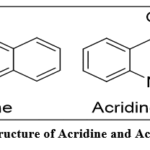 |
Figure 1: Structure of Acridine and Acridine-9-one Click here to View Figure |
Pharmacological Activities of Acridine Derivatives
Anticancer Activity12
Acridine derivatives are extensively studied for their anticancer properties, primarily due to their ability to intercalate DNA and inhibit enzymes such as topoisomerase I and II, which are essential for DNA replication and cell division. One of the most well-known examples is Amsacrine (m-AMSA) (Figure 2), a clinical topoisomerase II inhibitor used in the treatment of acute myeloid leukemia (AML). Amsacrine exerts its cytotoxic effects by stabilizing the DNA-topoisomerase complex, leading to DNA breaks and apoptosis. Another notable compound is DACA (N-[2-(dimethylamino)ethyl]acridine-4-carboxamide) (Figure 2), which combines DNA intercalation with topoisomerase I inhibition and shows promising results in preclinical studies against solid tumors. Furthermore, Acriflavine (Figure 2), a mixture of proflavine and trypaflavine, has gained attention for its ability to inhibit HIF-1α (hypoxia-inducible factor 1-alpha), a protein involved in tumor survival and metastasis, making it a potential anti-angiogenic agent in cancer therapy.
Photodynamic Therapy (PDT): Acridine derivatives such as acridine orange (Figure 2) are used as photosensitizers in cancer treatment. Under light activation, they produce reactive oxygen species (ROS) to kill tumor cells13.
G-quadruplex binding agents: Certain acridine derivatives can stabilize G-quadruplex DNA structures, inhibiting telomerase activity, a key enzyme in cancer cell immortality. E.g., BRACO-1913 (Figure 2).
Histone deacetylase (HDAC) inhibitors: Hybrid molecules combining acridine with HDAC-inhibiting motifs are being studied for epigenetic modulation in cancer14.
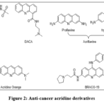 |
Figure 2: Anti-cancer acridine derivatives Click here to View Figure |
Antimalarial and Antiparasitic Activity
The acridine scaffold has a long history in antimalarial therapy, with Quinacrine (also known as mepacrine) (Figure 3) being one of the first synthetic antimalarials6. Quinacrine functions by intercalating into DNA and inhibiting plasmodial phospholipase A2, thereby disrupting parasite replication. It was widely used during World War II for malaria prophylaxis. Despite being largely replaced by newer drugs, quinacrine has seen renewed interest for its activity against chloroquine-resistant strains of Plasmodium falciparum15. Another example is Pyronaridine (Figure 3), an acridine-based compound used in combination with artesunate (in the combination drug Pyramax) to treat acute uncomplicated malaria. Its antimalarial activity stems from inhibition of hemozoin formation, which is toxic to the parasite. Additionally, acridine derivatives have shown activity against Leishmania spp. and Trypanosoma brucei, the causative agents of leishmaniasis and African sleeping sickness, respectively, making them valuable candidates in neglected tropical disease research16. Some acridine derivatives are designed to inhibit both heme detoxification and DNA replication, enhancing activity against resistant strains. Trypanocidal agents like 3-nitroacridines (Figure 3) are active against Trypanosoma cruzi (Chagas disease). Their mechanism includes redox cycling and DNA damage17.
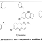 |
Figure 3: Antimalarial and Antiparasitic acridine derivatives Click here to View Figure |
Antimicrobial and Antiviral Activity
Acridine derivatives have demonstrated broad-spectrum antimicrobial activity against bacterial, fungal, and viral pathogens. For instance, Acriflavine, aside from its anticancer effects, is also an effective topical antiseptic and bacteriostatic agent, particularly against Gram-positive bacteria18. Its mechanism involves disruption of bacterial DNA synthesis via intercalation19. Other acridine analogs, such as ethacridine lactate (Figure 4), are used in obstetrics for intrauterine antisepsis and show potent antibacterial activity.20 Moreover, some derivatives exhibit antiviral activity. For example, acridine-based compounds have been investigated for inhibition of HIV integrase and SARS-CoV-2 replication (Acriflavine by inhibition of PLpro protease), suggesting that their intercalative and enzyme-targeting mechanisms could be repurposed in antiviral drug development20-22. These results highlight the scaffold’s versatility in targeting both prokaryotic and viral systems. Acridine derivatives like 9-aminoacridines (Figure 4) have shown inhibition of Mycobacterium tuberculosis, including drug-resistant strains23. Acridine derivatives can disrupt fungal mitochondrial function and inhibit fungal topoisomerases24.
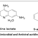 |
Figure 4: Antimicrobial and Antiviral acridine derivatives Click here to View Figure |
Neuroprotective and CNS Activity
In the realm of neurodegenerative diseases, acridine derivatives have gained interest as acetylcholinesterase (AChE) inhibitors for the treatment of Alzheimer’s disease (AD) 25. One of the most studied examples is Tacrine, the first FDA-approved drug for AD, although later withdrawn due to hepatotoxicity. Tacrine works by inhibiting AChE, thereby increasing synaptic levels of acetylcholine26. Its structure, based on acridine, has inspired the synthesis of several multitarget-directed ligands (MTDLs) that combine AChE inhibition with antioxidant or metal-chelating properties. Some newer hybrids link tacrine with other bioactive moieties such as coumarins or chalcones to enhance selectivity and reduce toxicity. Furthermore, acridine derivatives are being explored for their ability to inhibit monoamine oxidase (MAO A and B)27 enzyme useful in the treatment of Parkinson’s and depression. Some acridine hybrids may modulate glutamatergic signalling (NMDA anatagonism), a potential anti-seizure or neuroprotective agents28. Some hybride Acridine derivatives showed combine activity as AChE inhibition, metal-chelating, and antioxidant properties to address the multifactorial pathology of neurodegenerative diseases29.
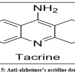 |
Figure 5: Anti-alzheimer’s acridine derivative Click here to View Figure |
Other Emerging Activities
Beyond well-established therapeutic areas, acridine-based compounds are being investigated for anti-diabetic, anti-inflammatory, antioxidant, and immunomodulatory effects30. Some derivatives exhibit significant inhibition of cyclooxygenase (COX) enzymes and lipoxygenase (LOX) pathways, offering potential in chronic inflammatory conditions31. In oxidative stress models, certain acridine analogs act as free radical scavengers, protecting cells from DNA and protein damage32. Additionally, acridine moieties have been incorporated into ligands for metal-based anticancer drugs, where they act as both targeting and chelating agents33. These emerging roles suggest that acridine’s therapeutic scope may extend far beyond its traditional applications.
Acridine-Based Hybrid Molecules and Multifunctional Agents
Hybrid molecules that combine the acridine scaffold with other bioactive pharmacophores have become a compelling strategy to develop multifunctional drugs. These conjugates aim to enhance efficacy, improve selectivity, and overcome drug resistance by simultaneously targeting multiple biological pathways. Acridine-based hybrids exploit the scaffold’s DNA intercalation ability alongside additional mechanisms contributed by the linked pharmacophores.
Acridine–Coumarin Hybrids
Coumarins exhibit antioxidant, anti-inflammatory, and anticancer activities. Their hybridization with acridine results in compounds that can intercalate DNA while inhibiting enzymes such as topoisomerases and kinases. For example, acridine–coumarin hybrids demonstrated significant anti-Alzheimer activity by inhibiting acetylcholinesterase enzyme34.
Acridine–Triazole Hybrids
Triazole rings are well-known for their antimicrobial and antifungal properties. Acridine–1,2,3-triazole (Figure 6) conjugates have been synthesized using click chemistry, resulting in potent antibacterial agents. The hybrid molecules combine the anti-bacterial activity of acridine with the enzyme inhibitory properties of triazoles, improving selectivity and reducing cytotoxicity35.
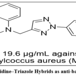 |
Figure 6: Acridine–Triazole Hybrids as anti-bacterial agents Click here to View Figure |
Acridine–Sulfonamide Hybrids
Sulfonamides are classical antibacterial agents that inhibit folate biosynthesis. Acridine–sulfonamide hybrids (Figure 7) have exhibited Alzheimer’s disease activity against AChE. These hybrids potentially inhibit AChE enzyme36.
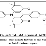 |
Figure 7: Acridine– Sulfonamide Hybrids as anti-bacterial agents as Ant-Alzheimers agents Click here to View Figure |
Acridine Hybrids Targeting Multiple Enzymes
Multifunctional acridine hybrids (Figure 8) targeting both acetylcholinesterase (AChE) and monoamine oxidase (MAO) are being developed as neuroprotective agents for treating neurodegenerative disorders. By simultaneously inhibiting these enzymes, these hybrids improve neurotransmitter balance and reduce oxidative stress. Recent acridine-based dual inhibitors have shown promising activity in vitro with improved BBB permeability37.
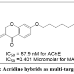 |
Figure 8: Acridine hybrids as multi-target agents Click here to View Figure |
Advantages and Challenges
The major advantage of acridine hybrids lies in their ability to act on multiple targets, potentially reducing the chance of drug resistance and adverse side effects by requiring lower doses. However, challenges include synthetic complexity, potential increased toxicity, and the need for thorough ADMET profiling to ensure drug-likeness.
Acridine Derivatives in the Drug Development Pipeline
In addition to approved acridine-based drugs, several novel acridine derivatives are currently under clinical and preclinical investigation, highlighting the scaffold’s ongoing relevance in drug discovery. Notably, C-1311 (Symadex®) (Figure 9), a synthetic acridine derivative, is in Phase II clinical trials as an anticancer agent targeting topoisomerase II with improved efficacy and safety profiles compared to older acridines38. Other promising pipeline candidates include 3,6-disubstituted acridines designed to selectively stabilize G-quadruplex DNA structures, exhibiting potent anticancer activity in preclinical models39. Additionally, acridine hybrids conjugated with kinase inhibitors and epigenetic modulators are being explored to overcome multidrug resistance in various malignancies. In the realm of infectious diseases, novel acridine analogs are under evaluation for enhanced antimalarial and antiviral activities, including inhibitors targeting emerging viruses such as SARS-CoV-240-41. These pipeline molecules underscore the continued innovation centered on the acridine core and its versatility in addressing unmet medical needs.
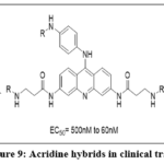 |
Figure 9: Acridine hybrids in clinical trails Click here to View Figure |
Acridine in Diagnostic and Imaging Applications
Fluorescent Properties of Acridine
Acridine derivatives are well known for their intrinsic fluorescence, which makes them valuable tools in biological imaging and diagnostic assays. Their planar structure allows them to intercalate with nucleic acids, resulting in changes to their fluorescence emission properties that can be exploited for DNA/RNA detection. This fluorescence is sensitive to microenvironment changes, enabling real-time monitoring of nucleic acid interactions and cellular processes42.
Acridine Orange as a Diagnostic Dye
One of the most widely used acridine compounds in diagnostics is Acridine Orange (AO), a cationic dye that intercalates into DNA and RNA, emitting green fluorescence when bound to DNA and red/orange when bound to RNA. AO staining is routinely employed in:
Fluorescence microscopy for differentiating live and dead cells based on membrane integrity.
Flow cytometry for cell cycle analysis and apoptosis detection.
Cancer diagnostics to identify malignant cells by staining nuclei with enhanced fluorescence intensity43
Applications in Microbial Identification
Acridine dyes, especially AO, are used in microbiology to stain bacteria and fungi for rapid detection under fluorescence microscopy. This enables early diagnosis of infections in clinical specimens. AO staining is also valuable in environmental microbiology for assessing microbial viability in water and soil samples44.
Use in Photodynamic Diagnosis (PDD)
Acridine derivatives are being explored in photodynamic diagnosis (PDD), where their fluorescence helps delineate tumor margins during surgery. For example, acridine-based probes can selectively accumulate in cancerous tissues and emit fluorescence upon excitation, assisting surgeons in real-time tumor visualization, improving resection accuracy45.
Emerging Imaging Modalities
Recent advances include conjugating acridine fluorophores with nanoparticles and antibodies to develop targeted imaging agents with higher specificity and sensitivity. These novel acridine-based probes are under investigation for use in in vivo imaging, biomarker detection, and theranostics combining diagnosis and therapy46.
Toxicological and Pharmacokinetic Considerations
Toxicity and DNA Damage
While acridine derivatives have demonstrated significant therapeutic potential, their interaction with DNA also raises concerns about toxicity and mutagenicity. Many acridine compounds, due to their DNA intercalation properties, can cause DNA strand breaks, mutations, and chromosomal aberrations, which may lead to genotoxic effects. For instance, Amsacrine, despite its anticancer efficacy, is associated with dose-limiting toxicities such as myelosuppression and cardiotoxicity47. Similarly, Tacrine, used for Alzheimer’s, showed hepatotoxicity leading to its withdrawal from clinical use48. This underscores the importance of balancing efficacy and safety in acridine drug development.
Pharmacokinetics and Metabolism
Acridine derivatives generally exhibit moderate to good oral bioavailability, but their pharmacokinetic profiles vary widely depending on substitution patterns and molecular size. They are metabolized primarily via hepatic pathways, including oxidation and conjugation. The metabolites may have altered activity or toxicity profiles. For example, Tacrine undergoes extensive first-pass metabolism to form hydroxylated metabolites, some of which contribute to liver toxicity49.
Strategies to Reduce Toxicity
Several approaches are employed to mitigate acridine-associated toxicity:
Targeted delivery systems: Nanoparticles, liposomes, and conjugation with targeting ligands enhance selective accumulation in diseased tissues, reducing systemic exposure50.
Prodrug design: Masking reactive groups to release active acridine moieties selectively at the target site minimizes off-target effects.
Structure modification: Reducing planarity or introducing bulky groups to decrease DNA intercalation strength and improve selectivity.
Hybrid molecules: Combining acridine with other moieties to balance activity and reduce toxic side effects.
ADMET Profiling and Drug-Likeness
Computational ADMET (Absorption, Distribution, Metabolism, Excretion, and Toxicity) modeling has become essential in acridine drug development to predict potential liabilities early. For example, tools assessing blood-brain barrier penetration, cytochrome P450 interactions, and mutagenic potential guide lead optimization. These predictions help prioritize candidates with favorable pharmacokinetic and safety profiles, accelerating clinical translation51.
Safety and Toxicity Profile of Acridine Derivatives
Acridine derivatives, while therapeutically promising, pose significant safety and toxicity challenges due to their inherent mode of action and chemical structure. The planar tricyclic structure allows acridines to intercalate into DNA, disrupting cellular processes, which is beneficial against target cells but may also affect normal tissues, raising concerns about genotoxicity and off-target toxicity.
Genotoxicity and Mutagenicity
Acridine compounds are well-documented mutagens and genotoxic agents. Their DNA intercalating property can cause frame-shift mutations and chromosomal breaks, leading to potential carcinogenicity52. Studies have shown that acridine derivatives induce DNA damage in bacterial, mammalian cell lines, and animal models53. For example, quinacrine and related compounds have demonstrated mutagenic effects in Ames tests and micronucleus assays, raising concerns for long-term safety54.
Organ-Specific Toxicity
Hepatotoxicity: Several acridine drugs exhibit liver toxicity, often through metabolic activation to reactive intermediates causing oxidative stress and hepatocellular damage. Quinacrine use has been associated with elevated liver enzymes and rare cases of hepatic dysfunction55.
Nephrotoxicity: Accumulation of acridine derivatives in the kidneys can induce tubular damage, particularly at higher doses or prolonged exposure56.
Neurotoxicity: Neurological side effects such as dizziness, headache, and cognitive impairment have been reported, especially with acridine antimalarials57.
Cardiotoxicity: Though less common, some acridine-based topoisomerase inhibitors have been linked to QT prolongation and arrhythmias in clinical settings58.
Phototoxicity and Photosensitization
Acridine derivatives, such as acridine orange, exhibit photosensitizing properties. Upon exposure to UV or visible light, they generate reactive oxygen species (ROS), which can induce cellular damage58. While exploited therapeutically in photodynamic therapy (PDT), unintended phototoxicity remains a safety concern in clinical use.
Approaches to Reduce Toxicity
To address these challenges, researchers have employed multiple strategies:
Structural modification: Altering substituents on the acridine ring to decrease DNA intercalation strength and reduce mutagenicity while retaining activity59.
Hybrid molecules: Combining acridine with other pharmacophores to increase target specificity and minimize off-target effects60.
Nanocarrier-based drug delivery: Using liposomes, polymeric nanoparticles, and other carriers to deliver acridine drugs selectively to diseased tissues, reducing systemic exposure and toxicity61.
Prodrug design: Developing acridine prodrugs activated only in the tumor microenvironment or specific pathological sites to limit toxicity62.
Regulatory Perspectives
Given the mutagenic potential, regulatory authorities require comprehensive genotoxicity and long-term carcinogenicity assessments for acridine derivatives. The FDA and EMA recommend a battery of tests including Ames, micronucleus, chromosomal aberration, and in vivo genotoxicity assays before approving acridine-containing drugs (ICH S2(R1) guidelines). Balancing therapeutic benefits against safety risks remains critical for clinical advancement63-64.
Challenges and Future Perspectives
Challenges in Acridine Drug Development
Despite the promising pharmacological activities of acridine derivatives, several challenges limit their clinical translation:
Toxicity and safety concerns: The intrinsic DNA intercalating property that underlies many acridine’s therapeutic effects also causes genotoxicity, mutagenicity, and off-target damage, which complicates safe dosing.
Drug resistance: Cancer cells and pathogens can develop resistance to acridine-based agents through mechanisms such as efflux pumps, DNA repair enhancements, and target mutations, reducing drug efficacy65.
Pharmacokinetic limitations: Many acridine derivatives suffer from poor solubility, rapid metabolism, or limited bioavailability, which restrict their therapeutic potential66.
Synthetic complexity: Designing and synthesizing multifunctional acridine hybrids with optimized activity and reduced toxicity requires sophisticated chemical approaches that can be costly and time-consuming67.
Emerging Strategies to Overcome Challenges
To address these challenges, ongoing research is exploring:
Targeted drug delivery: Using nanocarriers, antibody-drug conjugates, or prodrugs to enhance selective delivery to diseased tissues and reduce systemic toxicity.
Structure-based drug design: Utilizing computational modeling to design acridine derivatives with improved specificity for molecular targets such as topoisomerases or G-quadruplexes.
Multitargeted agents: Developing hybrid molecules that target multiple pathways simultaneously, potentially overcoming resistance and enhancing therapeutic efficacy68.
Biological evaluation using advanced models: Employing 3D cell cultures, organoids, and in vivo imaging to better predict clinical outcomes69.
Future Perspectives
The acridine scaffold continues to be a versatile platform for drug discovery, especially with advances in medicinal chemistry and molecular biology. Novel acridine derivatives designed through rational drug design and integrated with emerging technologies such as CRISPR gene editing and high-throughput screening may unlock new therapeutic possibilities. Furthermore, combining acridine compounds with immunotherapy and personalized medicine approaches could enhance treatment outcomes, especially in oncology and infectious diseases. Finally, expanding research into non-traditional applications such as epigenetic modulation, photodynamic therapy, and anti-inflammatory effects may broaden the clinical utility of acridine derivatives.
Conclusion
Acridine and its derivatives continue to be an exceptionally valuable class of compounds in medicinal chemistry, demonstrating a broad spectrum of biological activities including anticancer, antimalarial, antimicrobial, antiviral, and neuro-protective effects. Their unique ability to intercalate DNA and modulate various enzymes underpins much of their pharmacological potential. Importantly, the development of acridine-based hybrid molecules has further expanded their therapeutic scope by enabling multifunctional targeting and potentially reducing adverse effects. Despite significant advances, challenges such as toxicity, drug resistance, and pharmacokinetic limitations still hinder the full clinical potential of acridine derivatives. Innovative strategies, including targeted delivery systems, rational design, and multi-targeted agents, offer promising avenues to overcome these obstacles.
Looking forward, the integration of acridine chemistry with cutting-edge technologies like nanotechnology, computational modelling, and personalized medicine is likely to unlock novel therapeutic applications. Further research into epigenetic regulation, photodynamic therapy, and immunomodulation may also broaden the impact of acridine compounds. In summary, acridine remains a “magical molecule” in medicinal chemistry, whose continued exploration promises to yield new and effective therapeutic agents for a range of challenging diseases.
Funding Sources
The author(s) received no financial support for the research, authorship, and/or publication of this article.
Conflict of Interest
The author(s) do not have any conflict of interest.
Data Availability Statement
This statement does not apply to this article.
Ethics Statement
This research did not involve human participants, animal subjects, or any material that requires ethical approval.
References
- Prasher, P.; Sharma, M.; ; 2018, 9, 1589. doi:10.1039/C8MD00384J
CrossRef - Graebe, C.; Caro H.; Journal für Praktische Chemie., 1870, 2, 183. doi:10.1002/cber.18700030223
CrossRef - Baguley, B.C.; Wakelin, L.P.; Jacintho, J.D.; Kovacic, P.; Current Medicinal Chemistry, 2003, 1, 2643. doi:10.2174/0929867033456332
CrossRef - Jangir, D.K.; Dey, S.K.; Kundu, S.; Mehrotra, R.; Journal of Photochemistry and Photobiology B: Biology, 2012, 3, 38. doi:10.1016/j.jphotobiol.2012.05.005
CrossRef - Ketron, A.C.; Denny, W.A.; Graves, D.E.; Osheroff, N.; Biochemistry, 2012, 51, 1730. doi:10.1021/bi201159b
CrossRef - Ehsanian, R.; Van, W. C.; Feller, S.M.; Cell Communication and Signaling, 2011, 9, 1. doi:10.1186/1478-811X-9-13
CrossRef - Kumar, M.; Sarkar, A.; Scientia Pharmaceutica, 2022, 90, 12. doi:10.3390/scipharm90010012
CrossRef - Gensicka-Kowalewska, M., Cholewiński, G., Dzierzbicka, K., RSC Advances, 2017, 7, 15776. doi:10.1039/C7RA01026E
CrossRef - Goni, L.K.; Jafar, Mazumder, M.A.; Tripathy, D.B.; Quraishi, M.A.; Materials, 2022, 15, 7560. doi:10.3390/ma15217560
CrossRef - Luzzati, V.; Masson, F.; Lerman, L.S.; Journal of Molecular Biology, 1961, 3, 634. doi:10.1016/S0022-2836(61)80026-0
CrossRef - Lerman, L.S.; Proceedings of the National Academy of Sciences, 1963, 49, 94. doi:10.1073/pnas.49.1.94
CrossRef - Lou, Y.H.; Wang, J.S.; Dong, G.; Guo, P.P.; Wei, D.D.; Xie, S.S,; Yang, M.H.; Kong, L.Y.; Toxicology Research, 2015, 4, 1465. doi:10.1039/C5TX00096C
CrossRef - Marsh, K.L.; Willmore, E.; Tinelli, S.; Cornarotti, M.; Meczes, E.L.; Capranico, G.; Fisher L.M.; Austin, C.A.; Biochemical Pharmacology, 1996, 52, 1675. doi:10.1016/s0006-2952(96)00516-3
CrossRef - Lee, M.S.; Wang, J.C.; Beran, M.; Journal of Molecular Biology, 1992, 223, 837. doi:10.1016/0022-2836(92)90245-f
CrossRef - Tiberghien, F.; Loor, F.; Anticancer Drugs, 1996, 7, 568. doi:10.1097/00001813-199607000-00012
CrossRef - Patek, E.; Acta Obstetricia et Gynecologica Scandinavica, 1979, 58, 561. doi:10.3109/00016347909154619
CrossRef - Osuna, A.; Ruiz-Perez, L.M.; Gamarro, F.; Rodriguez-Santiago, J.I.; Castanys, S.; Sharples, D.; Galy, A.M.; Giovannangeli, G.; Galy, J.P.; Soyfer, J.C.; Barbe, J.; Chemotherapy, 1988, 34, 127. doi:10.1159/000238559
CrossRef - Tehlan, A.; Karmakar, B.C.; Paul, S.; Kumar, R.; Kaur, I.; Ghosh, A.; Mukhopadhyay, A.K.; Dhar, S.K.; FEMS Microbiology Letters, 2020, 367, 178. doi:10.1093/femsle/fnaa178
CrossRef - Piorecka, K.; Kurjata, J.; Stanczyk, W.A.; Journal of Medicinal Chemistry, 2022, 65, 11415. doi:10.1021/acs.jmedchem.2c00573
CrossRef - Jabri, T.; Khan, N.A.; Makhlouf, Z.; Akbar N.; Gul, J.; Shah, M.R.; Siddiqui, R.; Antibiotics, 2023, 12, 755. doi:10.3390/antibiotics1204075
CrossRef - Fujiwara, M.; Okamoto, M.; Watanabe, M.; Machida, H.; Shigeta, S.; Konno, K.; Yokota, T.; Baba, M.; Antiviral Research, 1999, 43, 189. doi:10.1016/s0166-3542(99)00045-5
CrossRef - Napolitano, V.; Dabrowska, A.; Schorpp, K.; Mourao, A.; Barreto-Duran, E.; Benedyk, M.; Botwina, P.; Brandner, S.; Bostock, M.; Chykunova Y.; Czarna, A.; Cell Chemical Biology, 2022, 29, 774. doi:10.1016/j.chembiol.2021.11.006
CrossRef - Medapi, B.; Meda, N.; Kulkarni, P.; Yogeeswari, P.; Sriram, D.; Bioorganic & Medicinal Chemistry, 2016, 24, 877. doi:10.1016/j.bmc.2016.01.011
CrossRef - Gabriel, I.; Molecules, 2020, 25, 1480. doi:10.3390/molecules25071480
CrossRef - Blackard, Jr W.G.; Sood, G.K.; Crowe, D.R.; Fallon, M.B.; Journal of Clinical Gastroenterology, 1998, 26, 57. doi:10.1097/00004836-199801000-00015
CrossRef - Lou, Y.H.; Wang, J.S.; Dong, G.; Guo, P.P.; Wei, D.D.; Xie, S.S.; Yang, M.H.; Kong, L.Y.; Toxicology Research, 2015, 4, 1465. doi:10.1039/C5TX00096C
CrossRef - Remya, R.S.; Ramalakshmi, N.; Nalini, C.N.; Amuthalakshmi, S.; Rasayan Journal of Chemistry, 2022, 15, 2318. doi:10.31788/RJC.2022.1547080
CrossRef - Barygin, O.I.; Gmiro, V.E.; Kim, K.K.; Magazanik, L.G.; Tikhonov, D.B.; Neuroscience Letters, 2009, 451, 29. doi:10.1016/j.neulet.2008.12.036
CrossRef - Sharma, A.; Piplani, P.; Current Topics in Medicinal Chemistry, 2023, 23, 1260. doi:10.2174/1568026623666230203141543
CrossRef - Rao, I.R.; Punitha, P.; Premalatha, B.; Prasad, T.S.; Suresh, M.; Next Research, 2024, 1, 100057. doi:10.1016/j.nexres.2024.100057
CrossRef - Chen, Y.L.; Lu, C.M.; Chen, I.L.; Tsao L.T.; Wang, J.P.; Journal of Medicinal Chemistry, 2002, 45, 4689. doi:10.1021/jm020102v
CrossRef - Raghu, P.S.; World Journal of Pharmaceutical Research, 2018, 7, 1739.
- Pal, C.; International Journal of Medical Biochemistry, 2023, 6, 150. doi:10.14744/ijmb.2023.47450
CrossRef - Hamulakova, S.; Janovec, L.; Soukup, O.; Jun, D.; Kuca, K.; International Journal of Biological Macromolecules, 2017, 104, 333. doi:10.1016/j.ijbiomac.2017.06.006
CrossRef - Aarjane, M.; Slassi, S.; Tazi, B.; Maouloua, M.; Amine, A.; Structural Chemistry, 2020, 31, 1523. doi:10.1007/s11224-020-01512-0
CrossRef - Ulus, R.; Esirden, İ.; Aday, B.; Turgut, G.Ç.; Şen, A.; Kaya, M.; Medicinal Chemistry Research, 2018, 27, 634. doi:10.1007/s00044-017-2088-2
CrossRef - Sun, Y.; Chen, J.; Chen, X.; Huang, L.; Li, X.; Bioorganic & Medicinal Chemistry, 2013, 21, 7406. doi:10.1016/j.bmc.2013.09.050
CrossRef - Isambert, N.; Campone, M.; Bourbouloux, E.; Drouin, M.; Major, A.; Yin, W.; Loadman, P.; Capizzi, R.; Grieshaber, C.; Fumoleau, P.; European Journal of Cancer, 2010, 46, 729. doi:10.1016/j.ejca.2009.12.005
CrossRef - Read, M.; Harrison, R.J.; Romagnoli, B.; Tanious, F.A.; Gowan, S.H.; Reszka, A.P.; Wilson, W.D.; Kelland, L.R.; Neidle, S.; Proceedings of the National Academy of Sciences, 2001, 98, 4844. doi:10.1073/pnas.081560598
CrossRef - Jones, T.; Monakhova, N.; Guivel-Benhassine, F.; Lepioshkin, A.; Bruel, T.; Lane, T.R.; Schwartz, O.; Puhl, A.C.; Makarov, V.; Ekins, S.; ACS Omega, 2023, 8, 40817. doi:10.1021/acsomega.3c05900
CrossRef - Guetzoyan, L.; Yu, X.M.; Ramiandrasoa, F.; Pethe, S.; Rogier, C.; Pradines, B.; Cresteil, T.; Perrée-Fauvet, M.; Mahy, J.P.; Bioorganic & Medicinal Chemistry, 2009, 17, 8032. doi:10.1016/j.bmc.2009.10.005
CrossRef - Limpouchová, Z.; Procházka, K.; Fluorescence Studies of Polymer Containing Systems, 2016, 16, 91. doi:10.1007/978-3-319-26788-3_4
CrossRef - Liu, M.; Li, R.; Tang, Y.; Chang, J.; Han, R.; Zhang, S.; Jiang, N.; Ma, F.; Oncology Letters, 2017, 13, 2221. doi:10.3892/ol.2017.5724
CrossRef - Boulos, L.; Prévost, M.; Barbeau, B.; Coallier, J.; Desjardins, R.; Journal of Microbiological Methods, 1999, 37, 77. doi:10.1016/S0167-7012(99)00048-2
CrossRef - Byvaltsev, V.A.; Bardonova, L.A.; Onaka, N.R.; Polkin, R.A.; Ochkal, S.V.; Shepelev, V.V.; Aliyev, M.A.; Potapov, A.A.; Frontiers in Oncology, 2019, 9, 925. doi:10.3389/fonc.2019.00925
CrossRef - Chen, H.; Zhang, W.; Zhu, G.; Xie, J.; Chen, X.; Nature Reviews Materials, 2017, 2, 1. doi:10.1038/natrevmats.2017.24
CrossRef - Morelli, M.B.; Bongiovanni, C.; Da, P.S.; Miano, C.; Sacchi, F.; Lauriola, M.; D’Uva, G.; Frontiers in Cardiovascular Medicine, 2022, 9, 847012. doi:10.3389/fcvm.2022.847012
CrossRef - Blackard, Jr W.G.; Sood, G.K.; Crowe, D.R.; Fallon, M.B.; Journal of Clinical Gastroenterology, 1998, 26, p. 57. doi:10.1097/00004836-199801000-00015
CrossRef - Madden, S.; Spaldin, V.; Park, B.K.; Clinical Pharmacokinetics, 1995, 28, 449. doi:10.2165/00003088-199528060-00003
CrossRef - Mitra, P.; Chakraborty, P.K.; Saha, P.; Ray, P.; Basu, S.; International Journal of Pharmaceutics, 2014, 473, 636. doi:10.1016/j.ijpharm.2014.07.051
CrossRef - Daoud, N.E.; Borah, P.; Deb, P.K.; Venugopala, K.N.; Hourani, W.; Alzweiri, M.; Bardaweel, S.K.; Tiwari, V.; Current Drug Metabolism, 2021, 22, 503. doi:10.2174/1389200222666210705122913
CrossRef - Arshad, R.; Farooq, S.; Iqbal, N.; Ali, S.S.; Letters in Applied Microbiology, 2006, 42, 94. doi:10.1111/j.1472-765X.2005.01819.x
CrossRef - Mortelmans, K.; Zeiger, E.; Mutation Research, 2000, 455, 29. doi:10.1016/s0027-5107(00)00064-6
CrossRef - Clarke, J.J.; Sokal, D.C.; Cancel, A.M.; Campen, D.B.; Gudi, R.; Wagner, V.O.; San, R.H.; Jacobson-Kram, D.; Mutation Research/Genetic Toxicology and Environmental Mutagenesis, 2001, 494, 41. doi:10.1016/S1383-5718(01)00178-4
CrossRef - Ciaccio, L.A.; Hill, J.L.; Kincl, F.A.; Contraception, 1978, 17, 231. doi:10.1016/0010-7824(78)90014-8
CrossRef - Patadiya, N.; Panchal, N.; Vaghela, V.; International Research Journal of Pharmacy, 2021, 12, 60. doi:10.7897/2230-8407.1206145
CrossRef - Grabias, B.; Kumar, S.; Expert Opinion on Drug Safety, 2016, 15, 903. doi:10.1080/14740338.2016.1175428
CrossRef - Patadiya, N.; Vaghela, V.; Asian Journal of Pharmaceutical Research, 2022, 12, 207. doi:10.52711/2231-5691.2022.00034
CrossRef - Osman, H.; Elsahy, D.; Saadatzadeh, M.R.; Pollok, K.E.; Yocom, S.; Hattab, E.M.; Georges, J.; Cohen-Gadol, A.A.; World Neurosurgery, 2018, 114, e1310. doi:10.1016/j.wneu.2018.03.207
CrossRef - Joshi, J.; Vala, B.; Singh, S.; Patel S.; Patadiya, N.; Research Journal of Pharmacognosy and Phytochemistry, 2025, 17,116. doi:10.52711/0975-4385.2025.00020
CrossRef - Zhang, Q.; Yu, X.; Current Topics in Medicinal Chemistry, 2021, 21, 1773. doi:10.2174/1568026621666210804115203
CrossRef - Patra, J.K.; Das, G.; Fraceto, L.F.; Campos, E.V.; Rodriguez-Torres, M.D.; Acosta-Torres, L.S.; Diaz-Torres, L.A.; Grillo, R.; Swamy, M.K.; Sharma, S.; Habtemariam, S.; Journal of Nanobiotechnology, 2018, 16, 1. doi:10.1186/s12951-018-0392-8
CrossRef - Denny, W.A.; European Journal of Medicinal Chemistry, 2001, 36, 577. doi:10.1016/s0223-5234(01)01253-3
CrossRef - ICH Harmonised Guideline, Genotoxicity Testing and Data Interpretation for Pharmaceuticals Intended for Human Use S2(R1), 2011, International Council for Harmonisation of Technical Requirements for Pharmaceuticals for Human Use.
- Garg, P.; Malhotra, J.; Kulkarni, P.; Horne, D.; Salgia, R.; Singhal, S.S.; Cancers, 2024, 16, 2478. doi:10.3390/cancers16132478
CrossRef - Sun, X.; Zhao, P.; Lin, J.; Chen, K.; Shen, J.; Cancer Drug Resistance, 2023, 6, 390. doi:10.20517/cdr.2023.16
CrossRef - Modh, D.H.; Kulkarni, V.M.; Mini Reviews in Medicinal Chemistry, 2024, 24, doi:10.2174/1389557523666230509123036
CrossRef - Cui, Z.; Li, X.; Li, L.; Zhang, B.; Gao, C.; Chen, Y.; Tan, C.; Liu, H.; Xie, W.; Yang, T.; Jiang, Y.; Bioorganic & Medicinal Chemistry, 2015, 24, 261. doi:10.1016/j.bmc.2015.12.011
CrossRef - Fang, Y.; Eglen, R.M.; LAS Discovery: Advancing Life Sciences R&D, 2017, 22, 456. doi:10.1177/1087057117696795
CrossRef
 PlumX:
  Views: 474 Views
  PDF Downloads: 113
 Accepted on: 05 Feb 2026
Second Review by: Dr. Arti Malviya
Final Approval by: Dr. Fozia Z. Haque


ISSN Online: 2231-5039