Formulation and Development of Metformin-Loaded Microspheres Using Eudragit S-100 as a Carrier Polymer
 , Alankar Shrivastav1, Vijay Sharma1, Mohd Junaid2
and Mariyam Khan3
, Alankar Shrivastav1, Vijay Sharma1, Mohd Junaid2
and Mariyam Khan31Faculty of Pharmacy, Pharmacy Academy, IFTM University, Moradabad, U.P, India.
2Department of Pharmacy, Mohammad Ali Jauhar University Rampur, U. P., India
3School of Pharmaceutical Sciences, IFTM University, Moradabad, U.P. India
Corresponding Author Email: Pawansingh690@gmail.com
DOI : http://dx.doi.org/10.13005/ojc/420318
Download this article as:
![]()
Metformin hydrochloride (MH), a member of the biguanide class of oral antidiabetic agents, is extensively used in the management of DM type II. Nonetheless, its therapeutic potential is limited because of its short half-life and relatively low oral absorption (approx. 55-60%), requiring multiple dosing. The purpose of the current study was to formulate and evaluate sustained release microspheres of Metformin hydrochloride by the solvent evaporation method to improve its bioavailability and effective duration of action. Microspheres were developed by the technique of solvent evaporation employing Eudragit S100 and Sodium CMC as polymeric carriers. After optimising polymer amounts, thirteen formulations (F1–F13) were prepared. The formulations were assessed based on % yield, % drug EE, surface morphology, scanning electron microscopy (SEM), FTIR pattern and in vitro drug release studies. There was no pronounced interaction between the drug and polymers, as observed from the FTIR spectra. SEM observation indicated that the higher the concentration of polymer, the smoother the microsphere surface. F6 was best among the formulations with a production yield of 96.78±0.005%, drug entrapment efficiency of 80.72±0.020%, and sustained drug release of 96.52±0.010%. Card Sort F6 Card Sort revealed that all formulations showed an efficient encapsulation and an economically controlled release profile; however, the F6 formulation was selected as the most optimised formulation. The optimised Metformin hydrochloride sustained release formulations E8, F6 and G3 were acceptable in terms of physicochemical properties andin vitro drug release pattern, out of which formulation F6 was considered as an ideal sustained-release delivery system of Metformin hydrochloride. The present plan of work demonstrates the feasibility of the solvent evaporation method for the preparation of controlled release microspheres to improve the bioavailability and lower the dosing frequency of anti-diabetic therapy.
KEYWORDS:Anti-diabetes; Bioavailability; Metformin Hydrochloride; Solvent evaporation
Introduction
Metformin Hydrochloride (MH) is an oral antidiabetic agent commonly used to treat type 2 diabetes mellitus (T2DM). It is in the biguanide class of drugs and acts on reducing glucose production in the liver and enhancing insulin sensitivity in peripheral tissue1. The use of microsphere technology is an attractive strategy to improve the clinical efficacy of MH by sustained and targeted delivery, which results in decreased dosing frequency, reduced fluctuation of drug level and alleviation of side effects2. Eudragit S 100 is a biocompatible and pH-responsive polymer and has been used in a large number of pharmaceutical preparations. Its solubility at pH levels higher than 7 also makes it eligible for a colon-specific delivery system, allowing for the release of MH in the lower intestine for enhanced bioavailability.
The goal of the present study is to prepare S100-coated MH microspheres for sustained and improved release of MH. Parameters including polymer concentration, drug-polymer ratio, and preparation method will be optimised to achieve microspheres with desirable properties, including targeted particle size, encapsulation efficiency, and release profile4. The in vitro release profile of MH from Eudragit S100 microspheres will be investigated in dissolution studies under simulated gastrointestinal conditions. Furthermore, stability and pharmacokinetic profiles of these microspheres will be evaluated to demonstrate their feasibility as an efficient dosage form for long-term MH treatment5. In conclusion, MH-loaded microspheres prepared by Eudragit S100 havethe potential to improve the clinical efficacy and reduce toxicity for diabetic patients by achieving the sustained release and targeted delivery of MH, and hence enhancing the therapeutic effect and reducing the side effects of MH.
Materials and Methods
Materials
Metformin hydrochloride was purchased from a Gift sample from Venus Biosciences Pvt Ltd, Baddi,Eudragit S100Gift sample from Alves Health Care, Baddi, Sodium carboxymethylcellulose, Tween 80, Methanol, HCl and other ingredients will be procured from CDH Laboratory.
Design of Experiment
A 32 Central composite design approachwas systematically implemented to optimise the formulation of microspheres, focusing on the influence of two key formulation factors: the concentration of sodium carboxymethyl cellulose (Sodium CMC) and Eudragit S100.6. These variables were optimised through the response surface methodology (RSM) for the nitrifier culture developed. A central composite design (CCD) was adopted for the experimental runs employing Eudragit S100 and Sodium CMC in different concentrations. In this way, a two-thirds full factorial design also permitted the consideration of both main and interaction effects of these independent variables on critical formulation responses.7The independent factors were Eudragit S100 (X₁) and Sodium CMC (X₂), the dependent responses evaluated were % DR and % EE of the microspheres. This approach allowed the selection of the best formulation parameters for achieving the requisite drug release kinetics. We want to have a model for the response that is optimized as well as empirically applicable to check the statistically made model by comparing the predicted values and the observed values8. Table 1 shows the 32 CCD configurations (patterns). The coded and actual values of the independent variables are listed in Table 2. 32CCD, response surface methodology, and polynomial equations were proposed as the trial application for Design Expert software9. The linear computer model equation that correlated the predicted value of each response will be shown in the following form:
Y=β0+β1X1+β2X2+β3X3+…+βnXn+ε
Y: Response variable
β₀: Intercept (constant term).
β₁, β₂, …, βₙ: Coefficients for the independent variables (X₁, X₂). These represent the effect of each factor on the response.
X₁, X₂, …, Xₙ: Independent variables or factors
ε: Experimental error or noise.
Preparation of optimised microspheres of metformin hydrochloride
Formulation S100-based microspheres were prepared by the emulsion solvent evaporation method. An appropriate quantity of Eudragit S100 was dissolved in 100 mL of methanol to obtain a homogeneous polymer solution. Metformin hydrochloride was then incorporated into this polymer solution and mixed well. The resulting drug–polymer mixture in the above ratio was poured into 250 mL of water based on sodium carboxymethyl cellulose mucilage (0.5% w/v containing 1% v/v of Tween 80), and stirring was continued at room temperature for 3 hrs to form spherical microspheres. In the present study, methanol was employed as a polymer solvent, while aqueous sodium CMC mucilage served as a microencapsulating medium and Tween 80 as a dispersing agent. The methanol evaporated in the course of the reflux. The microspheres were isolated by vacuum filtration, washed with distilled water and petroleum ether several times, and then dried at room temperature for 24 h to obtain free-flowing microspheres. The remaining microspheres were stored in a desiccator for the next analysis. The formulations were denominated as F1 to F13 according to the polymer concentration.
Table 1: The different variable parameters for the 32-factorial study with code value
|
Batch code |
Eudragit S100 |
Sodium CMC |
|
F-1 |
−1 | −1 |
| F-2 | 0 |
−1 |
|
F-3 |
1 | −1 |
| F-4 | −1 |
0 |
|
F-5 |
0 | 0 |
| F-6 | 1 |
0 |
|
F-7 |
−1 | 1 |
| F-8 | 0 |
1 |
|
F-9 |
1 | 1 |
| F-10 | 0 |
0 |
|
F-11 |
0 | 0 |
| F-12 | 0 |
0 |
|
F-13 |
0 |
0 |
Table 2: The different variable parameters for the 32 factorial study as independent Variables and Their Levels
|
Formulation code |
-1 | 0 |
1 |
|
Eudragit S100 (mg) |
0.5 | 0.75 | 1 |
| Sodium CMC (mg) | 0.1 | 0.25 |
0.5 |
Table 3: Formulation of Various Batches ofMicrospheres of Metformin Hydrochloride.
|
Formulation code |
Eudragit S100 | Code
Value |
Sodium CMC | Code
Value |
Tween 80 (mL) | Methanol
(mL) |
Metformin hydrochloride (mg) | Speed (rpm) |
| F-1 | 0.5 | -1 | 0.1 | -1 | 1 | 10 | 50 |
1000 |
|
F-2 |
0.75 | 0 | 0.1 | -1 | 1 | 10 | 50 | 1000 |
| F-3 | 1 | +1 | 0.1 | -1 | 1 | 10 | 50 |
1000 |
|
F-4 |
0.5 | -1 | 0.25 | 0 | 1 | 10 | 50 | 1000 |
| F-5 | 0.75 | 0 | 0.25 | 0 | 1 | 10 | 50 |
1000 |
|
F-6 |
1 | +1 | 0.25 | 0 | 1 | 10 | 50 | 1000 |
| F-7 | 0.5 | -1 | 0.5 | +1 | 1 | 10 | 50 |
1000 |
|
F-8 |
0.75 | 0 | 1 | +1 | 1 | 10 | 50 | 1000 |
| F-9 | 1 | +1 | 0.5 | +1 | 1 | 10 | 50 |
1000 |
|
F-10 |
0.75 | 0 | 0.25 | 0 | 1 | 10 | 50 | 1000 |
| F-11 | 0.75 | 0 | 0.25 | 0 | 1 | 10 | 50 |
1000 |
|
F-12 |
0.75 | 0 | 0.25 | 0 | 1 | 10 | 50 | 1000 |
| F-13 | 0.75 | 0 | 0.25 | 0 | 1 | 10 | 50 |
1000 |
Characterization of Microspheres
Micromeritic Properties of Microspheres
After the preparation of microspheres, the prepared microspheres were subjected to determination of their micrometric properties like bulk density and tapped density 10-11.
Percentage Drug Entrapment
The %EE was calculated based on the percentage of the drugs contained within the polymer. Several elements can influence the trapping efficiency of the drugs into microspheres. Some of these factors include the drug nature, polymer concentration, drug-to-polymer ratio, stirring rate, surfactant concentration, etc.12 The drug entrapment efficiency for each batch was expressed as % PDE by the following equation:
PDE = Practical drug content /Theoretical drug content × 100
Particle Size Analysis
Dynamic light scattering (DLS) Method Particle size and size distribution of the prepared microspheres were measured using a Zetasizer (Malvern Instruments Ltd., UK). A small quantity of microsphere sample was dispersed in distilled water and sonicated for 2–3 min to provide a homogeneous sample as well as prevent condensation ofthe sample before mixing the experimental sample by sonication with a 2–3 min duration. Then, the sample was put into the cuvette, and measurements were conducted at 25 ± 1 °C in ambient conditions. The data were expressed in terms of Z-average diameter (d.nm), polydispersity index (PDI), and intensity-based size distribution13.
Fourier Transformed Infrared (FTIR) Studies
FT-IR blank and drug-loaded formulations were studied to investigate the existence of any drug-polymer interaction, by using FTIR (Fourier Transform Infra-red spectrometer (Bruker Alpha, Germany) in the frequency range 4000-400cm-1.14.
Differential Scanning Calorimetry (DSC)
Physicochemical interactions and thermal behaviour were determined based on the Differential Scanning Calorimetry (DSC) thermograms of the drug, polymer, and the formulated microspheres. The DSC thermograph for the microspheres indicated some broadening and small shift in the characteristic endothermic peak of the drug, which suggested that part of the drug had become amorphous and was encapsulated successfully within the polymer matrix, without any significant drug–polymer interaction15.
X-ray Diffraction (XRD) Studies
The drug and excipient crystallinity effects on formulation were investigated by XRD analysis. XRD The blank and drug-loaded microparticles were examined by an X-ray Diffractometer (Rigaku-Ultima IV, Japan) and collected by Cu radiation at a 40-kV voltage and 30-mA current. The scanning rate was 2 °/min with a diffraction angle of 10 ° to 90°16.
Scanning Electron Microscopy (SEM)
The morphological and dimensional analyses of the microspheres were performed by scanning electron microscopy (SEM). Microsphere size and shape distribution were determined by SEM photomicrographs,analyzing about 500 microspheres17.
In-Vitro Drug Release
The studies were performed with microspheres containing 50 mg of MH to demonstrate the possibility of achieving the required controlled release drug delivery. Release profiles of the drugs were studied using the USP XXIII basket dissolution rate test apparatus (M2A314) (100rpm, 37±1 °C) for 2 h. 0.5% SLS) – pH 6.8 phosphate buffer pH =1.2, 8 hrs. 6.8 phosphate buffer (0.5% SLS). Sampling was carried out, and fresh medium was inoculated with an equal amount at intervals of different times (5ml at each time)18.
The sample was determined for Metformin directly or suitably diluted with pH 6.8 Phosphate buffer spectrophotometrically at 232 nm against a reagent blank using a UV/ VIS spectrophotometer. If the absorbance (conc.) of a released drug was outside the calibrated range, the dilution of the collected dissolution sample was also carried out, and mixing them after diluting by the addition of an appropriate volume (5-50ml) of dissolution medium. This was the dilution factor that was used in the calculation of the % cumulative drug release.
Stability studies
Accelerated stability testing was conducted according to ICH Q1A(R2) guidelines by storing the microspheres at 40 ± 2°C and 75 ± 5% RH for three months. Samples were evaluated at regular intervals for particle size, zeta potential, drug content, entrapment efficiency, and in vitro release to assess physicochemical changes and predict formulation shelf-life.
Results and Discussion
Percentage Yield Determination
The production yield of Metformin hydrochloride microspheres prepared with different concentrations of the polymer at 500 rpm is shown in Table 4. The production yield decreased with an increase in the composition ratio of drug and polymer.
Percentage Drug Entrapment (PDE)
The Entrapment Efficiency (EE) can be described as the amount of drug retention by the polymer; different parameters involving the EE of the drug in these microspheres. These include the type of drug, polymer concentration, drug polymer weight ratio, stirring speed, and emulsifier conc. All the developed formulations met the specifications as per the Pharmacopoeial limits of drug content. The drug entrapment efficiency in the prepared microcapsule was between 72.25 – 80.72.
Table 4: Composition of Microspheres of Metformin Hydrochloride
|
Formulation code |
Eudragit S100 | Code Value |
Sodium CMC | Code Value |
Production Yield (%) | Percentage Drug Entrapment (%) | %Drug Release |
| F-1 | 0.5 | -1 | 0.1 | -1 | 53.12±0.005 | 72.25±0.015 | 99.83±0.015 |
| F-2 | 0.75 | 0 | 0.1 | -1 | 65.22±0.010 | 75.42±0.023 | 98.14±0.015 |
| F-3 | 1 | +1 | 0.1 | -1 | 70.34±0.009 | 79.37±0.030 | 95.26±0.009 |
| F-4 | 0.5 | -1 | 0.25 | 0 | 90.64±0.010 | 75.19±0.020 | 96.45±0.009 |
| F-5 | 0.75 | 0 | 0.25 | 0 | 91.74±0.015 | 78.68±0.030 | 95.16±0.009 |
| F-6 | 1 | +1 | 0.25 | 0 | 96.78±0.005 | 80.72±0.020 | 96.52±0.010 |
| F-7 | 0.5 | -1 | 0.5 | +1 | 90.81±0.005 | 73.76±0.20 | 95.12±0.005 |
| F-8 | 0.75 | 0 | 0.5 | +1 | 89.91±0.005 | 77.14±0.015 | 91.62±0.009 |
| F-9 | 1 | +1 | 0.5 | +1 | 93.42±0.005 | 79.24±0.015 | 94.15±0.009 |
| F-10 | 0.75 | 0 | 0.25 | 0 | 91.74±0.015 | 78.68±0.030 | 95.16±0.009 |
| F-11 | 0.75 | 0 | 0.25 | 0 | 91.74±0.015 | 78.68±0.030 | 95.16±0.009 |
| F-12 | 0.75 | 0 | 0.25 | 0 | 91.74±0.015 | 78.68±0.030 | 95.16±0.009 |
| F-13 | 0.75 | 0 | 0.25 | 0 | 91.74±0.015 | 78.68±0.030 | 95.16±0.009 |
*n=3
Optimization data analysis
The dissolution profiles of nine formulations of metformin hydrochloride microspheres were analyzed using a few of the models by the software of Design‑Expert, version trial 9.0.1 (Stat‑Ease, Inc., USA). It was found that the quadratic models fitted the studied responses, % drug release and % entrapment efficiency, satisfactorily. The quadratic equations of the responses derived were as follows:
Yield=+91.00+0.5000A+12.00B-3.50AB-0.5000A2 -14.00B2 +3.00A2B+4.50 AB2+0.0000A2B2
DE=+78.68+2.77A+0.8600B-0.4100AB-0.7250A2 -2.40B2 +0.3850AB2 +0.6000A2B2
DR=+94.67-2.45A-0.7700B+0.6475AB-1.78A2 +1.81B2 -0.1825A2B+0.3675AB2+1.24A2B2
X1 and X2 are the coded values of the Eduragit S100 and Sodium CCM, respectively. The sign of a factor in the above equations signifies the amplification or diminution of the corresponding response. R2, s, % CV and ANOVA results are shown in Tables 3 and 4. An R2 and ANOVA output of the independent variables in the model indicated that it was fit to the observed response variable. for which classical power calculations are not instructive. Feed it the “signal” and the “noise,” and the graph will tell you how much of the design region you can estimate with that precision. In general, a FDS > 80% is fine, so most of your design space is ‘good enough’ for your purposes.
Evaluation of Optimized Formulation
The 3D surface and contour plots demonstrate the effect of two formulation factors, Eudragit S100 (X1) and Sodium CMC (X2), on three critical responses, like yield, drug entrapment, and drug release. The yield plot suggests that the production yield increased with the increase of Eudragit S100 and Sodium CMC concentrations, and the highest yield was obtained with higher levels of both components. Similarly, the drug entrapment plot indicates that the drug entrapment efficiency increases with increasing concentrations of the variables and reaches its maximum when the two variables are at high levels, indicating the strong polymeric encapsulation and matrix stability.On the contrary, the drug release plot indicates a descending tendency in the realised polymer concentration, especially in Eudragit S100, suggesting that the more compressed matrix gives more tabletting resistance and a longer period of sustained release. The overlay plot represents the optimal formulation zone, which imparts the proper balance in all three response parameters, drug entrapment and drug release. The yellow region inside the overlay plot corresponds to the feasible design space where the target requirements for all the responses are concurrently achieved, indicating the best formulation conditions. Overall, it can be concluded that the combination of high levels of Eudragit S100 and moderate levels of Sodium CMC satisfies the best relative balance among the three responses for cost-effective, optimum production, good drug entrapment and controlled release behaviour.
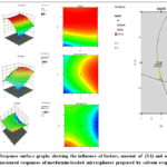 |
Figure 1: Response surface graphs showing the influence of factors, amount of (X1) and amount of (X2), on measured responses of metformin-loaded microspheres prepared by solvent evaporation |
Determination of Bulk Density
The bulk density of microsphere formulations F1–F13 was evaluated to assess the packing and flow characteristics of the powders, which are critical parameters influencing formulation development, particularly during capsule filling or tabletting processes. The observed bulk density values ranged from 0.38 ± 0.31 g/cm³ (F4) to 0.49 ± 0.17 g/cm³ (F7). Most formulations exhibited bulk densities between 0.39 and 0.46 g/cm³, suggesting relatively uniform powder characteristics across batches.
Formulations F1, F2, F4, and F5 showed comparatively lower bulk densities (≤0.41 g/cm³), indicating a more porous structure and lower packing efficiency, which may contribute to improved buoyancy and sustained release behaviour in gastroprotective systems. In contrast, formulations F7 (0.49 ± 0.17 g/cm³) and F8 (0.48 ± 0.19 g/cm³) demonstrated higher bulk densities, suggesting denser microspheres with potentially reduced floating capacity but improved handling during processing.
Formulation F6 exhibited a moderate bulk density of 0.42 ± 0.12 g/cm³, placing it within the optimal range for both flowability and buoyancy, making it a promising candidate for further development. Overall, the bulk density values across all formulations fall within acceptable pharmaceutical limits, reflecting the suitability of the formulation process for producing microspheres with desirable physical properties for oral drug delivery applications.
Table 5: Bulk Density of Metformin Hydrochloride Microsphere
|
Formulation code
|
Bulk Density
|
| F-1 | 0.39 ± 0.060 |
| F-2 | 0.41 ± 0.12 |
| F-3 | 0.43± 0.05 |
| F-4 | 0.38 ± 0.31 |
| F-5 | 0.39±0.14 |
| F-6 | 0.42±0.12 |
| F-7 | 0.49±0.17 |
| F-8 | 0.48±0.19 |
| F-9 | 0.46±0.21 |
| F-10 | 0.45±0.13 |
| F-11 | 0.44±0.15 |
| F-12 | 0.41±0.11 |
| F-13 | 0.43±0.14 |
Determination of Tapped Density
Tapped density is a key parameter used to evaluate the compressibility and packing behaviour of powdered formulations under standardized tapping or mechanical vibration. For microsphere formulations F1–F13, the tapped density values ranged from 0.38 ± 0.06 g/cm³ (F3) to 0.49 ± 0.13 g/cm³ (F7), indicating variability in packing efficiency among different batches.
Formulation F3 exhibited the lowest tapped density (0.38 ± 0.06 g/cm³), suggesting the presence of loosely packed particles with a high degree of porosity, which may be advantageous for maintaining buoyancy in gastroprotective systems. In contrast, F7 recorded the highest tapped density (0.49 ± 0.13 g/cm³), indicating better particle packing and reduced interparticle voids, which may enhance powder flow but potentially compromise floating ability.
Intermediate tapped density values were observed in formulations such as F6 (0.46 ± 0.14 g/cm³), F11 (0.47 ± 0.13 g/cm³), and F13 (0.46 ± 0.12 g/cm³), reflecting a balanced profile between flowability and compressibility. These formulations may offer suitable mechanical stability during processing without significantly affecting gastroprotective behaviour.
Overall, the tapped density results demonstrate that the microsphere formulations possess varied packing characteristics, which could influence not only flow and compressibility but also the floating behaviour and release kinetics of the drug. The data support the formulation’s adaptability to manufacturing conditions while retaining the physicochemical attributes desirable for oral controlled-release delivery systems.
Table 6: Tapped Density of Metformin Hydrochloride Microsphere
|
Formulation code |
Tapped Density |
| F-1 | 0.41 ± 0.060 |
| F-2 | 0.44 ± 0.3 |
| F-3 | 0.38± 0.06 |
| F-4 | 0.41 ± 0.3 |
| F-5 | 0.44±0.12 |
| F-6 | 0.46±0.14 |
| F-7 | 0.49±0.13 |
| F-8 | 0.45±0.16 |
| F-9 | 0.45±0.11 |
| F-10 | 0.43±0.10 |
| F-11 | 0.47±0.13 |
| F-12 | 0.42±0.09 |
| F-13 | 0.46±0.12 |
Particle Size Analysis
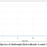 |
Figure 2: Zeta Spectra of Metformin Hydrochloride-Loaded Microspheres Click here to View Figure |
The Dynamic Light Scattering (DLS) analysis of the microspheres demonstrated a narrow and well-defined peak in the intensity distribution curve, confirming the presence of a monodisperse system. The Z-average diameter of the microspheres was found to be 1591 nm (1.591 µm), with a polydispersity index (PDI) of 1, suggesting moderate to broad size distribution and some degree of heterogeneity. The peak mean size by intensity was recorded at 1251 nm (1.251 µm), which reflects the dominant particle population contributing to light scattering. Additionally, the peak area under intensity was 100%, indicating that all detected particles fell within this size range. The derived mean count rate was 939.1 kcps, demonstrating a strong signal intensity. These findings confirm that the microspheres were successfully fabricated within the micron size range, which is favourable for controlled drug delivery applications.
By Fourier Transformed Infrared (FTIR) Spectroscopy
Fourier Transformed Infrared Spectroscopy Analysis of Metformin Hydrochloride
FT-IR spectra were recorded to assess the compatibility of the pure drug with other excipients present in the formulation. The main peak of Metformin hydrochloride bands was N-H(stretching) at 3366.53 cm-1, N-(CH3)2 at 3144.77 cm-1, C-N at 1635.54 cm-1and N-H(bending) at 1576.69 cm-1 was present in drug spectra. The FTIR spectra of the drug are shown in Figure 3.
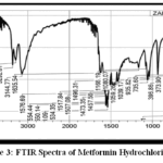 |
Figure 3: FTIR Spectra of Metformin Hydrochloride Click here to View Figure |
Fourier Transformed Infrared Spectroscopy Analysis of Eudragit S100
The FTIR spectra of Eudragit S100 show the characteristics of the peak of OH stretching, which occurred at 2986.89 cm-1, the peak of CH Stretching (Aromatic) at 2951.00 cm-1, CH Stretching (Aliphatic) at 1733.86 cm-1, C=O at 1730.27 cm-1, C=C at 1718.07 cm-1, Al-CH-bend at 1235.09 cm-1and the Ar-CH (In-plane Bending) at 1144.66 cm-1. The FTIR spectra of the Eudragit S100 are shown in Figure 4.
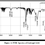 |
Figure 4: FTIR Spectra of Eudragit S100 Click here to View Figure |
Fourier Transformed Infrared Spectroscopy Analysis of Metformin Hydrochloride Microspheres
The main peak of metformin hydrochloride microspheres bands was N-H(stretching) at 2950.28 cm-1, N-(CH3)2 at 2925.17 cm-1, C-N at 2723.81 cm-1and N-H(bending) at 1448.23 cm-1 was present in the metformin hydrochloride microspheres spectra. The FTIR spectra of the metformin hydrochloride microspheres are shown in Figure 5.
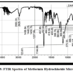 |
Figure 5: FTIR Spectra of Metformin Hydrochloride Microspheres Click here to View Figure |
By DSC Analysis
DSC thermograms were utilised to investigate the thermal behaviour and drug-polymer interactions in the developed microsphere formulations. The thermogram of the pure drug revealed a sharp endothermic peak at 238.34 °C with an enthalpy change (ΔH) of 826.018 J/g, indicative of its crystalline nature. In contrast, the microsphere formulation displayed thermal transitions at 289.01 °C and 387.17 °C with reduced ΔH values of 1.489 J/g and 627.833 J/g, respectively, suggesting partial amorphisation of the drug and successful incorporation within the polymer matrix. Another formulation showed a peak at 408.91 °C with a ΔH of 802.776 J/g, implying high thermal stability due to strong matrix integrity. A distinct DSC profile with multiple peaks, including one at 193.17 °C (ΔH = 438.602 J/g) and a decomposition peak at 312.83 °C (ΔH = -795.139 J/g), was also observed, indicating complex drug-polymer interactions and phase transitions. Collectively, these findings confirm the transformation of the drug from a crystalline to an amorphous or molecularly dispersed state, enhancing the stability and performance of the microsphere system.
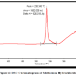 |
Figure 6: DSC Chromatogram of Metformin Hydrochloride Click here to View Figure |
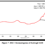 |
Figure 7: DSC Chromatogram of Eudragit S100 Click here to View Figure |
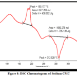 |
Figure 8: DSC Chromatogram of Sodium CMC Click here to View Figure |
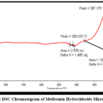 |
Figure 9: DSC Chromatogram of Metformin Hydrochloride Microspheres Click here to View Figure |
X-ray Diffractometer (XRD) Analysis
The X-ray diffraction (XRD) pattern of formulation F-6 exhibits a broad diffuse halo in the 2θ range of approximately 15° to 25°, indicative of an amorphous structure. The absence of distinct and sharp diffraction peaks confirms the non-crystalline nature of the formulation. This amorphisation is likely attributed to the entrapment of the drug within the polymeric matrix during microsphere preparation, possibly via techniques such as solvent evaporation or spray drying, which inhibit the formation of a well-ordered crystalline lattice. The observed maximum intensity, reaching approximately 850 counts, further supports the presence of an amorphous phase, as crystalline materials typically generate higher-intensity, well-defined peaks. The amorphous nature of the formulation is advantageous, as it is associated with enhanced solubility and improved dissolution characteristics, which can lead to increased bioavailability of poorly water-soluble drugs. Therefore, the XRD analysis confirms the successful development of an amorphous microsphere system in formulation F-6, which may contribute to improved therapeutic performance.
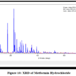 |
Figure 10: XRD of Metformin Hydrochloride Click here to View Figure |
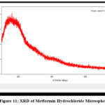 |
Figure 11: XRD of Metformin Hydrochloride Microspheres Click here to View Figure |
Scanning Electron Microscopy (SEM)Analysis
The SEM images (A–D) provide a detailed morphological analysis of the prepared microspheres. Image A, captured at low magnification (X25), shows multiple spherical microspheres with a smooth surface and uniform size distribution, indicating good particle formation and stability without signs of aggregation or collapse. Image B, at a higher magnification (X60), presents a single microsphere with a slightly rough surface and minor cracks, which suggest potential channels for drug release while still maintaining structural integrity. Image C, at magnification X200, reveals a fractured microsphere, exposing an internal porous and layered structure that hints at matrix-type polymer encapsulation, possibly enhancing controlled drug release. Finally, Image D, at high magnification (X1000), shows a compact, irregular surface texture with minimal pores, suggesting uniform polymer dispersion and effective film formation, contributing to sustained drug release. Collectively, these SEM images confirm that the microspheres are well-formed, structurally stable, and exhibit surface and internal characteristics suitable for controlled drug delivery systems.
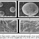 |
Figure 12: Representative scanning electron photomicrographs of taste-masked MT Loaded microspheres (F6 batch) at magnifications 25 (A), 60 (B), 100 (C), and 1000 (D). Click here to View Figure |
In-Vitro Drug Release
Drug release pattern for formulation F6 demonstrate a controlled and sustained release behaviour over 16 hours. Initially, at the 1st hour, F6 released 4.48 ± 0.97% of the drug, followed by a gradual increase to 9.61 ± 0.86% at the 2nd hour. As time progressed, the release continued steadily with 15.02 ± 0.66% at the 3rd hour and 21.23 ± 0.28% at the 4th hour. The mid-phase release profile showed 28.23 ± 0.72% at 5 hours and 35.44 ± 1.12% at 6 hours, indicating consistent polymer hydration and matrix erosion. The formulation continued its slow release, reaching 43.24 ± 0.90% at 7 hours and 51.26 ± 0.61% at 8 hours. By the 9th and 10th hours, F6 achieved 59.44 ± 0.78% and 96.52 ± 0.92% cumulative drug release, respectively. Compared to other formulations, F6 exhibited a moderate and steady release pattern, slower than F1, F4, and F7, but faster than F3. This profile suggests that F6 contains an appropriate balance of polymer and excipient concentration, contributing to its controlled release. Such a formulation is ideal for maintaining therapeutic levels over an extended period without a rapid initial burst, supporting its potential use in sustained-release oral dosage forms.
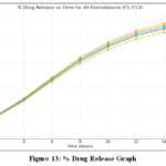 |
Figure 13: % Drug Release Graph Click here to View Figure |
Stability studies analysis:
The accelerated stability data demonstrated minimal variation in microsphere size, percent drug entrapment, and drug release over six months. All values remained nearly identical to the initial measurements, indicating excellent physical and chemical stability of the optimized formulation.
Table 7: Evaluation Parameters of Stability studies analysis
| Conditions | Duration | PDE (%) | MS (µm) | DR (%) |
| Zone II | 1 month | 77.84 | 1.034 | 96.33 |
| Accelerated (40 °C ± 2 °C / 75% RH ± 5% RH) | 3 months | 77.83 | 1.033 | 96.33 |
| Accelerated (40 °C ± 2 °C / 75% RH ± 5% RH) | 6 months | 77.83 | 1.033 | 96.32 |
Conclusion
The purpose of the present investigation was to prepare sustained release microspheres of Metformin hydrochloride to prolong the duration of drug action, to reduce the frequency and to improve patient compliance. Metformin, an oral antihyperglycemic agent characterised by a short half-life and high solubility, is an ideal candidate for CR formulations, which can offer improved bioavailability and prolonged effect. The solvent evaporation with higher yield was selected for the development of microspheres since it was suitable for drug encapsulation. The drug was characterised using FTIR, melting point, solubility and standard curve, and DSC showed no incompatibility with the polymer.
Nine different formulations (F1–F9) were prepared with different concentrations of Metformin hydrochloride using polymers and evaluated for % yield, entrapment efficiency, particle size, morphology and in vitro release. F6 proved to be best of all the developed formulations with 96.78% yield, 80.72% drug content and 91.14% entrapment efficiency. Sustained-release behaviour was also confirmed, over 16 h, and almost complete drug release was observed.
The accelerated stability study confirmed that the optimized microsphere formulation remained physically and chemically stable over six months, with negligible changes in microsphere size, entrapment efficiency, and drug release. These results indicate strong formulation robustness and suggest that the microspheres can maintain their performance and integrity under elevated temperature and humidity conditions.
These results reveal that solvent evaporation has been proven to be an efficient way to prepare sustained-release microspheres of Metformin hydrochloride. The developed formulation provides sustained release, improved bioavailability and better control of type 2 diabetes mellitus, with potential for being exploited in microsphere-based delivery systems.
Acknowledgement
The authors are thankful to IFTM University, Moradabad, Uttar Pradesh, India.
Funding Sources
The author(s) received no financial support for the research, authorship, and/or publication of this article.
Conflict of Interest
The author(s) do not have any conflict of interest.
Data Availability Statement
This statement does not apply to this article.
Ethics Statement
This research did not involve human participants, animal subjects, or any material that requires ethical approval.
References
- Wang Y, Song W, Li Y, Zhou Z, Li C, Yu W, He Y. Mater Today Bio. 2023;23(12):100874.1016/j.mtbio.2023.100874
CrossRef - Allam AN, Mehanna MM. Formulation, physicochemical characterisation and in vivo evaluation of ion-sensitive metformin-loaded biopolymeric beads. Drug Dev Ind Pharm. 2016;42(3):497-505.3109/03639045.2015.1058815
CrossRef - Jain AK, Sahu P, Mishra K, Jain SK. Repaglinide and metformin-loaded Amberlite resin-based floating microspheres for the effective management of type 2 diabetes. Curr Drug Deliv. 2021;18(5):654-68.2174/1567201817666201026105611
CrossRef - Sharma HK, Mohapatra J, Nath LK. Preparation and evaluation of starch microspheres by cross-linking technique. Pak J Pharm Sci. 2013;26(1):17-22.1016/j.carbpol.2008.03.005
- Mai NH, Do HH, Tran PHY, Nguyen CP, Nguyen VH, Nguyen NPN, Ngo KD, Nguyen DT, Le MQ. Optimization of controlled-release microspheres containing vitexin and isovitexin through experimental design and evaluation of their hypoglycemic effects. Pharmaceutics. 2025;17(7):819.3390/pharmaceutics17070819
CrossRef - Jiang C, Gong G, Xiao S, Zhang S, Chen D, Song S, Mechanical and biological properties of 3D-printed porous titanium scaffolds coated with composite growth factors. BMC Oral Health. 2025;25:808.1186/s12903-025-05596-8
CrossRef - Shan X, Yuan X, Wu X. An electrospun DFO-loaded microsphere/SAIB system orchestrates angiogenesis–osteogenesis coupling via HIF-1α activation for vascularized bone regeneration. Polymers. 2025;17(11): 1538.3390/polym17111538
CrossRef - He N, Wu D, Luo R, Cao Z, Shan S, Fei Q, Development of pH-sensitive chitosan-based microspheres for controlled release of metformin in diabetes management. J Microencapsul. 2025;42(1):1-20.1080/02652048.2025.2515840
CrossRef - Yetilmezsoy K, Kiyan E, Ilhan F. Synthesis of agro-industrial waste/sodium alginate/bovine gelatin-based polysaccharide hydrogel beads: characterization and application as controlled-release microencapsulated fertilizers. Int J Biol Macromol. 2024;279(Pt 3):135382.1016/j.ijbiomac.2024.135382
CrossRef - Berhe HE, Mezgebo DT, Abrha S, Haile TG, Molla F. Extraction, characterization, and evaluation of Lepidium sativum Linn. mucilage as a mucoadhesive polymer. Adv Pharmacol Pharm Sci. 2023;2023:5535344.1155/2023/5535344
CrossRef - Longre S, Rana D, Rangra S, Jindal AB, Salave S, Vitore J, Benival D. Quality-by-design based development of doxycycline hyclate-loaded polymeric microspheres for prolonged drug release. AAPS PharmSciTech. 2024;25(3):49.1208/s12249-024-02760-7
CrossRef - Chaerunisaa AY, Wardhana YW, Dewi MK, Putri ME, Rahmania FJ. Exploring the potential of cellulose nanocrystals originated from ramie (Boehmeria nivea L. Gaud) in formation of microspheres for enhanced solubility of furosemide. 2025;17(13):1879.10.3390/polym17131879
CrossRef - Gao F, Liu B, Liu Y, Xing L, Zhang Y. Preparation and enhanced oil recovery mechanisms of Janus-SiO₂-reinforced polymer gel microspheres. Gels. 2025;11(7):506.3390/gels11070506
CrossRef - Sushma R, Nagarajan S. Preparation and evaluation of floating microspheres of repaglinide. Int J Adv Pharmaceutics. 2013;3(1):30-6.
- Wang K, Song Z, Xu Z, Xi Y, Cui Y, Zhou H. Synthesis of quaternized magnetic chitosan and adsorption performance for methyl orange from aqueous solution. RSC Adv. 2025;15(26):21121-32. 1039/D5RA02862K
CrossRef - Nataren-Rodríguez F, Pacheco-Molina J, Gracia-Vásquez SL,Spray-dried polymeric microspheres for lipophilic drugs: formulation design, physicochemical characterization, and in vitro release evaluation. Pharmaceuticals. 2025;18(7):1020.3390/ph18071020
CrossRef - Gupta S, Dubey S, Patel SK, Lakra AP, Minz S. Chitosan-coated CMC-Na and carbopol hydrogel beads for controlled release of metformin in diabetes management. J Appl Pharm Res. 2025;13(2):73-85.69857/joapr.v13i2.1006
CrossRef - Mahajan S, Choukse R, Jain S, Shukla K. Floating microspheres of metformin hydrochloride: formulation and evaluation. Int J Pharm Sci Med. 2019;4(9):1-30.22270/ijpsm.v4i9.281
Accepted on: 09 Jan 2026
Second Review by: Dr .B. Manjunath
Final Approval by: Dr. B.K Sharma


![]()


