Assessment of the Antiulcer Potential of Ethanolic Extract of Medicago sativa L. in Pylorus Ligation–Induced Ulcer Model in Albino Rats
 , Anuj Kumar2, Alankar Shrivastav3, Mohd Fareed2, Md Zeeshan2, Shivam Kumar4, Renu Yadav5, Mohd Haider6
1School of Pharmaceutical Sciences,Faculty of Pharmacy, IFTM University, Moradabad, U.P. India
2Department of Pharmacy, Madhav University, Bharja, Abu Road, Pindwara, Rajasthan, India.
3Faculty of Pharmacy, Pharmacy Academy, IFTM University, Moradabad, U.P. India.
4Maharaja Agrasen Himalayan Garhwal University, Pauri Garhwal, Uttarakhand, India
5Umapati Mahadev College of Pharmacy, Vasudev Rd, Fazalpur, Amroha, Mohammadpur Jati, U.P. India
6Faculty of Pharmacy, Swami Vivekanand Subharti University, Meerut, U.P. India.
Corresponding Author E-mail:1106sonamsingh2909@gmail.com
DOI : http://dx.doi.org/10.13005/ojc/420304
Download this article as:
![]()
Phlogosis (inflammation) is a pathological response of active tissue to a wound, characterised by localised accumulation of blood cells and plasma. An ulcer is an erosion of the gastrointestinal tract caused by corrosive gastric acid secretion. Inflammation occurs in disorders such as rheumatoid arthritis, gout, and allergic reactions. Methods:Although synthetic anti-ulcer drugs are widely used, they often cause adverse effects, including gastric irritation and fluid retention, which further aggravate ulcers and may induce hepatotoxicity and nephrotoxicity. Coarsely powdered Medicago sativa was extracted using petroleum ether in a Soxhlet apparatus, followed by ethanol partitioning. Phytochemical screening revealed carbohydrates, nitrogenous bases, polyphenols, saponins, flavonoids, phytosterols, and fatty oils. Results:Ulcers were induced in 24-hour-fasted albino rats using pylorus ligation and ethanol models. Animals were divided into five groups: control, standard (pantoprazole 40 mg/kg), and three test groups. Ethanolic extract of Medicago sativa (0.1, 0.2, and 0.4 g/kg) significantly abridged ulcer formation. Conclusion:The extract demonstrated dose-dependent antiulcer activity comparable to that of the standard drug. Gastric parameters, including acidity, lesion index, and percentage ulcer inhibition, were evaluated to confirm efficacy.
KEYWORDS:Antiulcer activity; M. sativa; Pylorus ligation; Ulcer index
Introduction
An ulcer is an inflammatory lesion of the alimentary mucosal membrane.Peptic ulcer disease occurs when aggressive factors outweigh mucosal defence mechanisms. Duodenal ulcers are most common (≈95%), while gastric ulcers occur less frequently in the stomach lining(Adomi, Sakai, Ishikawa, Obara, & Huybrechts, 2026). The gastric mucous membrane is perpetually exposed to potentially noxious agents such as the acid and pepsins of the stomach, bile salts, dietary irritants, microbial toxins (especially those related to H. pylori), and numerous drugs (Canto et al., 2026). These agents promote gastric ulceration by increasing acid and pepsin secretion and inhibiting prostaglandin synthesis, cell proliferation, and gastric blood flow(Al Sayed, Abdelglil, Mohamed, Baiomy, & Mohamed, 2026). Traditional herbal medicine has been used worldwide as a complementary healing system for thousands of years. I hadn’t heard that either, and apparently, those kinds of treatments are still in high demand, particularly in third-world nations where the majority of people don’t have access to modern medicine(Perry, Pillarisetti, Gelfman, & Agrawal, 2025). Ten percent of people worldwide suffer from peptic ulcers, one of the most common gastrointestinal conditions (Bhakta et al., 2026). According to the most recent WHO data released in 2018, 55,560 people in India died from peptic ulcer disease, accounting for 0.63% of all deaths. Erosion in the duodenum or stomach lining is known as an ulcer(D’Urso, Anastasia, Toscano, Patti, & de Bartolomeis, 2018). Really, again, it is simply a sore on the skin or mucous membrane at the opening of your digestive system. Dysregulation of the offensive (acid, pepsin, bile acids, and H. pylori) and defence factors is responsible for gastric ulcers, a major gastrointestinal disorder. Nowadays, we have 2 main ways to treat peptic ulcers: reduce the production of gastric acid or increase defences in the mucosa of the stomach (Pudipeddi et al., 2026).
Although many medicinal plants are used in the traditional Indian system to treat various ailments,systematicpharmacological studies are mainly limited(Machado-Alba, Atehortua-Otero, & Cortes-Mejia, 2018). This study aimed to evaluate the acute toxicity and antiulcer activity of selected medicinal plants. Preliminary phytochemical screening confirmed the presence of key secondary metabolites, especially tannins and flavonoids, known for antiulcer activity(Danakumara et al., 2024).
Medicago sativa Linn. (Family: Leguminosae), widely known as alfalfa and frequently cited as “father of all foods,” is a perennial herb(Chandra et al., 2023). Grown as a fodder plant all over the world, this is the oldest plant. Since European settlers arrived in America, Medigo sativa has been widely grown. Medigo sativa has been cultivated for a number of reasons, including medical applications, animal feed, and soil enhancement(Chand et al., 2023, Bora & Sharma, 2011).
Medigo sativa has long been handed down in homoeopathic and Ayurvedic medicine to treat a variety of illnesses, including problems of the digestive and central nervous systems(Ren et al., 2025). However, this plant species has received only limited investigation. The traditional use and phytopharmacological potential of Medigo sativa are highlighted in this review(Danakumara et al., 2024).Only limited pharmacological studies have been conducted to validate the traditional claims of M. sativa.This review aims to highlight its unexplored potential among natural product researchers worldwide.Evaluating existing literature is essential to provide a solid foundation and direction for future studies on this plant(Chand et al., 2023, Zhang, Zheng, Wang, & Zhu, 2025).
Methods
All scientific publications (novel research, examinations, and brief messages), files, and reports from globally recognised records were thoroughly searched and carefully gathered. A medium-sized armed deciduous plant was found behind Ashish Jyot Flats, Bhavnagar-364002, Gujarat, India, in the eastern section of Green Era Foods & Nutraceuticals. Dr Sunita Garg of the NSICAR Department of Botany authenticated the plant using the authentication number NSICPR/RHMD/Consult/2023/4493-94.
Drugs and chemicals
The pharmaceutical company ESKAYEF INDIA LIMITED provided pantoprazole. The remaining chemicals were analytical grade and came from Merck in Darmstadt, Germany. All chemicals and reagents used were of analytical grade and obtained from Molychem in India. The study employed precoated TLC plates (Silica gel 60 F-254) made in Germany.
Extraction procedure
The polar-to-non-polar solvent system is the foundation of the plant extraction process. After being shade-dried, the Medigo sativa leaves were minced in a mortar and pestle until they were a coarse powder. Ethanol and petroleum ether were used to extract the resulting powdered substance. The resulting extracts were evaporated at 40 °C to obtain a semi-solid bulk after distillation to remove excess solvent. The phytochemical assays that were performed on theextracts are detailed below.(Ghorbel Koubaa, Jdidi, Chaabane, Aoiadni, & El Feki, 2026)
Phytochemical analysis
The area of chemistry known as phytochemistry studies the chemical composition of plants and plant-derived products (the chemistry of natural products). Numerous chemical components found in plants, such as Preliminary phytochemical screening discovered the occurrence of nitrogenous bases (alkaloids), glucides (carbohydrates), polyphenolic astringents (tannins), isoprenoid compounds (terpenoids), glycosidic surfactants (saponins), bioflavonoids (flavonoids), phytosterols (steroids), and non-volatile fatty oils. The conventional methods were used for qualitative phytochemical analysis(Shi, Wang, Wang, & Zhu, 2024).
Flavonoids
The extract solution (500 μL) was mixed with a few drops of NaOH solution and diluted with HCl. The presence of flavonoids was indicated by the solution turning yellow and eventually colourless(Khairi et al., 2025).
Alkaloids
Three reagents, Dragendorff’s, Wagner’s, and Mayer’s, were used in a rummage sale to examine for the presence of alkaloids. A few drops of the reagent were added to the 500μL extract solution. Alkaloids were detected by a reddish-brown precipitate(Cong et al., 2026).
Phenols and tannins
A few drops of FeCl3 were added to the 500 μL test solution. The production of a blue or blue-green coloured solution (500μL) indicated the occurrence of phenols and tannins(Andrade et al., 2026).
Proteins
The extract (500 μL) was mixed with a few drops of 4% NaOH, then with 1% CuSO4. The presence of proteins was revealed by a violet or pink solution(da Silva et al., 2026).
Carbohydrates
One to two millilitres of Anthrone reagent were added to the 500-microliter extract solution. The presence of carbohydrates was revealed by the production of the green solution(Dominguez-Delgado et al., 2026).
Saponins
The extract solution (500 μL) was shaken for 5 minutes after a few drops of Na2HCO3 were added. The occurrence of saponins was shown by the development of froth or foam(Yan et al., 2022).
Glycosides
The extracts (500μL) were mixed with a few drops of aqueous NaOH. The presence of glycosides was shown by a yellow-coloured solution(Laskar, Mazumder, & Talukdar, 2023).
Steroids
The extract solution (500 μL) was mixed with chloroform, and then concentrated H2SO4 was added gradually along the sides of the test tube. The occurrence of steroids was detected through the upper layer turning reddish orange, and the lower sulfuric acid portion turning brownish yellow(Bolleddu, Venkatesh, Narasimhaji, & Hazra, 2021).
Thin Layer Chromatography
To create a 4 mg/mL stock solution for the TLC solvent system, 40 mg of extract was dissolved in 10 mL of methanol.TLC was performed in a Petri plate-glass beaker chamber, and samples were applied using a pointed glass microcapillary. Developed plates were dried, photographed, and the Rf values of separated bands were calculated. Pilot TLC of the methanol extract used ethyl acetate, methanol, and chloroform (1:5:5 v/v/v), followed by modification with ethyl acetate to reduce tailing. The mobile phase presentation, optimal separation was designated for HPTLC fingerprinting, and Rf values were calculated using Equation 1(Ciesla, Kowalska, Oleszek, & Stochmal, 2013).
Rf value = solute migration distance/solvent front migration distance.
HPTLC Conditions and Instrument
I took 20 mg of extract and dissolved it in 5 mL of methanol, yielding a 4 mg/mL solution. I carried out HPTLC fingerprinting for the extract that worked best at the Department of Pharmaceutical Sciences, Jamia Hamdard, New Delhi. The mobile phase that gave the clearest TLC fingerprint was used for HPTLC under the following conditions: Stationary phase: Silica gel 60 F254 (E. Merck KGaA) (10 x 10 cm). Sample application: CAMAG Linomat-5; Detection: CAMAG TLC Scanner 3; Lamp: D2 & W; Measurement type: Remission; Measurement mode: Absorption; Optical filter: Second order; Data filtering: Savitsky-Golay 7. I applied 6 tracks (10, 20, 30, 40, 50, and 80 μl) of the 4 mg/mL ethanolic extract of M. sativa. The most active one is on the TLC plate. After developing the plate with the chosen mobile phase, I scanned it using a CAMAG scanner under visible light and at 254 nm and 366 nm(Ibrahim et al., 2020).
Pylorus ligation model
In this study, rats were divided into five groups, with each group containing six animals. Group 1 received normal saline and served as the control group. Group 2 received Pantoprazole (40 mg/kg) orally and served as the reference group. Groups 3, 4, and 5 received low, medium, and high doses of the extract, respectively, and the test drug was administered orally at different doses for 7 days in their respective groups. The control group received 1% (w/v) CMC in purified water, and the albino rats were fasted for 18–24 hours prior to the experiment. The standard drug and vehicle were administered orally one hour before pyloric ligation. (Figure 1).
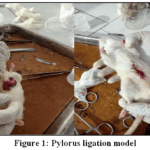 |
Figure 1: Pylorus ligation model Click here to View Figure |
Under ether anaesthesia, the pylorus was ligated through a small midline incision without disturbing blood vessels, and the abdomen was sutured. 4 hours later, animals were sacrificed, and gastric contents were collected for volume, pH, acidity, and pepsin analysis. The stomach was examined macroscopically, and ulcer lesions were scored based on severity(Chandra et al., 2023).
Macroscopic Estimation of the Stomach
The stomachs were unlocked lengthwise, the superior curvatures were removed, and the gastric contents were gently washed out using normal saline. The extent of ulceration was assessed with a 10× magnifying lens. Ulcer number and severity remained single-minded according to the technique described by Kulkarni, where lesions were scored as follows: 0.0 = normal mucosa, 0.5 = mucosal redness, 1 = superficial (spot) ulcers, 2 = deep ulcers, and 3 = perforated ulcers(Ortiz et al., 2021).
Determination of Gastric Volume and pH
The gastric contents were composed, centrifuged at 1000 rpm for 10 minutes, and the volume was measured. The pH was measured using a pH meter, and 1 mL of gastric juice was diluted with 1 mL of purified water for analysis (Hungate, Reichl, & Prins, 1971).
Determination of Total Acidity
An aliquot of gastric juice (1 mL) was diluted with 1 mL of distilled water in a conical flask. Phenolphthalein indicator remained additional, and the solution was titrated with 0.01 N NaOH until a persistent pink endpoint appeared. The volume of NaOH expended was recorded, and gastric acidity was calculated and expressed as mEq/L. equation:(Gunawardhana, Ching, Noothalapati, & Sampath Udeni Gunathilake, 2026).
Where V= Volume and N= Normality
Result and Discussion
Phytochemical Screening
The phytochemicals of the Medicago sativa plant indicate the occurrence of Flavonoids, alkaloids, glycosides, Carbohydrates, tannins, & phenolics, saponins, and steroids (Table 1).
Table 1: Chemical test results of Medicago sativa
|
S. No. |
Phytochemical Parameters | Result |
| 1.0 | Flavonoids |
+ |
|
2.0 |
Alkaloids | + |
| 3.0 | Glycosides |
+ |
|
4.0 |
Protein | – |
| 5.0 | Carbohydrates |
+ |
|
6.0 |
Tannin & Phenolic | + |
| 7.0 | Saponins |
+ |
|
8.0 |
Steroids | + |
| 9.0 | Starch |
– |
(-) represents Absence; (+) represents Presence.
Thin-layer chromatography
Standardised thin-layer chromatography is an effective method for screening, analysing, and assessing the quality of plants and herbal products. Due to its simplicity, speed, and sensitivity, TLC was used to select a mobile phase from extracts that showed strong, dose-dependent inhibition. Various solvent systems containing polar and nonpolar solvents were evaluated.Optimal separation was achieved using trichloromethane: methyl alcohol: ethyl ethanoate (1:5:5), producing distinct TLC spots (Figure 2).
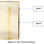 |
Figure 2: TLC of Plant Extract Click here to View Figure |
The chromatographic analysis of Medicago sativa plant extract showed two spots with different Rf values. Spot 1 had an Rf value of 0.83, while Spot 2 showed an Rf value of 0.57, indicating the presence of two different compounds in the extract.
HPTLC Analysis
Aliquots of 10–80 µL from a 4 mg/mL solution were applied as six separate tracks on the HPTLC plate.Chromatographic development was performed using trichloromethane: methyl alcohol: ethyl ethanoate (1:5:5) as the mobile phase.Subsequently, the plate was developed, air-dried and scanned at 254 nm and 366 nm.Nine well-resolved peaks were observed at 254 nm, while eight peaks appeared at 366 nm in the methanolic extract of Medicago sativa (Figure 3) (Table 2).
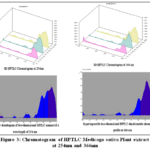 |
Figure 3: Chromatogram of HPTLC Medicago sativa Plant extract at 254nm and 366nm Click here to View Figure |
Table 2: Rf & AUC of HPTLC Medicago sativa Plant extract at 254nm and 366nm
| Peak | Rf | AUC | % Area | |
| 1 | 0.13 | 522.2 | 0.69 | |
| 2 | 0.17 | 208.8 | 0.35 | |
| 3 | 0.34 | 514.3 | 0.87 | |
| 4 | 0.50 | 1203.8 | 2.05 | |
| 5 | 0.55 | 1276.5 | 2.17 | |
| 6 | 0.65 | 812.6 | 1.38 | |
| 7 | 0.69 | 14430.0 | 24.52 | |
| 8 | 0.81 | 13773.5 | 23.41 | |
| 9 | 0.88 | 26100.1 | 44.36 | |
| 10 | 366nm | 0.01 | 442.8 | 0.68 |
| 11 | 0.13 | 622.0 | 0.96 | |
| 12 | 0.24 | 633.4 | 0.98 | |
| 13 | 0.48 | 1909.3 | 2.94 | |
| 14 | 0.59 | 210.4 | 0.32 | |
| 15 | 0.67 | 922.9 | 1.42 | |
| 16 | 0.72 | 14644.9 | 22.55 | |
| 17 | 0.84 | 45565.6 | 70.15 |
HPTLC is an advanced form of TLC that offers advantages such as densitometric scanning, method optimisation, minimal sample requirements, and high selectivity. It is well-suited for generating chromatographic profiles of complex matrices, including pharmaceuticals and natural products. HPTLC fingerprinting of Medicago sativa exposed nine distinct peaks at 254 nm with Rf values ranging from 0.13 to 0.88. At 366 nm, eight well-defined peaks were observed with Rf values between 0.01 and 0.84 using a chloroform: methanol: ethyl acetate (1:5:5) mobile phase. This fingerprint serves as a reliable tool for standardisation, quality control, and evaluation of M. sativa–based herbal formulations.
Pylorus Ligation
Oral administration of Medigo sativa at doses of 0.1, 0.2, and 0.4 g/kg significantly reduced ulcer index, gastric volume, free acidity, and total acidity in the pyloric ligation model. The extract showed dose-dependent gastric safety compared to the control group. Protection indices of 38.41%, 52.98%, and 60.46% were observed at 0.1, 0.2, and 0.4 g/kg, respectively. Pantoprazole (standard drug) produced a protection percentage of 63.58%. The control group showed severe gastric mucosal damage, marked by a high ulcer index, low pH, and increased gastric volume and acidity. Pantoprazole (40 mg/kg) significantly reduced ulcer index, gastric volume, free acidity, and total acidity while increasing gastric pH.EEMS treatment produced a dose-dependent gastroprotective effect, with progressive inhibition of ulceration. Medium and high doses of EEMS markedly suppressed gastric acid secretion, increasing pH and reducing gastric volume. Higher doses also significantly lowered free and total acidity compared to the control. The highest EEMS dose (0.4 g/kg) showed anti-ulcer effects comparable to those of pantoprazole, likely via cytoprotective and antisecretory mechanisms(Table 3, Table 4, Table 5).
Table 3: Result of ethanolic extract (EE) of Medicago sativa on Ulcer Index, Ulcer Score, % Inhibition of Ulceration, and percent ulcer in Pylorous Ligation Model
| S.No. | Treatment | Ulcer index | % Inhibition of Ulceration |
| 1 | Blank | 15.10±.81 | — |
| 2 | Pantoprazole (40 mg/kg) | 5.50±0.17*** | 63.58 |
| 3 | EEMS (0.1gm/kg) | 9.30±0.45 | 38.41 |
| 4 | EEMS (0.2gm /kg) | 7.10±0.38* | 52.98 |
| 5 | EEMS (0.4gm/kg) | 5.12±0.19** | 60.46 |
Table 4: The Effect of Ethanolic Extract of Medicago sativa on Gastric pH and Gastric Volume in Pylorus Ligation Model
| S.No. | Group | Dose | Gastric pH | Gastric Volume (ml) | Parameter | Description |
| 1 | Control | – | 1.25±0.24 | 4.15±1.17 | Data expression | Mean ± SEM (n = 6) |
| 2 | Standard (Pantoprazole) | 40 mg/kg | 6.78±0.71*** | 2.05±0.31*** | Statistical test | One-way ANOVA |
| 3 | Low dose (0.1gm/kg) | EEMS | 3.15±0.23 | 2.85±0.75* | Post-hoc test | Tukey’s multiple comparison test |
| 4 | Medium dose (0.2gm /kg) | EEMS | 4.83±1.1* | 2.99±0.35* | Significance level | *P < 0.01, **P < 0.001 vs. control |
| 5 | High dose (0.4 gm/kg) | EEMS | 6.55±.0.74*** | 2.85±0.23** | – | – |
Table 5: The Effect of Ethanolic Extract of Medicago sativa L. on Free Acidity and total Acidity in Pylorus Ligation Model
| S.No. | Group | Dose | Free Acidity (meq/ltr) | TotalAcidity (meq/ltr) | Parameter | Description |
| 1 | Control | – | 18.12±0.15 | 20.75±0.75 | Data expression | Mean ± SEM (n = 6) |
| 2 | Standard (Pantoprazole) | 40 mg/kg | 7.05±0.74*** | 8.82±0.59*** | Statistical test | One-way ANOVA |
| 3 | Low dose (0.1 gm /kg) | EEMS | 11.95±0.11 | 13.11±0.45 | Post-hoc test | Tukey’s multiple comparison test |
| 4 | Medium dose (0.2gm/kg) | EEMS | 9.15±0.42* | 11.12±0.75* | Significance level | *P < 0.01, **P < 0.001 vs. control |
| 5 | High dose (0.4gm /kg) | EEMS | 7.51±0.81** | 9.01±0.91** | – | – |
Results are expressed as mean ± SEM for 6 animals per group.Data remained investigated using one-way ANOVA followed by Tukey’s post hoc test, with significance set at P < 0.01 and *P < 0.001 vs control.
Histopathology examination
Histological examination clearly revealed gastric lesions and mucosal damage in rat stomachs. The control group showed chronic lymphocyte-induced inflammation, while Group B exhibited submucosal inflammation with chronic inflammatory cells. Group C showed intact mucosa with extensive epithelial regeneration, whereas Group D had focal mucosal ulcers with limited regenerated cells.Group E demonstrated normal gastric architecture through undamaged epithelium, lamina propria, and muscularis mucosa (Figure 4, Figure 5).
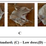 |
Figure 4: (A)– Control; (B) – Standard; (C) – Low dose;(D) – Medium dose ;(E)- High dose Click here to View Figure |
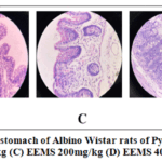 |
Figure 5: Histopathology of the stomach of Albino Wistar rats of Pylorus ligation model (A) Control Group (B) EEMS 100mg/kg (C) EEMS 200mg/kg (D) EEMS 400mg/kg (E) PTZ 40 mg/kg. |
Conclusion
The study demonstrated a strong antiulcer effect of the ethanolic extract of Medicago sativa in pylorus–ligated albino rats. The extract presented dose-dependent increases in cutting-edge gastric pH and reductions in ulcer index, gastric volume, and acidity. The highest dose (400 mg/kg) produced protective effects comparable to pantoprazole (40 mg/kg). Phytochemical screening and histopathology confirmed mucosal protection and epithelial regeneration, supporting antioxidant and cytoprotective mechanisms. These findings validate the traditional use of M. sativa and highlight its potential as a natural antiulcer agent, warranting further mechanistic and formulation studies.
Acknowledgement
The authors are thankful to IFTM University, Moradabad. Uttar pradesh, India.
Funding Sources
The author(s) received no financial support for the research, authorship, and/or publication of this article.
Conflict of Interest
The author(s) do not have any conflict of interest.
Data Availability Statement
This statement does not apply to this article.
Ethics Statement
This research did not involve human participants, animal subjects, or any material that requires ethical approval.
References
- Adomi, M., Sakai, T., Ishikawa, T., Obara, T., & Huybrechts, K. F. (2026). Medication use during pregnancy in Japan from 2005 to 2019: Trends and safety considerations. Int J Gynaecol Obstet. doi: 10.1002/ijgo.70860
CrossRef - Al Sayed, H. Y. I., Abdelglil, M. I., Mohamed, H. R. H., Baiomy, A. A. A., & Mohamed, A. S. (2026). Pomegranate peel extract nanoparticles enhance insulin production and antioxidant activity of diabetic rats. Tissue Cell, 101, 103423. doi: 10.1016/j.tice.2026.103423
CrossRef - Andrade, J., de Almeida-Apolonio, A. A., Dantas, F., Santos, J., Sangalli, A., Negri, M., . . . de Oliveira, K. M. P. (2026). Phytochemical and Biological Evaluation of Leaves, Stems, and Roots of Schinus weinmanniifolia Mart. Ex Engl. Chem Biodivers, 23(3), e02974. doi: 10.1002/cbdv.202502974
CrossRef - Bhakta, P., Raja, M., Arumugam, S., Yellurkar, M. L., Prasanna, V. S., Bhar, K., . . . Dutta, A. (2026). Anti-Ulcer Effect of Ornithogalum umbellatum at Various Potencies in Indomethacin-Induced Gastric Ulceration in Sprague-Dawley Rats: A Pre-clinical Study. Homeopathy. doi: 10.1055/a-2708-3244
CrossRef - Bolleddu, R., Venkatesh, S., Narasimhaji, C. V., & Hazra, J. (2021). Pharmacognostical and phytochemical studies of Atibala (Abutilon indicum [Linn.] sweet) fruit. Ayu, 42(3), 138-142. doi: 10.4103/ayu.AYU_264_20
CrossRef - Bora, K. S., & Sharma, A. (2011). Phytochemical and pharmacological potential of Medicago sativa: a review. Pharm Biol, 49(2), 211-220. doi: 10.3109/13880209.2010.504732
CrossRef - Canto, P. A., Montoro, J., Balaguer-Rosello, A., Villalba, M., Chorao, P., Louro, A., . . . Sanz, J. (2026). Chronic graft-versus-host disease in the era of post-transplant cyclophosphamide. Haematologica. doi: 10.3324/haematol.2025.289266
CrossRef - Chand, S., Roy, A. K., Singh, T., Agrawal, R. K., Yadav, V. K., Kumar, S., . . . Yadava, D. K. (2023). Twenty-four years lucerne (Medicago sativa L.) breeder seed production in India: a retrospective study. Front Plant Sci, 14, 1259967. doi: 10.3389/fpls.2023.1259967
CrossRef - Chandra, P., Kaleem, M., Sachan, N., Pathak, R., Alanazi, A. S., Alsaif, N. A., . . . Kabra, A. (2023). Gastroprotective evaluation of Medicago sativa L. (Fabaceae) on diabetic rats. Saudi Pharm J, 31(11), 101815. doi: 10.1016/j.jsps.2023.101815
CrossRef - Ciesla, L., Kowalska, I., Oleszek, W., & Stochmal, A. (2013). Free radical scavenging activities of polyphenolic compounds isolated from Medicago sativa and Medicago truncatula assessed by means of thin-layer chromatography DPPH rapid test. Phytochem Anal, 24(1), 47-52. doi: 10.1002/pca.2379
CrossRef - Cong, R., Kim, J. W., Park, J. S., Park, J. S., Kang, K., & Shim, S. M. (2026). Systematic evaluation of permeability-related behavior and physicochemical determinants of structurally diverse phytochemicals using a Caco-2 cell model. Food Funct. doi: 10.1039/d5fo05099e
CrossRef - D’Urso, G., Anastasia, A., Toscano, E., Patti, S., & de Bartolomeis, A. (2018). Aripiprazole-induced atrial fibrillation in a patient with concomitant risk factors. Exp Clin Psychopharmacol, 26(5), 509-513. doi: 10.1037/pha0000219
CrossRef - da Silva, M. V. F., Silva, J., de Oliveira, V. M., da Rocha, M. N., de Morais, S. M., & Marinho, E. S. (2026). Bioactive flavonoids from Anacardium occidentale as promising natural inhibitors of Cryptococcus neoformans: a computational perspective on secondary metabolites against critical fungal pathogens. Arch Microbiol, 208(5). doi: 10.1007/s00203-026-04814-9
CrossRef - Danakumara, T., Kumar, N., Patil, B. S., Kumar, T., Bharadwaj, C., Jain, P. K., . . . Varshney, R. K. (2024). Unraveling the genetics of heat tolerance in chickpea landraces (Cicer arietinum L.) using genome-wide association studies. Front Plant Sci, 15, 1376381. doi: 10.3389/fpls.2024.1376381
CrossRef - Dominguez-Delgado, C. L., Guadarrama-Lopez, M. M., Cruz-Narvaez, Y., Puente-Lee, R. I., Ojeda-Piedra, S. A., & Zambrano-Zaragoza, M. L. (2026). Casimiroa edulis Leaf Extract-Loaded PLGA Nanoparticles: Untargeted Phytochemical Profiling and Wound-Healing-Oriented Antioxidant/Occlusive Characterization. Pharmaceutics, 18(2). doi: 10.3390/pharmaceutics18020249
CrossRef - Ghorbel Koubaa, F., Jdidi, H., Chaabane, M., Aoiadni, N., & El Feki, A. (2026). Medicago sativa alleviates behavioral impairments and oxidative stress in the cerebrum and cerebellum of ovariectomized mice. Reprod Fertil Dev, 38(2). doi: 10.1071/RD25113
CrossRef - Gunawardhana, G., Ching, Y. C., Noothalapati, H., & Sampath Udeni Gunathilake, T. M. (2026). pH-responsive onion-structured hydrogel beads based on chitosan-spider silk-graphene quantum dots for controlled drug delivery. Int J Biol Macromol, 150384. doi: 10.1016/j.ijbiomac.2026.150384
CrossRef - Hungate, R. E., Reichl, J., & Prins, R. (1971). Parameters of rumen fermentation in a continuously fed sheep: evidence of a microbial rumination pool. Appl Microbiol, 22(6), 1104-1113. doi: 10.1128/am.22.6.1104-1113.1971
CrossRef - Ibrahim, R. S., Khairy, A., Zaatout, H. H., Hammoda, H. M., Metwally, A. M., & Salman, A. M. (2020). Chemometric evaluation of alfalfa sprouting impact on its metabolic profile using HPTLC fingerprint-efficacy relationship analysis modelled with partial least squares regression. J Pharm Biomed Anal, 179, 112990. doi: 10.1016/j.jpba.2019.112990
CrossRef - Khairi, N., Nursamsiar, N., Utami, N. F., Marwati, M., Nur, S., Indrisari, M., & Kursia, S. (2025). Phytochemical profiling and enzyme inhibitory activity of Sterculia populifolia DC stem bark extract and fractions against elastase and tyrosinase. Narra J, 5(3), e1778. doi: 10.52225/narra.v5i3.1778
CrossRef - Laskar, Y. B., Mazumder, P. B., & Talukdar, A. D. (2023). Hibiscus sabdariffa anthocyanins are potential modulators of estrogen receptor alpha activity with favourable toxicology: a computational analysis using molecular docking, ADME/Tox prediction, 2D/3D QSAR and molecular dynamics simulation. J Biomol Struct Dyn, 41(2), 611-633. doi: 10.1080/07391102.2021.2009914
CrossRef - Machado-Alba, J. E., Atehortua-Otero, M. A., & Cortes-Mejia, D. A. (2018). Profile of antiretroviral agents use in Colombia. Biomedica, 38(4), 527-533. doi: 10.7705/biomedica.v38i4.3885
CrossRef - Ortiz, M., Jaime, F., Ortiz, L., Carrasco, R., Orellana, M. J., Torres, J., . . . Harris, P. R. (2021). [Normal values of eosinophils in gastric and duodenal mucosa of children referred to upper gastrointestinal endoscopy]. Andes Pediatr, 92(5), 683-689. doi: 10.32641/andespediatr.v92i5.2552
CrossRef - Perry, S., Pillarisetti, L., Gelfman, T., & Agrawal, D. K. (2025). Gut-Brain Axis in Inflammatory Bowel Disease: Pathogenesis and Therapeutics. Arch Intern Med Res, 8(4), 339-345. doi: 10.26502/aimr.0227
CrossRef - Pudipeddi, A., Chaemsupaphan, T., Arzivian, A., Kim, Y. S., Paramsothy, S., Kariyawasam, V., & Leong, R. W. (2026). Vedolizumab in inflammatory bowel disease: pharmacokinetics and the role of immunomodulator co-therapy. Therap Adv Gastroenterol, 19, 17562848251414825. doi: 10.1177/17562848251414825
CrossRef - Ren, N., Liu, J., Wang, H., Liu, Z., Liu, X., & Li, G. (2025). Combined transcriptomic and proteomic analysis reveals the response mechanisms of alfalfa to freezing stress. Front Plant Sci, 16, 1682825. doi: 10.3389/fpls.2025.1682825
CrossRef - Shi, B., Wang, Y., Wang, L., & Zhu, S. (2024). Genome-Wide Identification of the Brassinosteroid Signal Kinase Gene Family and Its Profiling under Salinity Stress. Int J Mol Sci, 25(15). doi: 10.3390/ijms25158499
CrossRef - Yan, H., Ni, W., Yu, L. L., Xiao, L. G., Ji, Y. H., & Liu, H. Y. (2022). Parisvaniosides A-E, five new steroidal saponins from Paris vaniotii. Steroids, 177, 108949. doi: 10.1016/j.steroids.2021.108949
CrossRef - Zhang, Y., Zheng, C., Wang, S., & Zhu, F. (2025). Variations in Nodule Microbial Communities and Their Association with Root-Colonizing Arbuscular Mycorrhizal Fungi in Medicago Sativa. Microb Ecol, 89(1), 36. doi: 10.1007/s00248-025-02687-x
CrossRef
Accepted on: 10 Apr 2026
Second Review by: Dr. Asif Khan
Final Approval by: Dr. Luigi Campanella


ISSN Online: 2231-5039

![]()


