Phytosome-Mediated Delivery of Passiflora vitifolia Extract: Optimization, Characterization, and Anti-Inflammatory Assessment
 and Shaweta Sharma2
1Department of Pharmaceutics, School of Pharmaceutical Sciences, Shri Guru Ram Rai University, Dehradun, Uttarakhand, India.
2Department of Pharmacy, School of Medical and Allied Sciences, Galgotias University, Greater Noida, Uttar Pradesh, India.
Corresponding Author E-mail:jpandey1112@gmail.com
DOI : http://dx.doi.org/10.13005/ojc/420238
Download this article as:
![]()
This research aimed to create and improve a phytosomal delivery system of Passifloravitifoliahydroethanolic leaf extract for enhanced anti-inflammatory potential. Passifloravitifolia leaves were authenticated and subjected to extraction with various solvents; the hydroethanolic extract (PV-ex) exhibited the highest extractive value (9.54% w/w), TPC (34.68± 0.56 mg GAE/g) and TFC (25.47±0.83 mg RUT/g), and was therefore selected for formulation. Pharmacognostical parameters i.e., ash values, moisture content and heavy metal analysis confirmed acceptable quality and safety, with Pb, Zn, Cr, As and Cd within WHO/FAO limits and mercury was not detected. Thin-film hydration was utilized to incorporate PV-ex into phospholipid-based phytosomes (PV-ex-phyto), which were then optimized using a Box-Behnken design with incubation time, temperature and phospholipid content as independent variables. The optimized formulation (PV‑ex‑phytoOPT) showed particle size of 184.05 ± 0.21 nm, entrapment efficiency of 79.89 ± 0.18% and zeta potential of −18.52 ± 0.10 mV. TEM and FTIR confirmed spherical nano‑vesicles with non‑covalent complexation between extract and phospholipids. Rutin release was sustained in vitro in phosphate buffer pH 7.4, reaching 89.25±1.73% after 12 hours.Egg albumin denaturation assay was utilized to determine an anti-inflammatory action. The results showed concentration-dependent inhibition, with PV-ex-phytoOPT achieving approximately 88–89% inhibition at 200 µg/mL, better than the plain extract and on par with diclofenac sodium. These findings suggest that P. vitifoliaphytosomes represented a promising, safe and effective nanocarrier system to enhance dermal delivery and anti‑inflammatory efficacy of Rutin‑rich herbal extracts.
KEYWORDS:Anti-Inflammatory Activity; Box-Banken Factorial Design; Optimized Formulation; Passifloravitifolia
Introduction
Passiflora, the genus of tropical plants that includes the passion fruit, is the largest and most widely distributed. This genus has over 400 species, which are separated into 21 subgenera.1,2 In South America’s tropical regions and jungles, over 350 species have been identified, 60 of which are edible.1,3,4 Because of its edible fruits, decorative uses, and therapeutic qualities, passion fruit is a significant fruit crop in many exotic and subtropical nations. Few species (P. Quadrangularis, P. edulisand P. ligularis) are mainly cultivated to produce fruit juice. Some species are known for their ethnobotanical uses, and P. incarnata is noted for its sedative qualities.5 In current years, there has been a substantial increase in interest in natural products as a possible source of novel medications around the world. Approximately 40% of current medications are derived from natural sources. Due to the indiscriminate use of commercial chemical antimicrobial medications that are commonly used to treat human illnesses, human pathogenic bacteria are now evolving multiple drug resistance. Over the last three centuries, much research has been conducted to identify clinically beneficial antibacterial medicines.6,7 The discovery of novel therapeutic compounds could result from the increased interest in traditional ethnomedicine.
The plant P. vitifolia is known as the Crimson Passion Flower. P. vitifolia grows in Colombia, namely in Victoria a (Caldas), El Cerrito (Valle del Cauca) and Ibagué (Tolima).8,9 The plant is a type of evergreen vine. It is a tropical lowland plant. The plant needs full light and protection from the hot sun.10,11 The P. vitifolia includes a number of bioactive chemicals like as glycosidically bound volatiles.12 phenols, alkaloids, glycosyl flavonoids and cyanogenic mixtures that have distinct therapeutic properties.13,14
Phytosomes are molecular complexes formed by the interaction of phospholipids with naturally derived phytochemicals, typically achieved by reacting phosphatidylcholine or other polar head groups with plant extracts in an aprotic solvent.15,16 Compared to conventional formulations, these systems demonstrate enhanced pharmacokinetic and pharmacological performance. In phytosomes, the lipid-compatible phosphatidyl component surrounds and stabilizes the hydrophilic phytoconstituent–choline complex. Key advantages of phytosomal systems include improved bioavailability, greater chemical stability due to the formation of bonds between the polar head of the phospholipid and the phytoconstituent, and efficient drug encapsulation.17,18 Enhanced absorption of active compounds allows for reduced dosing while maintaining therapeutic efficacy, particularly for polar phytochemicals. Thus, “Formulation, optimization and assessment of anti-inflammatory effect of Passifloravitifolia extract loaded Phytosomes” was the focus of the current study.
Material and Methods
Rutin (standard), glycerylmonostearate, and all other reagents and solvents were brought from reputed companies. Each solvent and reagent utilized was of analytical grade.
Plant Material Collection
Passifloravitifolia leaves were collected as crude plant material from Doiwala, Dehradun’s Rajeswari Nursery. After leaves were thoroughly dried, whole plant specimen’s herbarium was conveyed to the Botanical Survery of India (BSI), a Botany Discipline of Forest Research Institute in Dehradun, for taxonomic analysis. The results showed that the plant was Passifloravitifolia, with voucher sample number BSI/NRC/Tech./Herb(Ident.)/2022-23/1191.
Preparation and Characterization of Passifloravitifolia (PV) Leaves Extract
Preparation of PassifloravitifoliaPlant Extract (PV-ex)
Freshly harvested Passifloravitifolia (PV) leaves were cleaned and rinsed with filtered water to get rid of anything unwanted. Under a shade cloth, the leaves were sun-dried for four to five days until they were completely dry. A mechanical grinder was used to treat the leaves after they had dried in order to remove additional material. Dried and coarsely crushed PV leaves were packed separately for each solvent (pet ether and hydro alcohol) in a Soxhlet device. Each extract was kept at -20°C after being fully dried under low pressure. The extract was made into a sticky, dark material. The extract was retained and utilized for extractive value evaluation and photochemical screening.19
Characterization of PassifloravitifoliaExtract (PV-ex)
The produced extracts of Passifloravitifolia leaves (PV-ex) were used to determine extractive value, phytochemical screening, TPC, and TFC.
Assessment of Extractive value
The extractive value detection is important in understanding chemical contents nature and determining certain constituent value in a particular solvent utilized in procedure of extraction. In this method, different Passifloravitifolia extracts were filtered, evaporated until become dry, with crude plant material traces accumulating at bottom of a China dish at 50°C. The extractive values of several solvent extracts were determined.
Ash Value
When a medication or herbal product is burned at a high temperature, an inorganic residue known as ash value remains after. Purity, quality and safety are assessed by measuring the total mineral content of product. The values of the different extracts water soluble ash, total ash and acid insoluble ash were identified.20
Foreign Organic Matter Analysis
Finding and quantifying organic contaminants in a sample is known as foreign organic matter (FOM) analysis. To remove dust particles from the fruit or leaf surface, whole plant was washed thoroughly with water. To allow any extra water to drain, the mixture was spread out on filter paper. Thoroughly cleaned and properly drained entire plant material (500 gm) was gathered and evenly spread as a thin layer over a clean white cotton cloth. The sample was carefully examined both visually and with the aid of a 6× magnifying lens to detect and separate any extraneous materials. After that, the amount of the separated foreign material was determined by weighing it and expressed as gm per 100 grams of original sample.21 The process was applied in a total of three sets. The equation displays the formula for calculating the foreign organic materials.
![]()
M= Weight of an unoccupied dish in grams
M1= Weight of a dish containing foreign materials in grams
M2= Sample weight in gram entire plant material)
Fluorescent Based Analytical Technique
Fluorescent analysis is an analytical chemistry branch which employs fluorescence to identify and measure compounds. The phenomenon known as fluorescence occurs when a material absorbs light at a specific wavelength and then produces light at longer wavelength. This property can be utilized for the identification of compounds, as different molecules exhibit characteristic fluorescence patterns. In present study, several chemical reagents were applied to the powdered drug sample such as hydrochloric acid (HCl), diluted sulfuric acid, diluted nitric acid, sodium hydroxide, and antimony trichloride. Then treated samples were observed under ultraviolet, visible light, and normal daylight to observe their fluorescence behavior. The extract of Passifloravitifolia exhibited the most fluorescence effect.22
Loss on Drying
Loss on drying (LOD) is a quantitative analytical procedure that identifies the volatile matter amount and moisture content in sample. An accurately measured 1 gram sample was placed in a hot air oven and dried at 100°C until a constant weight was achieved. Reduction in weight was then recorded and expressed as the loss on drying.23 The following equations were utilized to determine moisture content and loss during drying:
![]()
Phytochemical Determination
Phytochemical screening of plant extracts was performed by following process discussed by Birudu RB et al., 2015.24 Preliminary phytochemical screening was performed on all generated plant leaf extracts to determine quinines, alkaloids, fixed oils, resins, tannins, saponins, flavanoids, lipids, proteins, carboxylic acids amd phenolic compounds presence.
Characterization of Passifloravitifoliaextract(PV-ex)
The Passifloravitifolia extract (PV-ex) was freeze-dried for 5 days at -100°C utilizing a ScanvacCoolSafe Touch 100-9 system (Labogene). PV-ex that had been freeze-dried was kept at -20°C for later utilization. Then it was evaluated for rutin, total flavonoids (TF) and total phenolics (TP).
Estimation of Total Phenolic content (TPC)
The Passifloravitifolia leaf extract’s total phenolic content (TPC) was determined using the Folin-Ciocalteu reagent method. The results were expressed in milligrams of gallic acid equivalent per gram of dried extract, with gallic acid serving as the measuring reference. Calibration curve for gallic acid was made by mixing Folin-Ciocalteu reagent 5.0 ml, sodium carbonate solution 4.0 ml (75 g/l), and 1 ml aliquots of standard solutions at 20, 40, 60, 80 and 100 µg/ml. After then, samples (1 mg/100 ml) were prepared with ethanol as the solvent. The resulting sample solutions (approximately 1 ml) were mixed separately using the same reagents. After being wrapped in parafilm, the tubes were left to stand for half an hour. The absorbance was measured at 760 nm after 30 minutes of room temperature incubation by using Shimadzu UV-1800 spectrophotometer.19
Total Flavonoid content (TFC) Estimation
Crude extracts’ TFC was measured utilizing aluminum chloride colorimetric technique [25], with minor adjustments. To extract solution 1 ml (0.2 mg/mL prepared in methanol), 200 μL of 5% (w/w) sodium nitrite and distilled water 4 ml were added. Following a reaction time of six minutes, 200 μL of 10% (w/w) aluminum chloride was added, and the mixture was left to stand for five more minutes. After that, 2 mL of 10% (w/w) sodium hydroxide was mixed, and methanol was used to bring the total volume to 10 mL. The absorbance of resulting solution was recorded at 425 nm after 30 minutes. The Rutin calibration curve was utilized to determine TFC by using rutin (2-26 μg/mL) as a reference. Rutin equivalents in milligrams per gram of sample are used to display the results. The samples were examined in triplicate.25
Estimation of Rutin
A series of Rutin sample solutions were prepared and analyzed at a wavelength of 376 nm, covering a concentration range of 2–10 μg/mL.26
Heavy Metal Screening method
For the analysis of heavy metals, extract 1.00 g was initially mixed with double-distilled water (2 mL). Next, 8 mL of a 1:1 solution of perchloric acid and nitric acid and concentrated sulfuric acid (H₂SO₄) 5 ml were added. After that, the mixture was heated to 200°C for about half an hour to produce a clear solution with white fumes. The solution was allowed to cool before being diluted with double-distilled water to a final volume of 50 mL and placed in a PET container that had been previously cleaned for additional metal analysis.
Passifloravitifolia (PV-ex) Leaves Hydroethanolic Extract Fingerprinting Analysis Using Thin Layer Chromatography (TLC)
TLC is a easy and better way for identifying and measuring secondary metabolites in plant extracts. TLC fingerprinting of the herbal extract was performed on Silica gel 60 F254 (Merck) plates as the stationary phase in order to ascertain the quantity of components present, identify particular compounds, and assess chemical purity. By observing production of products or the elimination of reactants, TLC can also be used to track the development of reactions [27]. The different ratios of solvent system utilized to conduct TLC study are shown in Table 1.
Table 1: TLC analysis of Passifloravitifolia (PV-ex) leaf hydroethanolic extract using different solvent ratios
|
S. No. |
Solvent System | Ratio |
| 1 | Chloroform/Methanol |
1:9 |
|
2 |
Chloroform/Methanol | 2:8 |
| 3 | Chloroform/Methanol |
3:7 |
|
4 |
Chloroform/Methanol |
4:6 |
Preparation of Passifloravitifolia leaves Extract loaded phytosomes (PV-ex-PS)
Passifloravitifoliaextract phytosomes (PV-ex-PS) were created by using thin-layer hydration technique.38,39 PV-ex was carefully measured and dissolved at 60°C under reflux with magnetic stirring in a round-bottom flask containing 100 mL of a chloroform-ethanol mixture until a homogenous suspension was obtained. Glycerylmonostearate was added gradually to the solubilized PV-ex after being dissolved in 25 ml of 100% ethanol. For 2 hours, the mixture was agitated at 90 rpm at 600°C while in reflux. A rotary evaporator operating at lower pressure was then used to extract the solvent. To eliminate any residual solvent, the viscous, adhesive, semi-transparent brown substance was additionally dried under stream of nitrogen, serving as a cryoprotective step.28
To reduce particle size, the resulting dry film was sonicated in a water bath for half an hour after being rehydrated with 80 mL of water. The phytosomal formulations were characterized by evaluating parameters such as particle size, zeta potential, encapsulation efficiency, polydispersity index, surface morphology, stability, drug loading capacity, and in vitro drug release behavior. The prepared phytosomes were freeze-dried at −100°C for about five days using the ScanvacCoolsafe Touch 100-9 system (Labogene) for long-term storage. Mannitol was used as a lyoprotectant to preserve structural integrity.
Experimental Design
Table 2 shows the three levels of a factorial independent variable (A, B, and C; phospholipid content, temperature and incubation duration). The variables response (R; dependent variables) were analyzed using Box-Behnken design of experiments (DoE), and their surfaces were developed by utilizing Design Expert® 13.0, Stat-Ease Inc., Minneapolis, to create two-dimensional (2 & 3-D) models and quadratic equations. The following is the generated quadratic polynomial equation;
![]()
Here, Y represents response variable (entrapment efficiency and particle size) corresponding to each combination of factors. The term b₀ denotes the intercept, while b₁ to b₃₃ represent the regression coefficients derived from experimental response values of Y. The variables X1, X2, and X3, along with their and square value (X1², X2², X3²) and interaction terms (X1X2, X1X3, X2X3), were used to determine both an individual and combined effects of factors on response.
Fitness & Analysis of Response Model
Analysis of Variances (ANOVA) was used to establish the obtained quadratic polynomial equation for statistical validation. To identify the optimal formulation within the studied experimental domain, response surface models (both 2D and 3D) were employed. These models demonstrated statistically significant coefficients along with satisfactory R² values, as validated by triplicate center points across 17 experimental runs (Table 2). The responses’ practical versus theoretical projected values were quantitatively examined using linear regression plots between predicted and actual response values.
Table 2: The Box-Behnken design independent variables and responses in the formulation of PV-extract loaded phytosomes (PV-ex phytosomes)
|
Independent Variable |
Levels | ||
| Level (-1) | Level 0 |
Level (+1) |
|
|
A-Phospholipid concentration |
100 | 200 | 300 |
| B-Temperature (°C) | 50 | 40 |
60 |
|
C-Reaction time (h) |
1.5 | 1 | 2 |
| Response |
Desirability constraints |
||
|
Y1-Particle size |
Minimum | ||
| Y2-Entrapment Efficiency (%) |
Maximize |
||
Evaluation of Prepared Phytosomes of PassifloravitifoliaExtract (PV-ex-phyto)
The prepared phytosomes of Passifloravitifolia extract (PV-ex-phyto) were examined for several characteristics like as zeta potential, particle size, in-vitro drug release, entrapment efficiency and anti-inflammatory effectiveness.
Particle Size
The particle size of phytosomal formulation of PV-ex-phyto was measured using DLS and a Zeta sizer (Malvern Zetamaster ZEM 5002, Malvern, UK).29 The formulation’s particle size was assessed following two rounds of mixing with distilled water and 5 minutes of vortexing.
Entrapment Efficiency (EE)
Free rutin was separated using an Amicon Ultra 100 kDa membrane filter by centrifugation at 14,000×g for half an hour to evaluate the encapsulation efficiency of the phytosomal formulation. The previously described UV-visible technique was then used to quantify Rutin quantity in filtrate. EE was measured by utilizing quantity of PV-ex phytosomes equal to 20 mg Rutin. Mixture was continuously mixed by utilizing magnetic stirrer for four hours, followed by a one-hour settling period. The clarified liquid was carefully decanted and subjected to centrifugation for 15 minutes at 5000 rpm. After centrifugation, resulting supernatant was passed through a 0.45 µm Whatman filter paper for further purification. Using a UV spectrophotometer to measure the absorbance at the appropriate wavelength after adequate dilution, the Rutin concentration was determined. Every measurement was done 3 times.29
The following formula was utilized to determine EE (%):
![]()
Where, T- Rutin total amount, S- Rutin amount in filtrate. In the current study, % EE and vesicle size were chosen as dependent variables with the ability to influence phytosome formulation. The chosen parameters were examined using ANOVA.
Determination of Zeta Potential
Zeta potential was determined using the Malvern zetasizer. A diluted system was put into a zeta potential measurement cell in order to measure the electric potential of phytosomes, including the zeta potential of their Stern layer.29
Optimization Study
Analysis of variance (ANOVA) was used to determine selected independent variables: phospholipid concentration is a Factor A, reaction time (h) is a Factor C and temperature °C is a Factor B. Based on the obtained results, the software recommended a checkpoint batch, that measured the optimized formulation for additional assessment. This checkpoint batch was selected for detailed assessment of evaluation parameters by comparing the predicted outcomes with the experimental findings. The optimized batch was examined for different parameter.
Formulation and Assessment of PV-ex Phytosomes Optimized Batch (PV-ex-phytoOPT)
An optimal batch was selected with the predetermined aim of reducing vesicle size while enhancing EE. Optimized PV-ex phytosomes batch (PV-ex-phytoOPT) was made using the formula recommended by the design expert. The PV-ex-phytosomes optimized batch was subsequently analyzed for % yield, TEM, particle size, zeta potential, EE, and drug release in vitro. An optimized formula was obtained by fitting the dependent response values in optimization software and analyzing them using ANOVA.
Transmission Electron Microscopy (TEM)
The enhanced PV-ex-phytosome shape and aggregation were studied using a TEM, the JEOL-JEM-1011. A single drop of the phytosome sample was applied to a grid coated with carbon after being mixed with distilled water. A 1% phosphotungstic acid solution was used for negative staining to enhance contrast. Sample was subsequently left to air-dry at room temperature for approximately 15 minutes before observation.30
PV-ex-phytoOPT In-vitro Release Investigation
Dialysis bag method was utilized to study the drug in-vitro release of PV-ex phytosomes. The dialysis bag process entailed placing pre-weighed samples of PV-ex phytosomes (equal to Rutin dosage) into two pre-soaked dialysis bags. Magnetic stirrer was utilized to continuously stir phytosomes at 100 rpm while they were dispersed in pH 7.4 phosphate-buffered saline (PBS) 100 mL and kept at 37 ±0.5 °C. Samples were gathered at specified time points (0, 0.5, 1, 2, 4, 6, 8, 12 hours), and each withdrawn volume was immediately replenished with an equal amount of fresh PBS to preserve sink conditions. UV-visible spectrophotometry was used to measure the amount of rutin present in the samples. Experiments were conducted in triplicate, and all outcomes are presented as mean ± standard deviation (SD).
In vitro Evaluation of Anti-inflammatory Effect by Utilizing an Egg Albumin Denaturation Method
The main goal of the egg albumin denaturation assay is to find substances or agents that, in certain situations, could interfere with or stop egg albumin from denaturing. Denaturation occurs when structures of proteins are altered, resulting in biological action loss. In this research, an egg albumin is used as representative protein and is exposed to conditions such as elevated temperatures, specific pH ranges, or chemical agents that induce denaturation. This process disrupts its native structure, leading to changes in its physical properties and resulting in biological action loss. The egg albumin denaturation assay evaluates a drug or chemical’s ability to prevent or reduce egg albumin denaturation in order to assess its anti-inflammatory qualities. For the experiment, a reaction system of 5 mL was prepared by combining 0.2 ml of freshly obtained hen’s egg albumin with phosphate-buffered saline 2.8 ml (PBS, pH 7.4) and varying concentrations of PV-ex-phytoOPT. This resulted in final concentrations of 50, 100, 150 and 200 µg/mL. The test sample was substituted with an equivalent volume of double-distilled water to create a control. The prepared mixtures were first heated for five minutes at 70°C, and then they were incubated in a BOD incubator for fifteen minutes at 37±2°C. After cooling to room temperature, each sample absorbance was determined at 660 nm. The standard drug utilized was Diclofenac sodium. The protein denaturation percentage inhibition was calculated by utilizing the formula provided below.
![]()
Result
In current research, Passifloravitifolia leaf extract was loaded into phytosomes to improve extract penetration, thus increasing bioavailability and anti-inflammatory effect. The plant material has been validated by BSI Dehradun (Ass No. 1190). The formulation and procedure conditions were optimized to generate the necessary phytosomes, which were then turned into gel.
Characterization of Passifloravitifolia Leaves Extract (PV-ex)
The various extracts of PV with various solvents were characterized, and the following findings were observed, that are summarized in table 3.
Table 3: Summary of characterization of different solvent-based Passifloravitifolia leaf extract
|
Extract |
Extract colour | Odour | Consistency | Touch sense | Plant material weight (g) | Extract amount
(gm) |
%Extractive Value (w/w) |
| Petroleum ether | Light Yellow | Characteristics | Semisolid | Sticky | 15 | 0.50 |
6.34 |
|
Hydro- ethanol |
Dark Green | Characteristics | Semisolid | Sticky | 15 | 1.13 | 9.54 |
| Acetone | Dark Green | Characteristics | Semisolid | Sticky | 15 | 0.89 |
5.94 |
|
Ethyl Acetate |
Green | Characteristics | Semisolid | Sticky | 15 | 0.49 | 3.27 |
| Chloroform | Light Green | Characteristics | Semisolid | Sticky | 15 | 0.31 |
2.07 |
|
n-hexane |
Light Green | Characteristics | Solid | Powder | 15 | 0.19 |
1.27 |
The hydroethanolic extract was assessed to have highest extractive value when compared to other extracts, hence it was utilized to determine other parameters.
Ash Value
Table 4 shows the ash values for PV leaves. According to Pal R. et al. (2020), Passifloravitifolia (PV) leaves contain 6.5% total ash, 6.5 ± 0.3% acid-soluble ash, 1.0 ± 0.1% acid-insoluble ash, and 4.1 ± 0.26% water-soluble ash. Our results closely matched the reported results.
Table 4: Ash Content ofPassifloravitifoliaplant
|
Parameter |
% w/w |
| Total Ash |
5.85% |
|
Water Soluble Ash |
3.45% |
| Acid Insoluble Ash |
1.05% |
Foreign Organic Matter Estimation
Any visible signs of contamination from organic foreign elements, like molds, insects, or other animal contamination, must be absent from a medicinal plant material [21]. No foreign organic materials were identified in the powdered material of Passifloravitifolia.
Fluorescent Examination of Passifloravitifolia
Fluorescence analysis is a useful technology for biochemical analyses as well as medical diagnostic procedures. A variety of fluorescence detection techniques have been used to attain high sensitivity [22]. Table 5 summarizes the florescent analysis of Passifloravitifolia plants.
Table 5: Fluorescent determinationofPassifloravitifolia (PV)plant
|
Sample |
Day light | Fluorescence light |
| Powder sample | Greenish brown to dull dark green |
Bright yellowish green |
|
Treated with dilute nitric acid |
Yellowish brown to brownish green | Yellow to yellowish orange |
| Treated with sodium hydroxide in water | Dark green to greenish brown |
Strong yellowish green / greenish |
|
Treated with hydrochloric acid |
Brownish green to dark brown |
Dull yellowish brown |
|
Treated with dilute sulphuric acid |
Dark brown to blackish brown |
Reddish brown to dull orange |
|
Treated with antimony trichloride |
Yellowish green to greenish brown |
Bright greenish yellow / yellow |
Loss on Drying or Moisture Content
Moisture content of Passifloravitifolia leaves was identified to be 6-7% w/w in dry leaves and approximately 75.16% in fresh leaves.
Phytochemical Analysis
Characterization of Passifloravitifolia Extract (PV-ex)
The extract of Passifloravitifolia (PV-ex) was analyzed to quantify its rutin content, TFC and TPC, and results are presented in the following section.
Rutin (RUT) Content Determination
Rutin (RUT) content was assessed by utilizing standard calibration curve of RUT to identify TFC. TFC in Passifloravitifolia leaves was found to be 25.47±0.83 mg/g dry plant, only the qualitative estimation was done by previous researchers.
TPC Determination
Hydroethanolic extract of PV TPC was quantified by utilizing the Folin–Ciocalteu (FC) technique. The TPC was calculated based on calibration equation y = 0.02x − 0.002 (R² = 0.9999), where y corresponds to the measured absorbance and x denotes the concentration of gallic acid (mg/mL). The results were displayed as mg GAE/g, or milligrams of gallic acid equivalents per gram of extract. The Passifloravitifolia (PV-ex) hydroethanolic extract TPC was calculated and depicted in Table 7 and Figure 1.
TFC Determination
Standard RUT was used to estimate the TFC of PV-ex hydroethanolic extract. TFC values were determined using calibration curve y=0.0163x-0.0032, R²=0.9999, where c is RUT solution concentration (mg/mL) and represented as mg RUT/g dried extract and y represents absorbance. Flavonoid content of Passifloravitifolia (L.) (PV-ex) hydroethanolic extract was measured and shown in Figure 1 and Table 6.
Table 6: TPC, TFC and IC50 values of various extract of PV
|
Extract of PV |
TFC (mg RUT/g) | TPC (mg GAE/g) | IC50 Value (μg/ml) |
| Hydroethanolic extract | 25.47 ± 0.83 mg/g | 34.68 ± 0.56 mg/g |
7.36 µg/ml |
|
Petroleum ether |
3.4 ± 0.2 mg/g | 8.1 ± 0.1 mg/g | 982.21 µg/ml |
| Acetone | 34.89 ± 0.78 mg/g | 15.20 ± 0.25 mg/g |
1098.56 µg/ml |
|
Ethyl Acetate |
35.20 ± 0.47 mg/g | 16.26 ± 0.65 mg/g | 1115.27 µg/ml |
| Chloroform | 5.90 ± 0.51 mg/g | 110.18 ± 0.32 mg/g |
58.98 µg/ml |
|
n-hexane |
25.16 ± 0.36 mg/g | 28.90 ± 0.52 mg/g |
968.60 µg/mL |
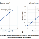 |
Figure 1: Calibration curve of Rutin (A) and Gallic acid (B) for TFC and TPC of hydroethanolic extract of Passifloravitifolia (PV-ex) determination Click here to View Figure |
The total flavonoid and phenolic content of several extracts of Passifloravitifolia (PV-ex) were estimated. The total flavonoid content of ethyl acetate was 35.20 ± 0.47 mg RUT/g, whereas total phenolic content of chloroform was 110.18 ± 0.32 mg GAE/g, the highest value. The hydroethanolic extract has total flavonoid and phenolic levels of 34.68 ± 0.56 mg GAE/g and 25.47 ± 0.83 mg RUT/g.
Heavy Metal Screening
Leaf extracts from Passifloravitifolia include lead (2.45 µg/ml), chromium (0.68 µg/ml), zinc (1.38 µg/ml), cadmium (0.002 µg/ml) and arsenic (0.72 µg/ml). Recent research on Passiflora species and associated medicinal plants found zinc (1.29-1.43 µg/ml), lead (2.30-2.87 µg/ml), chromium (0.47-0.76 µg/ml), cadmium (0.002-0.003 µg/ml) and arsenic (0.75-0.85 µg/ml), in acid digests or extracts, but no mercury was found. These values are within the WHO/FAO permitted range for medicinal plants, with accumulation determined by site-specific soil and environmental circumstances. Our investigation found comparable values, indicating the safety of pharmacological use.
TLC Fingerprint Profiling of Hydroethanolic Leaf Extract of Passifloravitifolia (PV-ex)
TLC fingerprinting was used to identify the compounds, count the components in a mixture, and verify the compounds’ purity. Monitoring the development of a reaction can also be done by observing the appearance of a product or the elimination of a reactant. TLC study was performed using the solvent system listed below in different ratios (Table 7 Figure 2).
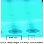 |
Figure 2: Schematic diagram of TLC analysis of Passifloravitifolia extract (PV-ex) with standard Rutin® Click here to View Figure |
Table 7: Hydroethanolic extract of Passifloravitifolia(PV-ex) TLC analysis
|
Mobile Phase |
Observed results |
|
Chloroform/ Methanol (1:9) |
Not suitable |
| Chloroform/
Methanol (2:8) |
Not suitable |
|
Chloroform/ Methanol (3:7) |
Not suitable |
| Chloroform/
Methanol (4:6) |
Most suitable |
Rf value = 0.92 (mean)
Distance travelled by Mobile Phase: 5 cm
Spot distance = 4.6
Different combinations of mobile phase were utilized to determine the best ratio for detecting Rutin in the hydroethanolic extract. It was discovered that the mobile phase (4:6) was the best ratio for detecting rutin in the extract. Rutin’sRf value was 0.92±0.12 in a hydroethanolic extract of Passifloravitifolia.
Preparation and Characterization of hydroethanolic extract loaded phytosomes of Passifloravitifolia (PV-ex ps)
The hydroethanolic extract-loaded Passifloravitifoliaphytosomes (PV-ex ps) were successfully synthesized utilizing thin-film hydration process. Prepared phytosomes were examined for a variety of criteria.
Analysis of Design
For 17 batches, the observed response ranges (R1 & R2) were 143.67-394.42 nm (R1, Particle size) and 40.21-80.05% (R2, Entrapment efficiency). The F10 batch had the smallest particle size (143.67 nm), while the F5 batch had the highest entrapment effectiveness (80.05%). Table 8 depicts the obtained responses.
Table 8: Multiple runs under various experimental circumstances
|
Batch |
Factor A (Phospholipid concentration, mg) | Factor B
(Temperature, degree C) |
Factor C (Reflux time, hr) | Response 1 (Particle size, nm) | Response 2
(Entrapment Efficiency, %) |
| F1 | 100 | 40 | 1.5 | 393.92 |
44.35 |
|
F2 |
200 | 60 | 2 | 190.71 | 75.74 |
| F3 | 300 | 60 | 1.5 | 233.82 |
78.45 |
|
F4 |
100 | 60 | 1.5 | 150.85 | 70.97 |
| F5 | 300 | 50 | 2 | 198.82 |
80.05 |
|
F6 |
100 | 50 | 1 | 205.87 | 58.88 |
| F7 | 200 | 40 | 1 | 394.42 |
40.21 |
|
F8 |
300 | 50 | 1 | 343.72 | 63.22 |
| F9 | 200 | 50 | 1.5 | 341.61 |
75.82 |
|
F10 |
200 | 60 | 1 | 143.67 | 64.98 |
| F11 | 300 | 40 | 1.5 | 347.45 |
55.63 |
|
F12 |
100 | 50 | 2 | 354.27 | 67.36 |
| F13 | 200 | 40 | 2 | 350.82 |
53.75 |
The ANOVA findings for a regression model for EE and particle size are summarized in Table 9, which aids in understanding how different factors contribute to response variable variability. The significance of the whole regression model suggests that a considerable degree of response variable variability can be explained by a combination of factors (F-value = 20.24, p-value = 0.0010). Temperature (B) significantly affected particle size (p-value<0.001). There were also significant interactions between phospholipid concentration and reflux time (p-value=0.0023). Overall, models efficiently capture important influences on response variables, with temperature as the primary significant factor for particle size.
Table 9: ANOVA for a regression model for Particle size and EE
|
Source |
Particle size (nm) | EE (%) | |||
| F-value | p-value | F-value | p-value |
|
|
|
Model |
20.24 | 0.0010 | 466.12 | 0.0001 |
significant |
|
A-Phospholipid concentration |
0.0534 | 0.8248 | 343.25 | 0.0003 | |
| B-Temperature | 88.15 | < 0.0001 | 2479.89 |
< 0.0001 |
|
|
C-Reflux time |
0.0072 | 0.9351 | 659.51 | 0.0001 | |
| AB | 5.01 | 0.0664 | 7.74 |
0.0689 |
|
|
AC |
25.74 | 0.0023 | 37.37 | 0.0088 | |
| BC | 2.46 | 0.1679 | 4.14 |
0.1347 |
|
|
A² |
27.78 | 0.0133 | |||
| B² | 602.50 |
0.0001 |
|||
|
C² |
180.02 |
0.0009 |
|||
The ANOVA findings for a particle size regression model are summarized in Table 9, which aids in understanding the various factors that contribute to response variable variability. The total model is highly significant (F-value=466.12, p-value=0.0001), according to the ANOVA analysis, suggesting that it sufficiently explains the variability in the response variable. Significant impacts were observed for phospholipid concentration (p-value=0.0003), temperature (p-value<0.0001), and reflux duration (p-value=0.0001). In response 2, significant interactions include phospholipid and reflux time (p value=0.0088). Furthermore, quadratic effects for phospholipid (p=0.0133), temperature (p=0.0001), and reflux time (p=0.0009) were significant, showing a nonlinear relationship. These findings show all three characteristics as important predictors, with strong main and interaction effects. Figure 3 depicts the 3D surface plot and 2D contour plot for particle size while figure 4 depicts the 2D contour plot and 3D surface plot for EE.
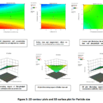 |
Figure 3: 2D contour plots and 3D surface plot for Particle size Click here to View Figure |
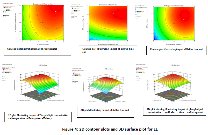 |
Figure 4: 2D contour plots and 3D surface plot for EE Click here to View Figure |
Zeta Potential
Zeta potential values of Passifloravitifolia phytosomes (PV-ex phyto) were found to range between -18.5 mV and -42.5 mV.
Optimization and Validation of Developed Model
The formulation parameters for PV phytosome preparation were refined using a numerical optimization approach. This technology suggested alternate methods and optimal conditions for producing PV phytosomes. The conditions were selected based on criteria like maximum EE and minimum particle size. Produced phytosomes of Passifloravitifolia (PV-ex phyto) showed particle sizes ranging from 143.67 to 394.42 nm and entrapment effectiveness varying from 40.21 to 80.05%.
Table 10: Actual responses vs. predicted responses
|
Formulation (PV-ex-phytoOPT) |
Phospholipid Concentration
(mg) |
Temperature
°C |
Reflux time | Particle size (nm) | Entrapment efficiency (%) |
| Predicted | 300.0 | 59.447 | 2 hr | 172.01 |
80.05 |
|
Actual |
300.0 | 59.447 | 2 hr | 184.05 |
79.89 |
It was found that the particle size was 184.05 nm, that was close to projected value of 172.01 nm. Similarly, the optimized batch had an entrapment efficiency of 79.89%, compared to 80.05% for the predicted solution (Table 10). The observed response values for dependent variables fell within 95% confidence interval, validating accuracy and reliability of established model for the generation of PV-ex phytosomes.
Formulation and Evaluation of PV-ex Phytosomes Optimized Batch (PV-ex-phytoOPT)
The phytosomes formed by PV extract under ideal conditions were evaluated for a variety of properties. PV-ex-phytoOPT particles measured 184.05±0.11 nm (Figure 5), with a Rutin% EE of 79.89±0.15%. The efficiency of statistical design tools in refining formulation and processing variables was validated, as the optimized batch exhibited particle size and EE values closely aligned with those predicted by the factorial design model. PV-ex-phytoOPT’s zeta potential was determined to be −18.52 mV, which is within the acceptable range of −30 to +30 mV, which suggests that the formulated phytosomes possess adequate stability.
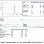 |
Figure 5: Particle size (A) and Zeta potential (B) of PV-ex-phytoOPT Click here to View Figure |
FTIR spectra
FTIR detects molecular interactions via chemical fingerprints of complexation, while TEM confirms nanoscale architecture, morphology and vesicle integrity. When combined, these orthogonal methods confirm the quality of formulation and phytosome formation. This combination approach supports claims of improved stability and delivery performance of well-developed phytosomes by combining structure-function assessment (TEM) and composition-interaction analysis (FTIR). By measuring diagnostic band shifts like P=O, P-O-C, C=O, and phenolic O-H stretching vibrations, FTIR can identify drug-phospholipid complexation. When comparing phytosomes to individual components or physical mixtures, the distinctive phosphatidylcholine band C=O stretching at ~1735 cm⁻¹, P=O near ~1249 cm⁻¹, P-O-C between 1057-1093 cm⁻¹, and the choline region around 970-985 cm⁻¹ serve as reference anchors to identify interaction-driven spectral changes. Complexation is characterized by shifts or broadening of the extract’s O-H band (~3400 cm⁻¹) and modifications in phospholipid C=O and P=O peaks, indicating the formation of phytosomes through hydrogen bonding or other intermolecular interactions. The improved formulation (PV-ex-phytoOPT) was tested for compatibility between excipients and extract by utilizing FTIR. Furthermore, absence of new peaks and significant shifts in characteristic bands verified the extract’s steady integration into the phytosome matrix. Figure 6 depicts the Rutin (pure) spectra of FTIR (A), formulation blend spectra of FTIR (B), optimized formulation FTIR spectra (C).
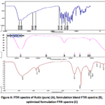 |
Figure 6: FTIR spectra of Rutin (pure) (A), formulation blend FTIR spectra (B), optimized formulation FTIR spectra (C) Click here to View Figure |
The FTIR spectra of pure Rutin were 3321, 3408, (O-H stretching), 2714 (C-H bonding), 2924, 2843 (CH2-stretching), 1383 (C-OH vibrations) and 1462 (C=O groups). The observed shifts in peaks at 1082 and 1074 cm⁻¹ suggest primary alcohol presence by C–O stretching vibrations, whereas the bands showing at 1043 cm⁻¹ and 1049 cm⁻¹ resemble to C–O–C stretching vibrations characteristic of strong anhydride groups. O-H stretch aromatic ethers were associated with the peaks at 1275 and 1112 cm-1, while secondary alcohol CO stretching and OH bending phenols were associated with peaks at 1401.49 and 1384 cm-1, respectively. While peaks at 2123 and 2849 cm-1 show alkanes C-H stretching, peaks at 1638 and 1605 cm-1 indicate β, α unsaturated ketone (alkene) presence. Alcohol’s O-H stretching was detected by the high peak at 3313.76 and 3412 cm-1.
TEM Evaluation
Usually, amphiphilic phospholipids make up phytosomes, that contain two nonpolar hydrophobic fatty acid chains along with a polar, positively charged hydrophilic head group. Complexation with phospholipids increases quercetin’s solubility by enhancing its molecular compatibility and interaction with the lipid matrix. Since TEM allows for the direct visualization of spherical morphology, aggregation and lamellarity behavior, it is crucial for evaluating vesicular architecture. It confirms that the complexes spontaneously organize into submicron or nano vesicular structures instead of forming irregular, amorphous aggregates [30].
TEM investigation of optimized phytosomes formulation (PV-ex-phytoOPT) revealed well-defined, mostly spherical vesicles with an average particle size of around 184.00 nm, which was consistent with the 184 nm size measured by zeta sizer measurements (Figure 7). This morphological consistency also helps successful phytosome development and structural stability.
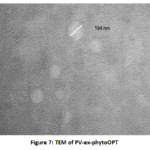 |
Figure 7: TEM of PV-ex-phytoOPT Click here to View Figure |
Study of In-vitro Drug Release
In-vitro release profile of PV-ex-phytoOPT for rutin was evaluated. The outcomes indicated that over 70% of the active phytoconstituents from the plant formulation were released within 8 hours (Figure 8). The released phytoconstituents amount was quantified by utilizing UV–visible spectrophotometric analysis. These outcomes indicated that generated phytosomes may improve anti-inflammatory activity of PV-ex-phytoOPT, making them suitable for developing a PV-based sustained drug delivery system. The % drug release from the phytosomal formulation was 89.25±1.73% after 12 hours.
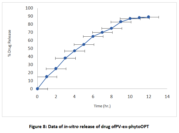 |
Figure 8: Data of in-vitro release of drug ofPV-ex-phytoOPT Click here to View Figure |
Assessment of the in-vitro anti-inflammatory effect of PV-ex-phytoOPT
Utilizing an egg albumin denaturation assay, anti-inflammatory action of PV-ex and PV-ex-phytoOPT formulations was assessed and compared with diclofenac sodium, a common anti-inflammatory medication. At a dosage of 100 μg/mL, diclofenac sodium inhibited by 86.25±0.62%.IPc-ex had an inhibition rate of 80.16±0.58%, whereas improved formulation demonstrated a concentration-dependent inhibitory action, with 88.50±0.60% at 200 μg/mL and 39.60±0.71% at 50 μg/mL maximum inhibition (Figure 9). Diclofenac sodium was more effective and served as reference standard, whereas improved formulation had substantial anti-inflammatory characteristics.
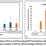 |
Figure 9: Protein denaturation analysis of PV-ex and PV-ex-phytoOPT formulations: (A) Absorbance measurement of the standard drug and test samples at 660 nm; (B) Percentage inhibition of proteinase activity |
Discussion
The results of this work show that Passifloravitifolia hydroethanolic leaf extract may be efficiently standardized, manufactured into phytosomes, and functionally positioned as a promising anti-inflammatory phytopharmaceutical. Among all solvent systems, the hydroethanolic extract had the highest extraction value of 9.54% w/w, indicating its suitability as the principal therapeutic fraction. The extract had TPC of 34.68 ± 0.56 mg GAE/g and TFC of 25.47 ± 0.83 mg RUT/g, indicating a flavonoid-rich matrix dominated by Rutin and other phenolics, which can support anti-inflammatory and antioxidant effects. Physicochemical parameters indicated raw material’s good quality, with water soluble ash 3.45% w/w, total ash 5.85% w/w, and acid insoluble ash 1.05% w/w, while moisture level of dried leaves remained within 6-7% w/w (fresh leaves 75.16%), limiting degradation and microbial growth concerns. Heavy metal analysis of Passifloravitifolia extract revealed lead 2.45 µg/mL, chromium 0.68 µg/mL, zinc 1.38 µg/mL, cadmium 0.002 µg/mL and arsenic 0.72 µg/mL. Mercury hadn’t been found, and all values were under WHO/FAO standards for medicinal plants, indicating toxicologically acceptable exposure from topical or transdermal usage. Fluorescence behavior, the absence of foreign organic matter, and reproducible TLC fingerprints (chloroform:methanol systems) confirmed the identity, purity, and batch-to-batch consistency of the standardized extract.
The comparison of solvent fractions revealed that antioxidant capability was highly impacted by both phenolic load and solvent polarity. The hydroethanolic extract displayed an IC₅₀ of 7.36 µg/mL, reflecting potent free radical scavenging at low concentrations, whereas non polar extracts like petroleum ether (TPC 8.10 ± 0.10 mg GAE/g, TFC 3.40 ± 0.20 mg RUT/g, IC₅₀ 982.21 µg/mL) and n-hexane (TPC 28.90 ± 0.52 mg GAE/g, TFC 25.16 ± 0.36 mg RUT/g, IC₅₀ 968.60 µg/mL) showed markedly weaker activity. The ethyl acetate and acetone fractions had higher TFC (35.20±0.47 mg RUT/g) and TPC (16.26±0.65 mg GAE/g), but the hydroethanolic fraction offered an optimal balance between yield, phenolic density, and bioactivity, making it ideal for phytosomal loading.
The Box-Behnken design allows for a systematic assessment of action of phospholipid content, temperature, and reaction time on important qualitative parameters of PV-ex phytosomes. Particle size ranged from 143.67 to 394.42 nm and entrapment efficiency from 40.21 to 80.05% over the design space, suggesting that formulation factors and their interactions had a significant impact on vesicle properties. The optimized batch (PV ex phytoOPT) met the pre-set desirability criteria of minimal particle size and maximal entrapment, achieving a mean vesicle size of 184.05±0.11 nm, entrapment efficiency of 79.89±0.15%, and zeta potential of -18.52 mV. These values are close to those predicted by the model and indicate physically stable nanosized carriers with efficient drug loading.
Morphological and spectroscopic analysis provided additional evidence for successful phytosome development. TEM scans of PV ex phytoOPT indicated primarily spherical, discrete vesicles in the sub 200 nm range, with low aggregation, which was consistent with DLS findings. FTIR spectra showed retention of characteristic bands of PV-ex and phospholipids, along with subtle shifts and broadening in hydroxyl and carbonyl regions, pointing to non-covalent interactions (hydrogen bonding and polar head group association), rather than chemical degradation of the marker constituents.
In-vitro release results showed that phytosomal encapsulation effectively regulated Rutin’s release profile. PV-ex phytoOPT exhibited a sustained pattern, with cumulative Rutin release approaching 89.25±1.73% at the end of 12 hours in phosphate buffer pH 7.4, compared to faster release from non-encapsulated extract. This suggests prolonged availability at the site of application and potential for once or twice daily dosing. The release curve, corroborated by 12 h data (≈80-90% drug release), suggests diffusion-controlled kinetics, which are ideal for maintaining stable anti-inflammatory levels without burst effect.
The egg albumin denaturation assay confirmed that Passifloravitifolia’s pharmacological potential increases with phytosomal loading. PV-ex phytoOPT inhibited protein denaturation at concentrations ranging from 88-89% at 200 µg/mL. This was higher than the free extract concentration and comparable to conventional diclofenac sodium. These findings showed that nanophytosomes enhanced the accessibility of Rutin and other flavonoids to biological targets, transforming the strong antioxidant profile (TPC and TFC values) into a more pronounced functional anti-inflammatory response. The data showed that Passifloravitifoliahydroethanolic leaf extract, when standardized and formulated as phytosomes had ≈180 nm size with ≈79% entrapment, provides a safe, stable, and effective platform for topical anti-inflammatory therapy. This optimized PV-ex phytoOPT system shows compliance with heavy metal limits, robust antioxidant activity, sustained Rutin release (89.25±1.73%/12 h), and high in-vitro inhibition of protein denaturation (≈88-89% at 200 µg/mL). Further in-vivo, permeation, and clinical studies are needed to translate this into a novel herbal dosage form for managing inflammatory conditions.
Conclusion
Encapsulating Passifloravitifoliahydroethanolic leaf extract into phospholipid‑based phytosomes successfully transformed a chemically rich but conventional extract into a standardized nanocarrier system with superior biopharmaceutical performance for anti-inflammatory therapy. The extract showed a high extractive value (9.54% w/w) along with substantial total phenolic and flavonoid contents (34.68 ± 0.56 mg GAE/g and 14.47 ± 0.83 mg RUT/g), validating P. vitifolia as a potent source of anti-inflammatory and antioxidant phytoconstituents. Pharmacognostic, physicochemical and heavy‑metal assessments confirmed that the raw material and extract complied with accepted pharmacopeial and WHO/FAO limits, establishing a robust safety and quality foundation for formulation development. Application of a Box–Behnken design enabled rational optimization of formulation and process variables, yielding an optimized phytosome batch (PV‑ex‑phytoOPT) with nano‑range particle size, high entrapment efficiency with satisfactory zeta potential corroborated by TEM and FTIR as stable, spherical vesicles formed via non‑covalent complexation. The optimized system provided sustained Rutin release and produced concentration‑dependent inhibition of protein denaturation approaching 88–89% at 200 µg/mL, outperforming plain extract and approaching the reference drug diclofenac sodium in-vitro. Overall, these outcomes indicate that P. vitifoliaphytosomes can effectively enhance dermal delivery and anti‑inflammatory efficacy of Rutin‑rich extracts while maintaining acceptable safety. Future work should focus on ex-vivo skin permeation, in-vivo anti‑inflammatory models and long‑term stability studies to translate this optimized system into a clinically relevant topical or transdermal phytopharmaceutical.
Acknowledgements
Authors are highly thankful to their Universities/Colleges to provide library facilities for the literature survey.
Reference
- Pereira Leal, A. E., de Lavor, É. M., de Menezes Barbosa, J., de MouraFontesAraújo, M. T., dos Santos CerqueiraAlves, C., de Oliveira Junior, R. G., de Lima, Á. A., & da Silva Almeida, J. R. (2022). Pharmacological activities of the genus Passiflora (Passifloraceae): A patent review. Current Topics in Medicinal Chemistry, 22(28), 2315–2328.
CrossRef - Franco-Mora, O., López-Sandoval, J. A., Castañeda-Vildózola, Á.,& Sánchez-Pale, J. R. (2022). Diversity of genus Passiflora L. in Mexico and a particular study on Passiflorabiflora Lam. fruit characteristics. Genetic Resources and Crop Evolution, 69(7), 2565–2574.
CrossRef - Drewinski, M. P., Corrêa-Santos, M. P., Lima, V. X., Lima, F. T., Palacio, M., Borges, M. E., Trierveiler-Pereira, L., Magnago, A. C., Furtado, A. N., Lenz, A. R., & Silva-Filho, A. G. (2024). Over 400 food resources from Brazil: Evidence-based records of wild edible mushrooms. IMA Fungus, 15(1), Article 40.
CrossRef - Gori, B., Ulian, T., Bernal, H. Y., &Diazgranados, M. (2022). Understanding the diversity and biogeography of Colombian edible plants. Scientific Reports, 12(1), Article 7835.
CrossRef - Thokchom, R., &Mandal, G. (2017). Production preference and importance of passion fruit (Passifloraedulis): A review. Journal of Agricultural Engineering and Food Technology, 4(1), 27–30.
- Yuan, H., Ma, Q., Ye, L., &Piao, G. (2016). The traditional medicine and modern medicine from natural products. Molecules, 21(5), Article 559.
CrossRef - Alara, J. A., &Alara, O. R. (2024). An overview of the global alarming increase of multiple drug resistance: A major challenge in clinical diagnosis. Infectious Disorders – Drug Targets, 24(3), 26–42.
CrossRef - Pal, R. (2021). A review on the pharmacological importance of crimson passion flower (Passifloravitifolia: Passifloraceae)—An endangered species. Acta Scientific Microbiology, 4(9), 92–98.
CrossRef - Patel, S. A., Sahoo, S., Kaushik, A., Rahvar, K., Patel, T., Kajal, C., &Khusboo, C. (2024). The morphology, chemical constituents and uses of passion flower: A review. Pharma Science Monitor, 15(2).
- Ni, Y. W., Lin, K. H., Chen, K. H., Wu, C. W., & Chang, Y. S. (2020). Flavonoid compounds and photosynthesis in Passiflora plant leaves under varying light intensities. Plants, 9(5), Article 633.
CrossRef - Silva, G. C., &Bottoli, C. B. G. (2015). Analyses of Passiflora compounds by chromatographic and electrophoretic techniques. Critical Reviews in Analytical Chemistry, 45(1), 76–95.
CrossRef - Casierra-Posada, F., &Jarma-Orozco, A. (2016). Nutritional composition of Passiflora species. In M. Simmonds & V. R. Preedy (Eds.), Nutritional composition of fruit cultivars (pp. 517–534). Academic Press.
CrossRef - Nikolova, K., Velikova, M., Gentscheva, G., Gerasimova, A., Slavov, P., Harbaliev, N., Makedonski, L., Buhalova, D., Petkova, N., &Gavrilova, A. (2024). Chemical compositions, pharmacological properties and medicinal effects of genus Passiflora L.: A review. Plants, 13(2), Article 228.
CrossRef - Sakalem, M. E., Negri, G., &Tabach, R. (2012). Chemical composition of hydroethanolic extracts from five species of the Passiflora genus. RevistaBrasileira de Farmacognosia, 22, 1219–1232.
CrossRef - Ingale, A. G., &Hivrale, A. U. (2010). Pharmacological studies of Passiflora sp. and their bioactive compounds. African Journal of Plant Science, 4(10), 417–426.
- Echeverry González, S. M., Santos, A. M., Júnior, C. C., Saravanan, S., Castellanos, L., Serafini, M. R., & Aragon, M. (2025). Natural therapies: A systematic review of the medicinal applications of Passifloraligularis. Phytochemistry Reviews. Advance online publication.
CrossRef - Barani, M., Sangiovanni, E., Angarano, M., Rajizadeh, M. A., Mehrabani, M., Piazza, S., Gangadharappa, H. V., Pardakhty, A., Mehrbani, M., Dell’Agli, M., &Nematollahi, M. H. (2021). Phytosomes as innovative delivery systems for phytochemicals: A comprehensive review of literature. International Journal of Nanomedicine, 16, 6983–7022.
CrossRef - , K. S., & K., A. (2021). Phytosome as a novel drug delivery system for bioavailability enancement of phytoconstituents and its applications: A review. Journal of Drug Delivery and Therapeutics, 11(3).
CrossRef - Rodríguez, Á. A., Arteaga, J. J., Arango, W. M., &Pabón, M. F. (2021). Vasodilator effect of ethanolic extracts of Passifloravitifolia and Passifloraedulis f. edulis seeds. Journal of Applied Pharmaceutical Science, 11(10), 61–69.
CrossRef - Rastogi, A., Yadav, P., & Mishra, M. K. (2022). Preparation and evaluation of new polyherbal formulation for wound healing activity. Journal of Pharmaceutical Negative Results, 13.
CrossRef - Singh, P., Khosa, R. L., Srivastava, S., Mishra, G., Jha, K. K., Srivastava, S., Verma, R. K., &Tahseen, M. A. (2014). Pharmacognostical study and establishment of quality parameters of aerial parts of Costusspeciosus—A well-known tropical folklore medicine. Asian Pacific Journal of Tropical Biomedicine, 4(6), 486–491.
CrossRef - Das, P. K., Vaghela, J. S., &Badore, N. (2021). Pharmacognostical, phytochemical and fluorescence analysis of the plant Bougainvillea spectabilis (Willd.). Research Journal of Pharmacy and Technology, 14(7), 3733–3738.
CrossRef - Razvi, S. Z., Kamm, I., Nguyen, T., Pellett, J. D., & Kumar, A. (2021). Loss on drying using halogen moisture analyzer: An orthogonal technique for monitoring volatile content for in-process control samples during pharmaceutical manufacturing. Organic Process Research & Development, 25(2), 300–307.
CrossRef - Birudu, R. B., Naik, J. M., Revana, S. D., Jilani, S. K., &Janardhan, M. (2015). Phytochemical screening of ethanolic extract of Passiflorafoetida (Linn) and medicinal importance. Indian Journal of Research in Pharmacy and Biotechnology, 3(4), 324–327.
- Shraim, A. M., Ahmed, T. A., Rahman, M. M., &Hijji, Y. M. (2021). Determination of total flavonoid content by aluminum chloride assay: A critical evaluation. LWT, 150, Article 111932.
CrossRef - Srivastava, N., Bansal, A., Aggarwal, K., &Nagpal, K. (2024). Development and validation of UV spectrophotometric method for the quantitative estimation of quercetin in bulk followed by its solubility studies. Journal of Applied Spectroscopy, 91(3), 700–708.
CrossRef - Phamiwon, Z. A., & Sheila, J. (2017). Phytochemical analysis and thin layer chromatographic studies of Passifloraedulis leaf extracts. International Journal of Food Science and Nutrition, 2, 38–41.
- Jain, A., Verma, R., & Mehta, P. D. (2023). Formulation and evaluation of phytosomes loaded with Pithecellobiumbijeninum leaf extract. Current Research in Pharmaceutical Sciences, 151–156.
CrossRef - Karpe, S., Chandewar, A., Kochar, N., &Shrirao, A. (2025). Formulation, optimization and characterization of Pongamiapinnataphytosomes for therapeutic potential. Indian Journal of Pharmaceutical Education and Research, 59(3), 949–959.
CrossRef - Tripathy, S., Patel, D., Baro, L., & Nair, S. (2013). A review on phytosomes, their characterization, advancement and potential for transdermal application. Journal of Drug Delivery and Therapeutics, 3, 147–152.
CrossRef
Accepted on: 25 Feb 2026
Second Review by: Dr. G.K. Radha
Final Approval by: Dr. B. K Sharma


![]()


