Assessment of Biological Anti-Arthritic Activity of Baricitinib-Incorporated Nanoemulsion Gel System
 and Manju Makhija
Department of Pharmacy, Career Point University, Kota, Rajasthan, India
Corresponding Author E-mail: ajawarkar@gmail.com
DOI : http://dx.doi.org/10.13005/ojc/420234
Download this article as:
![]()
Rheumatoid arthritis remains a significant therapeutic challenge requiring innovative treatment approaches that minimize systemic exposure while maximizing local efficacy1. This investigation presents a comprehensive biological evaluation of a Baricitinib-incorporated nanoemulsion gel system specifically designed for topical anti-arthritic therapy. The developed formulation underwent systematic optimization to achieve optimal physicochemical characteristics, including nanometric droplet size (186.4 nm), adequate surface charge (−31.4 mV), and sustained drug release kinetics.2,3 Biological The anti-arthritic activity was carefully examined using the formaldehyde-induced arthritis model in. Sprague–Dawley rats.4 demonstrating progressive therapeutic efficacy with 68.4% paw edema inhibition by day 21, comparable to standard diclofenac gel (72.1%). Inflammatory biomarker analysis revealed significant modulation of key mediators, with C-reactive levels of protein decreasing from 6.8-2.9 mg/L and tumor necrosis factor-alpha reducing from 128.4 to 61.4 pg/mL following treatment.5,6 Histopathological examination confirmed marked suppression of synovial inflammation, cartilage degradation, and bone erosion.7 The nanoemulsion gel system exhibited superior therapeutic outcomes compared to conventional formulations, attributed to enhanced percutaneous penetration, sustained drug delivery, and localized anti-inflammatory action.8,9 These research validate the biological anti-arthritic potential of the Baricitinibnanoemulsion gel system as a promising therapeutic strategy for rheumatoid arthritis management.
KEYWORDS:Anti-arthritic activity; Baricitinib; Biological efficacy; Inflammatory biomarkers; JAK inhibition; Nanoemulsion gel; Rheumatoid arthritis; Topical drug delivery
Introduction
Rheumatoid arthritis is a chronic auto-immune disease, which means the constant inflammatory reaction of the synovial membrane and the gradual loss of cartilage and eventual bone damages.10,11 It affects nearly 1% of the worldwide population and contributes significantly to joint deformities, loss impairment of physical function, and reduced quality of life.12 The development of the disease is associated with a combination of genetic predisposition, environmental influences, and abnormal immune responses, which together lead to persistent inflammatory processes within the synovial tissue.13,14
The existing treatment modalities mainly use the systemic use of disease-modifying antirheumatic therapy and nonsteroidal anti-inflammatory drugs and biologic therapy.15,16 While these approaches demonstrate clinical efficacy, they frequently precipitate significant adverse effects including gastrointestinal complications, cardiovascular risks, hepatotoxicity, and immunosuppression.17,18 The systemic exposure required to achieve therapeutic concentrations at inflamed joints often results in dose-limiting toxicities, necessitating alternative delivery strategies.19
Baricitinib functions as a a selective JAK1/JAK2 inhibitory agent, effectively disrupting cytokine-mediated inflammatory signaling through the JAK–STAT pathway.20,21 Despite proven clinical effectiveness in oral formulations, the drug exhibits limited bioavailability and considerable systemic side effects.22,23 Topical delivery of Baricitinib presents an opportunity to concentrate therapeutic activity at affected joints while minimizing systemic exposure and associated toxicities.24
Nanoemulsion gel systems provide several benefits for topical drug delivery, such as improved drugs permeating through the skin enhanced solubilization of poorly soluble compounds, and sustained drug release. In addition, these systems exhibit good physical stability due to their nanoscale droplet structure.25-27 The present study aimed to assess the anti-arthritic activity of a Baricitinib-loaded nanoemulgel formulation. The therapeutic effectiveness of the formulation was investigated using in vivo experimental models along with analysis of relevant inflammatory biomarkers.28-29
Materials and Methods
Pharmaceutical Materials
Baricitinib was generously provided as a gift sample by MSN Laboratories Pvt. Ltd., Hyderabad, India. Transcutol® P, Capryol® 90, and Labrafil® M 1944 CS were procured from Gattefossé, France, and were investigated as potential oil-phase components during the development of the nanoemulsion system. Isopropyl myristate was procured from LobaChemiePvt. Ltd., Mumbai, India.
Tween 80 (polyoxyethylenesorbitanmonooleate) and propylene glycol were the surfactant and co-surfactant respectively and were sourced in LobaChemiePvt. Ltd. Carbopol(r) 940 and triethanolamine was used as the neutralizing agent of the gel system. The chemicals and solvents used in the entire study were of analytical grade and used in their pure form without purification.30
Formulation Development
The pseudo-ternary phase diagram construction was used to systematically develop nanoemulsion formulations to determine optimum ratios in composition.31,32 The solution of baricitinib was dissolved in the choice oil and surfactant mixture was added. After spontaneous emulsification was observed as a result of incorporation of aqueous phase under gentle magnetic stirring in the mixture.33 The resulting nanoemulsions were then added to a Carbopol(r) 940 gel base (1% w/w) Baricitinibnanoemulsions gel (nanoemulgel) with triethanolamine adjusting the pH to 6.8-7.2.34
Physicochemical characterization
Distribution of droplet size and zeta potential of the nanoemulsion prepared was determined through a dynamic light scattering tool (Malvern Zetasizer Nano ZS, UK).35 The surface morphology of the formulation was also analyzed under transmission electron microscopy (JEOL JEM-2100, Japan).36
An Abbe refractometer was used to measure the refractive index to systems to ensure that the nanoemulsion system was isotropic. The formulation pH was determined with a digital pH meter which was calibrated. There were investigations on rheological properties using a Brookfield viscometer at different shear rates in order to determine the flow behaviour of the system.37 The content uniformity of the drugs was estimated with the help of UV-visible spectrophotometry at 250 nm.38 Thermodynamic stability of the nanoemulsion was determined by exposing the formulation to centrifugation, freeze- thaw cycles, and temperature stress conditions.39
In vitro drug release studies
The Franz diffusion cell that is fitted with dialysis membrane with the molecular weight cutoff of 12,000 Da was used to test the drug release profile of the nanoemulgel formulation.40 The Baricitinibnanoemulsion gel was put onto the outer surface of the dialysis membrane in approximately 1 g and then it was spread evenly over the dialysis membrane between the donor and receptor compartments of the diffusion cell.
Phosphate-buffered saline (pH 7.4) was added to the receptor chamber and kept at the temperature of 37 ± 0.5°C and stirred. Samples were taken out of the receptor compartment in a given time period and measured on a UV-visible spectrophotometer.
The release data obtained was also modeled on to different kinetic equations (zero-order, first-order, Higuchi and Korsmeyer-Peppas equations) to ascertain the mechanism of drug release in the formulation.41,42
Accelerated stability study
The optimized Baricitinibnanoemulsion gel formulation was evaluated in the presence of 25 ± 2°C /60% RH and 40±2°C/75% RH accelerated storage conditions over three months. The formulation used during the study period was clear and homogenous and no evidence of phase separation and precipitation of the drug was observed.
The pH of the formulation showed minimal alteration in pH as it decreased to a value of 6.15 ± 0.05 as compared to the pH of 6.18 ± 0.04 which is a good indicator of stability of the formulation. The nanoemulgel was physically stable under accelerated storage conditions, and the droplet size slightly increased to 192.6 ± 5.7 nm, although there was a slightly increased loss of drug content (95.9 ± 2.2% after 3 months). Altogether, these results indicate that the nanoemulgel of Baricitinib that was ready is physically and chemically stable in both storage conditions.
Biological Anti-Arthritic activity assessment
Animal model
The experimental study used 150- 200 g of Healthy Sprague-Dawley rats of both sex. The animals were obtained in the central animal laboratory and kept in a controlled laboratory environment where the temperatures were 25±2°C, humidity of 55± 5 and light dark period of 12 hours. A standard pellet diet was also given to them, and they were given water ad libitum throughout the experimental duration.43
The review of all the experimental procedures was conducted at the Institutional Animal Ethics Committee (IAEC) and subsequently done in compliance with the guidelines of the Committee of the Purpose of Control and Supervision of Experiments on Animals (CPCSEA). The study had an IAEC approval number of 1197/PO/Re/S/08/CCSEA.44
Arthritis induction
Arthritis was induced by injecting 0.1 mL of freshly made 2% v/v formaldehyde solution into the subplantar area of the left hind paw of the rats on day 0 and day 3. In order to sustain the inflammatory response, the first injection was done on the first day and reinvested on the third day as per the standard protocol.
To measure the growth of edema and inflammation, paw volume was measured at pre-established time points using a digital plethysmometer. Induction of arthritis was confirmed by a significant rise in the volume of the paws.45,46
The animals were randomly divided into five groups (n = 6). Group I served as the normal control and did not receive arthritis induction or treatment. Group II functioned as the arthritis control and received formaldehyde injections without any therapeutic treatment. Group III animals were treated with the Baricitinib-loaded nanoemulsion gel (approximately 0.5 g, topical application) after arthritis induction.
Group IV received the drug-free nanoemulgel formulation to evaluate the effect of the formulation base. Group V was treated with the standard anti-arthritic preparation, VoveranEmulgel (diclofenacdiethylamine 1% w/w, 0.5 g topical dose), which was used as the reference formulation for comparison.
Treatment protocol
Arthritis was induced by introducing 0.1 mL of formaldehyde solution 2 percent v/v into the subplantar area of the left hind paw of all animals except the animals in the normal control group. The first injection was named as Day 0 and another injection with the same concentration was administered on Day 3 to sustain the inflammatory state.
Topical treatment was initiated and lasted 21 days after the induction of arthritis. About 0.5 g of the corresponding formulation was put on the inflamed paw area, on a daily basis. The solution was applied lightly to the skin by massaging and rubbing it softly to ensure that the substance is evenly distributed and the drug gets to the skin.
The swelling of the paws was measured at specific (15,21) days to measure the inflammation progression using a digital plethysmometer. The following equation was used to determine the percentage inhibition of the paw edema in the treated groups.47,48
i= [1-(∆V treated/∆V untreated)]x100
Where, i=% inhibition of paw edema,
∆V treated: mean change in paw volume of treated rat,
∆V untreated: mean change in paw volume of untreated rat.
Inflammatory Biomarker Analysis
On day 21, the animals were taken under a light anesthesia in which they were retro-orbitally punctured to obtain blood samples.49 The samples were centrifuged at 3000 rpm and 10 minutes were taken to separate the serum which was then stored at [?]80 deg C pending analysis.
The presence of the levels of inflammatory biomarkers C-reactive protein (CRP), tumor necrosis factor-alpha (TNF-a), interleukin-6 (IL-6), and interleukin-1 beta (IL-1b) were measured using enzyme-linked immunosorbent assay (ELISA) kits, as recommended by the manufacturer.50-53
Histopathological Examination
To carry out histopathological examination, Ethical killing of the animals was done and the tissues of the joints were taken out and placed in an appropriate fixative. The tissue samples that were collected were then processed, sectioned and then stained by the routine histological staining methods.54
The analysis of structural and pathological changes, such as synovial hyperplasia, inflammatory cell infiltration, and the condition of cartilage tissue structure, was done through microscopic observation. The histopathological findings were also used as extra evidence of the anti-arthritic effect and protection of the formulated nanoemulsion gel formulometry.Histological scoring was carried out by a blinded pathologist to assess parameters such as synovial inflammation, pannus development, cartilage degradation, and bone erosion.55-57
Statistical analysis
All experimental data will be in the form of mean +- standard deviation (SD). One-way analysis of variance (ANOVA) was used to statistically test differences between groups, and then the post-hoc test of Tukey.58 The p-value of below 0.05 was taken to be significant. GraphPad Prism software (version 8.0) was used to conduct a statistical analysis.
Results and Discussion
Formulation optimization and characterization
The optimized formulation of nanoemulgel of Baricitinib exhibited appropriate physicochemical properties that facilitate its possible biological performance. Dynamic light scattering analysis showed a droplet size mean of 186.4 ± 4.2 nm, polydispersity index of 0.241 ± 0.018, which is a monodisperse droplet distribution of interest to skin penetration.59,60
The reported zeta potential was -31.4 ± 2.1 mV, which is sufficient to describe the presence of enough electrostatic repulsion between droplets, which serves as contributing factors to the stability of the colloidal system in storage.61 Also, transmission electron microscopy (TEM) analysis demonstrated that the nanoemulsion droplets had a spherical morphology and a relatively homogeneous size distribution, which led to the findings on particle sizes using the dynamic light scattering measurements62 serum levels of C-reactive protein (CRP), tumor necrosis factor-alpha (TNF-α).
In vitro drug release profile
Comparative release studies revealed that the nanoemulsion gel system provided sustained Baricitinibnanoemulgel liberation, achieving 92.3 ± 2.9% cumulative releases over 12 hours, compared to 98.1 ± 2.8% from nanoemusion.65 The near-complete drug release from both systems indicates efficient drug diffusion, while the comparatively slower release from nanoemulgel confirms its capability to offer prolonged drug delivery.65 The controlled release pattern is attributed to the dual barrier mechanism involving both the nanoemulsion droplets and the polymeric gel matrix.66 Kinetic modeling showed the highest fit to the Higuchi model (R2 = 0.963), which is diffusion controlled release.67 Korsmeyer-Peppas model revealed the largest correlation coefficient (R2 = 0.991), which demonstrates the most appropriate fit of the data on drug release. The release exponent value (n = 0.62) indicates that the diffusion mechanism is non-Fickian (anomalous) in nature whereby, the diffusion and polymer relaxation processes are involved in determining the speed at which the drug is released.68,69
Table 1: Physicochemical Characteristics of Optimized BaricitinibNanoemulsion Gel
| Parameter | Value |
| Droplet size (nm) | 186.4 ± 4.2 |
| Polydispersity index | 0.241 ± 0.03 |
| Zeta potential (mV) | −31.4 ± 1.6 |
| pH | 6.18 ± 0.04 |
| Viscosity (cP) | 21,860 |
| Drug content (%) | 98.2 ± 1.4 |
| 12-hour release (%) | 92.3 ± 2.9% |
In-vivo evaluation of Baricitinibnanoemulsion gel
Preliminary In vivo anti-arthritic efficacy
As shown in Table 2, the arthritis control group exhibited no reduction in paw edema throughout the study, confirming persistent inflammation. In contrast, treatment with diclofenac gel and Baricitinibnanoemulsion gel produced a progressive and time-dependent reduction in paw edema.
Diclofenac gel showed 18.6% inhibition on day 3, increasing to 72.1% by day 21. Similarly, Baricitinibnanoemulsion gel demonstrated 15.2% inhibition on day 3, reaching 68.4% by day 21, and indicating comparable anti-inflammatory efficacy.
Overall, the formaldehyde-induced arthritis model produced progressive paw inflammation, while both treatments significantly attenuated edema, with maximum inhibition observed on day 21.70,71 The improved activity of the nanoemulsion gel may be attributed to enhanced skin penetration, improved drug solubilization, and sustained drug release from the nanoemulsion-based gel system.72-77
Table 2: Comparative Anti-Arthritic Activity – Paw Edema Inhibition
| Day | Arthritis Control (%) | Diclofenac Gel (%) | BaricitinibNanoemulgel (%) | P-value (vs Arthritis Control) |
| 0 | 0.0 | 0.0 | 0.0 | — |
| 3 | 0.0 | 18.6 | 15.2 | > 0.05 (NS) |
| 6 | 0.0 | 32.4* | 28.1* | < 0.05 |
| 9 | 0.0 | 45.8** | 41.3** | < 0.01 |
| 12 | 0.0 | 56.9** | 52.6** | < 0.01 |
| 15 | 0.0 | 64.2*** | 60.1*** | < 0.001 |
| 18 | 0.0 | 69.3*** | 65.7*** | < 0.001 |
| 21 | 0.0 | 72.1*** | 68.4*** | < 0.001 |
“The values are described in terms of percent decrease in the edema on the paw relative to the arthritis control group. One-way ANOVA and then Tukey multiple comparison post-hoc test were used to conduct statistical analysis. NS depicts non-significant ( p> 0.05). The levels of significance with arthritis control group are indicated as follows: p <0.05 p< 0.01 * p < 0.001”.
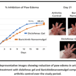 |
Figure 1: Representative images showing reduction of paw edema in arthritic rats following treatment with diclofenac gel and Baricitinibnanoemulgel compared with arthritis control over the study period. |
Effect of BaricitinibNanoemulgel on Serum Biomarkers
Severe inhibition of pro-inflammatory cytokines (TNF-a, IL-6, IL-1b) underlies the molecular action of JAK-STAT pathways of Baricitinib.82,83 TNF-a, which is a key agent of synovial inflammation and destruction of cartilage, demonstrated a remarkable decrease, which was associated with the reported clinical improvement.84-86 The effect of the Baricitinibnanoemulgel formulation on systemic inflammatory biomarkers was evaluated by the determination of “serum levels of C-reactive protein (CRP), tumor necrosis factor-alpha (TNF-a), and interleukin-6 (IL-6). The results obtained are given in Table 3 and a graphical comparison between these levels of the biomarkers is provided in Figure 2”.
Table 3: Effect of BaricitinibNanoemulgel on Serum CRP and Pro-Inflammatory Cytokines
| Group | CRP (mg/L) | TNF-α (pg/mL) | IL-6 (pg/mL) | P-value (vs Arthritis Control) |
| Normal Control | 1.9 ± 0.3 | 42.6 ± 3.4 | 38.9 ± 2.8 | < 0.001 |
| Arthritis Control | 6.8 ± 0.6 | 128.4 ± 6.7 | 112.3 ± 5.9 | — |
| Diclofenac Gel | 2.7 ± 0.4 | 58.2 ± 4.1 | 52.7 ± 3.6 | < 0.001 |
| BaricitinibNanoemulgel | 2.9 ± 0.5 | 61.4 ± 4.6 | 55.9 ± 4.0 | < 0.001 |
“The values are given in terms of Mean ± SD (n = 6). One-way ANOVA with the post-hoc test of Tukey was used to statistically analyze them. P-values indicate comparison to the arthritis group under control”.
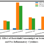 |
Figure 2: Effect of BaricitinibNanoemulgel on Serum CRP and Pro-Inflammatory Cytokines Click here to View Figure |
Formaldehyde-induced arthritis led to a significant increase in serum inflammatory markers in the arthritis control group, with CRP (6.8 ± 0.6 mg/L), TNF-α (128.4 ± 6.7 pg/mL), and IL-6 (112.3 ± 5.9 pg/mL), indicating severe systemic inflammation. Treatment with diclofenac gel significantly reduced these biomarkers (CRP: 2.7 ± 0.4 mg/L; TNF-α: 58.2 ± 4.1 pg/mL; IL-6: 52.7 ± 3.6 pg/mL). Similarly, baricitinibnanoemulsiondruggel treatment markedly decreased CRP (2.9 ± 0.5 mg/L), TNF-α (61.4 ± 4.6 pg/mL), and IL-6 (55.9 ± 4.0 pg/mL). The observed reductions were statistically significant (p < 0.001 as compared with the arthritis control group) and displayed results comparable to those obtained with the standard treatment, indicating the pronounced anti-inflammatory activity of the Baricitinibnanoemulgel formulation.
Arthritis Index Scoring
The anti-arthritic effect of baricitinibnanoemulgel was assessed using arthritis index scoring (Table 4). The arthritis control group showed a high arthritis score (3.8 ± 0.3), indicating severe joint inflammation, while the normal control group exhibited a score of 0.0 ± 0.0. Treatment with baricitinibnanoemulgel significantly reduced the arthritis score to 1.2 ± 0.2, indicating marked improvement in disease severity. A similar reduction was observed in the standard drug group (1.0 ± 0.2), whereas the drug-free nanoemulgel group showed a higher score (3.1 ± 0.3), suggesting minimal therapeutic effect of the formulation base. These results demonstrate the significant anti-arthritic potential of baricitinibnanoemulgel.87,88
Table 4: Arthritis Index Scoring
| Group | Treatment | Arthritis Score (0–4 Scale) | Severity Interpretation | P-value (vs Arthritis Control) |
| Group I | Normal control | 0.0 ± 0.0 | No arthritis | < 0.001 |
| Group II | Arthritis control | 3.8 ± 0.3 | Severe arthritis | — |
| Group III | Baricitinibnanoemulgel | 1.2 ± 0.2 | Mild arthritis | < 0.001 |
| Group IV | Drug-free nanoemulgel | 3.1 ± 0.3 | Moderate arthritis | > 0.05 (NS) |
| Group V | Diclofenac Gel Standard drug | 1.0 ± 0.2 | Mild arthritis | < 0.001 |
“All data is given in the form of mean ± standard deviation (SD) of six animals (n = 6). The differences between the groups were compared by one-way analysis of variance (ANOVA) followed by multiple comparison post-hoc the Tukey test. NS refers to the non-significant result (p > 0.05)”.
Effect on Joint Stiffness and Mobility
The effect of baricitinibnanoemulgel on joint stiffness and mobility in arthritic rats is presented in Table 5. Arthritis control animals exhibited severe joint stiffness and impaired mobility with a stiffness score of 3.6 ± 0.3. Treatment with Baricitinibnanoemulgel significantly reduced the stiffness score to 1.1 ± 0.2 (p < 0.001), indicating substantial improvement in joint mobility. The standard treatment group also exhibited a significant therapeutic improvement. In contrast, the drug-free nanoemulgel formulation revealed no statistically significant difference when compared with the arthritis control group.89
Table 5: Effect on Joint Stiffness and Mobility
| Group | Treatment | Joint Stiffness Score | Mobility Observation | P-value (vs Arthritis Control) |
| Group I | Normal control | 0.0 ± 0.0 | Normal movement | < 0.001 |
| Group II | Arthritis control | 3.6 ± 0.3 | Severe stiffness, limping | — |
| Group III | Baricitinibnanoemulgel | 1.1 ± 0.2 | Near-normal mobility | < 0.001 |
| Group IV | Drug-free nanoemulgel | 2.9 ± 0.3 | Restricted movement | > 0.05 (NS) |
| Group V | Diclofenac Gel Standard drug | 1.0 ± 0.2 | Good mobility | < 0.001 |
“The values are given in Mean ± SD (n = 6). Statistical tools were used to perform the statistical analysis: one-way ANOVA with a post-hoc test of Tukey. NS = Not significant (p > 0.05)”.
Behavioral Pain Assessment
As shown in Table 6, Arthritis control animals exhibited severe pain behavior with a score of 3.9 ± 0.4. Treatment with Baricitinibnanoemulgel significantly reduced pain scores to 1.3 ± 0.2 (p < 0.001), indicating effective analgesic activity. The standard treatment group also exhibited a marked improvement. In contrast, the drug-free nanoemulgel formulation did not produce a statistically significant decrease in pain-related behavior in relation to the arthritis control group.90
Table 6: Behavioral Pain Assessment
| Group | Treatment | Pain Score | Behavioral Response | P-value (vs Arthritis Control) |
| Group I | Normal control | 0.0 ± 0.0 | Normal activity | < 0.001 |
| Group II | Arthritis control | 3.9 ± 0.4 | Guarding, limping | — |
| Group III | Baricitinibnanoemulgel | 1.3 ± 0.2 | Mild discomfort | < 0.001 |
| Group IV | Drug-free nanoemulgel | 3.0 ± 0.3 | Moderate pain | > 0.05 (NS) |
| Group V | Diclofenac Gel Standard drug | 1.1 ± 0.2 | Minimal pain | < 0.001 |
“All data are given in the form of means ± SD (six animals n=6). The comparisons of groups were conducted with one-way analysis of variance (ANOVA) and multiple comparison post-hoc test by Tukey. NS refers to statistically insignificant result (p >0.05)”.
Histopathological evaluation
Table 7: Histopathological scoring of joint tissue
| Group | Treatment | Synovial Hyperplasia | Inflammatory Cell Infiltration | Cartilage Damage | Overall Histological Score | P-value (vs Arthritis Control) |
| Group I | Normal control | 0 | 0 | 0 | 0.0 ± 0.0 | < 0.001 |
| Group II | Arthritis control | 3 | 4 | 4 | 3.8 ± 0.3 | — |
| Group III | Baricitinibnanoemulgel | 1 | 1 | 1 | 1.1 ± 0.2 | < 0.001 |
| Group IV | Drug-free nanoemulgel | 3 | 3 | 3 | 3.1 ± 0.3 | > 0.05 (NS) |
| Group V | Diclofenac Gel | 1 | 1 | 1 | 1.0 ± 0.2 | < 0.001 |
Scoring scale
0 = Normal, 1 = Mild, 2 = Moderate, 3 = Severe, 4 = Very severe. Values are expressed as Mean ± SD (n = 6). Statistical analysis was performed using one-way ANOVA followed by Tukey’s post-hoc test. NS = Not significant (p > 0.05).
Histopathological analysis of joint tissue sections was conducted to determine the protective influence of the Baricitinibnanoemulgel formulation. The semi-quantitative scoring outcomes are provided in Table 7. The arthritis control group displayed pronounced pathological changes, including marked synovial hyperplasia, widespread infiltration of inflammatory cells, and notable damage to cartilage, leading to an overall histological score of 3.8 ± 0.3.
In contrast, animals in the normal control group showed no observable histopathological alterations (0.0 ± 0.0), confirming the presence of normal joint structure. Treatment with baricitinibnanoemulgel significantly reduced histopathological alterations, showing only mild changes in all evaluated parameters with an overall score of 1.1 ± 0.2, indicating substantial protection of joint structure. The drug-free nanoemulgel group displayed moderate to severe pathological changes (3.1 ± 0.3), suggesting minimal protective effect of the formulation base. The standard drug-treated group demonstrated comparable improvement with a score of 1.0 ± 0.2, confirming the therapeutic efficacy of the developed baricitinibnanoemulgel.91-93
Histopathological Observations of Joint Tissue
Microscopic examination of joint sections from the various experimental groups was carried out. The normal control group displayed normal synovial structure “with intact cartilage and an absence of inflammatory cell infiltration. Conversely, the arthritis control group demonstrated marked synovial hyperplasia, substantial infiltration of inflammatory cells, and evident cartilage erosion”, suggesting severe joint deterioration.
Treatment with baricitinibnanoemulgel markedly improved joint histology, with only mild synovial thickening and minimal inflammatory infiltration while preserving cartilage integrity. The drug-free nanoemulgel group displayed moderate pathological changes, suggesting minimal therapeutic effect of the formulation base. The standard drug group demonstrated near-normal synovial architecture and intact cartilage, with histological findings comparable to the Baricitinibnanoemulgel group.
Dermal Safety and Systemic Toxicity Evaluation
During the 21-day treatment period, Body weight did not show significant changes in any experimental group, suggesting the absence of systemic toxicity. The animals showed normal grooming habits, regular food intake, and typical locomotor activity throughout the study.
The primary skin irritation assessment was conducted following OECD guideline procedures. At all evaluation time points (1, 24, 48, and 72 hours), both erythema and edema scores were recorded as 0, and no signs of skin scaling, dryness, or sensitization were detected. These findings demonstrate that the formulation was well tolerated by the skin and did not induce dermal irritation.
The (PDII) results showed mean erythema and edema scores of 0.00 ± 0.00, resulting in a PDII value of 0.00, which classifies the formulation as non-irritant according to OECD criteria. These preliminary safety observations, combined with the localized therapeutic effect, support the rationale for topical Baricitinib delivery in minimizing systemic exposure while maintaining efficacy. These findings indicate that the developed Baricitinibnanoemulgel is safe and non-irritant, and well tolerated for topical application.94,95
Conclusion
The current research showed anti-arthritic effects of the developed Baricitinibnanoemulsion gel in formaldehyde induced arthritis model. The preparation resulted in a significant reduction of paw edema with an inhibition of 68.4%, that was equivalent to the control treatment. Moreover, inflammatory biomarker analysis revealed that there was an important decrease in “C-reactive protein, tumor necrosis factor-a and interleukins” which were the signs of successful inhibition of inflammatory pathways related to JAK-STAT signaling.Protection against erosion of the synovial inflammation, cartilage damage, and bone erosion in treated animals was also confirmed by histopathological analysis. The nanoemulsion gel presented had a better therapeutic performance than the conventional formulation and this could be explained by a higher penetration of the drug and the longer release of the drug at the site of inflammation.In general, these findings suggest that topical Baricitinibnanoemulsion gel can be a promising approach to the treatment of rheumatoid arthritis More clinical trials, such as the pharmacokinetic analysis, and additional clinical trials are needed to prove its therapeutic value.
Funding Sources
The author(s) received no financial support for the research, authorship, and/or publication of this article.
Conflict of Interest
The author(s) do not have any conflict of interest.
Data Availability Statement
This statement does not apply to this article.
Ethics Statement
This research did not involve human participants, animal subjects, or any material that requires ethical approval.
References
- McInnes IB, Schett G. The pathogenesis of rheumatoid arthritis. N Engl J Med. 2011;365(23):2205-2219.
CrossRef - Sonneville-Aubrun O, Simonnet JT, L’Alloret F. Nanoemulsions: a new vehicle for skincare products. Adv Colloid Interface Sci. 2004;108-109:145-149.
CrossRef - Gupta A, Eral HB, Hatton TA, Doyle PS. Nanoemulsions: formation, properties and applications. Soft Matter. 2016;12(11):2826-2841.
CrossRef - Newbould BB. Chemotherapy of arthritis induced in rats by mycobacterial adjuvant. Br J PharmacolChemother. 1963;21(1):127-136.
CrossRef - Arend WP, Dayer JM. Inhibition of the production and effects of interleukin-1 and tumor necrosis factor alpha in rheumatoid arthritis.Arthritis Rheum. 1995;38(2):151-160.
CrossRef - Firestein GS. Evolving concepts of rheumatoid arthritis.Nature. 2003;423(6937):356-361.
CrossRef - Mankin HJ, Dorfman H, Lippiello L, Zarins A. Biochemical and metabolic abnormalities in articular cartilage from osteo-arthritic human hips. II. Correlation of morphology with biochemical and metabolic data. J Bone Joint Surg Am. 1971;53(3):523-537.
CrossRef - Patel MR, Patel RB, Parikh JR, Solanki AB, Patel BG. Effect of formulation components on the in vitro permeation of microemulsion drug delivery system of fluconazole.AAPS PharmSciTech. 2009;10(3):917-923.
CrossRef - Sarkar R, Kanti Das P, Pall S, Bose S. Nanoemulsion-based drug delivery systems for pharmaceutical and biomedical applications. In: Andronescu E, Grumezescu AM, editors. Nanostructures for Drug Delivery.Elsevier; 2017. p. 667-700.
- Smolen JS, Aletaha D, McInnes IB. Rheumatoid arthritis. Lancet. 2016;388(10055):2023-2038.
CrossRef - Scott DL, Wolfe F, Huizinga TW. Rheumatoid arthritis.Lancet. 2010;376(9746):1094-1108.
CrossRef - Cross M, Smith E, Hoy D, et al. The global burden of rheumatoid arthritis: estimates from the Global Burden of Disease 2010 study. Ann Rheum Dis. 2014;73(7):1316-1322.
CrossRef - Klareskog L, Catrina AI, Paget S. Rheumatoid arthritis. Lancet. 2009;373(9664):659-672.
CrossRef - McInnes IB, Schett G. Cytokines in the pathogenesis of rheumatoid arthritis. Nat Rev Immunol. 2007;7(6):429-442.
CrossRef - Singh JA, Saag KG, Bridges SL Jr, et al. 2015 American College of Rheumatology Guideline for the Treatment of Rheumatoid Arthritis. Arthritis Rheumatol. 2016;68(1):1-26.
CrossRef - Smolen JS, Landewé RBM, Bijlsma JWJ, et al. EULAR recommendations for the management of rheumatoid arthritis with synthetic and biological disease-modifying antirheumatic drugs: 2019 update. Ann Rheum Dis. 2020;79(6):685-699.
CrossRef - Sostres C, Gargallo CJ, Lanas A. Nonsteroidal anti-inflammatory drugs and upper and lower gastrointestinal mucosal damage. Arthritis Res Ther. 2013;15Suppl 3(Suppl 3):S3.
CrossRef - Curtis JR, Westfall AO, Allison J, et al. Population-based assessment of adverse events associated with long-term glucocorticoid use. Arthritis Rheum. 2006;55(3):420-426.
CrossRef - Schett G, Emery P, Tanaka Y, et al. Tapering biologic and conventional DMARD therapy in rheumatoid arthritis: current evidence and future directions. Ann Rheum Dis. 2016;75(8):1428-1437.
CrossRef - Taylor PC, Keystone EC, van der Heijde D, et al. Baricitinib versus placebo or adalimumab in rheumatoid arthritis. N Engl J Med. 2017;376(7):652-662.
CrossRef - Fridman JS, Scherle PA, Collins R, et al. Selective inhibition of JAK1 and JAK2 is efficacious in rodent models of arthritis: preclinical characterization of INCB028050. J Immunol. 2010;184(9):5298-5307.
CrossRef - Shi JG, Chen X, Lee F, et al. The pharmacokinetics, pharmacodynamics, and safety of baricitinib, an oral JAK 1/2 inhibitor, in healthy volunteers.J ClinPharmacol. 2014;54(12):1354-1361.
CrossRef - Harigai M. Growing evidence of the safety of JAK inhibitors in patients with rheumatoid arthritis.Rheumatology (Oxford). 2019;58(Suppl 1):i34-i42.
CrossRef - Prausnitz MR, Langer R. Transdermal drug delivery. Nat Biotechnol. 2008;26(11):1261-1268.
CrossRef - McClements DJ. Nanoemulsions versus microemulsions: terminology, differences, and similarities. Soft Matter. 2012;8(6):1719-1729.
CrossRef - Shakeel F, Baboota S, Ahuja A, Ali J, Aqil M, Shafiq S. Nanoemulsions as vehicles for transdermal delivery of aceclofenac. AAPS PharmSciTech. 2007;8(4):E104.
CrossRef - Anton N, Benoit JP, Saulnier P. Design and production of nanoparticles formulated from nano-emulsion templates—A review. J Control Release. 2008;128(3):185-199.
CrossRef - Gurpreet K, Singh SK. Review of nanoemulsion formulation and characterization techniques. Indian J Pharm Sci. 2018;80(5):781-789.
CrossRef - Jaiswal M, Dudhe R, Sharma PK. Nanoemulsion: an advanced mode of drug delivery system. 3 Biotech. 2015;5(2):123-127.
CrossRef - Lawrence MJ, Rees GD. Microemulsion-based media as novel drug delivery systems. Adv Drug Deliv Rev. 2000;45(1):89-121.
CrossRef - Ravi Kumar MNV, Bakowsky U, Lehr CM. Preparation and characterization of cationic PLGA nanospheres as DNA carriers. Biomaterials. 2004;25(10):1771-1777.
CrossRef - Date AA, Nagarsenker MS. Design and evaluation of self-nanoemulsifying drug delivery systems (SNEDDS) for cefpodoximeproxetil. Int J Pharm. 2007;329(1-2):166-172.
CrossRef - Solans C, Izquierdo P, Nolla J, Azemar N, Garcia-Celma MJ. Nano-emulsions.CurrOpin Colloid Interface Sci. 2005;10(3-4):102-110.
CrossRef - Bruschi ML. Strategies to Modify the Drug Release from Pharmaceutical Systems. Woodhead Publishing; 2015.
- Bhattacharjee S. DLS and zeta potential – What they are and what they are not? J Control Release. 2016;235:337-351.
CrossRef - Williams DB, Carter CB. Transmission Electron Microscopy: A Textbook for Materials Science. 2nd ed. Springer; 2009.
CrossRef - Larson RG. The Structure and Rheology of Complex Fluids.Oxford University Press; 1999.
- Skoog DA, West DM, Holler FJ, Crouch SR. Fundamentals of Analytical Chemistry. 9th ed. Brooks/Cole; 2014.
- ICH Harmonised Tripartite Guideline. Stability Testing of New Drug Substances and Products Q1A(R2). International Conference on Harmonisation; 2003.
- Franz TJ. Percutaneous absorption on the relevance of in vitro data. J Invest Dermatol. 1975;64(3):190-195.
CrossRef - Higuchi T. Rate of release of medicaments from ointment bases containing drugs in suspension. J Pharm Sci. 1961;50(10):874-875.
CrossRef - YogeshMahadeo Amgaonkar1, NitinIndarchandKochar, Anil VishwanathChandewar.Boswellic Acid Loaded Nanoemulgel for Rheumatoid Arthritis: Formulation Design and Optimization by QbD, in vitro, ex vivo, and in vivo Evaluation. Ind. J. Pharm. Edu. Res., 2024; 58(2):546-554.
CrossRef - National Research Council. Guide for the Care and Use of Laboratory Animals. 8th ed. National Academies Press; 2011.
- Committee for the Purpose of Control and Supervision of Experiments on Animals (CPCSEA). Guidelines for Laboratory Animal Facility.Indian J Pharmacol. 2003;35:257-274.
- Sharma JN, Al-Omran A, Parvathy SS. Role of nitric oxide in inflammatory diseases. Inflammopharmacology. 2007;15(6):252-259.
CrossRef - Bendele A, McComb J, Gould T, et al. Animal models of arthritis: relevance to human disease. ToxicolPathol. 1999;27(1):134-142.
CrossRef - VelmuruganChinnasamy ,VetriselvanSubramaniyan eta la. Antiarthritic Activity of AchyranthesAspera on Formaldehyde – Induced Arthritis in Rats. Open Access Maced J Med Sci. 2019 Aug 30;7(17):2709–2714.
CrossRef - Winter CA, Risley EA, Nuss GW. Carrageenin-induced edema in hind paw of the rat as an assay for antiinflammatory drugs. ProcSocExpBiol Med. 1962;111:544-547.
CrossRef - Parasuraman S, Raveendran R, Kesavan R. Blood sample collection in small laboratory animals. J PharmacolPharmacother. 2010;1(2):87-93.
CrossRef - Pepys MB, Hirschfield GM. C-reactive protein: a critical update. J Clin Invest. 2003;111(12):1805-1812.
CrossRef - Feldmann M, Brennan FM, Maini RN. Role of cytokines in rheumatoid arthritis.Annu Rev Immunol. 1996;14:397-440.
CrossRef - Choy EH, Panayi GS. Cytokine pathways and joint inflammation in rheumatoid arthritis. N Engl J Med. 2001;344(12):907-916.
CrossRef - International Committee for Standardization in Haematology. Recommendation for measurement of erythrocyte sedimentation rate of human blood.Am J ClinPathol. 1977;68(5):505-507.
CrossRef - Bancroft JD, Gamble M. Theory and Practice of Histological Techniques. 6th ed. Churchill Livingstone; 2008.
- Rosenberg L. Chemical basis for the histological use of safranin O in the study of articular cartilage. J Bone Joint Surg Am. 1971;53(1):69-82.
CrossRef - Fischer AH, Jacobson KA, Rose J, Zeller R. Hematoxylin and eosin staining of tissue and cell sections. CSH Protoc. 2008;2008:pdb.prot4986.
CrossRef - van den Berg WB. Lessons from animal models of arthritis.CurrRheumatol Rep. 2002;4(3):232-239.
CrossRef - Tukey JW. Comparing individual means in the analysis of variance. Biometrics. 1949;5(2):99-114.
CrossRef - Tadros T, Izquierdo P, Esquena J, Solans C. Formation and stability of nano-emulsions. Adv Colloid Interface Sci. 2004;108-109:303-318.
CrossRef - Shafiq S, Shakeel F, Talegaonkar S, Ahmad FJ, Khar RK, Ali M. Development and bioavailability assessment of ramiprilnanoemulsion formulation. Eur J Pharm Biopharm. 2007;66(2):227-243.
CrossRef - Heurtault B, Saulnier P, Pech B, Proust JE, Benoit JP. Physico-chemical stability of colloidal lipid particles.Biomaterials. 2003;24(23):4283-4300.
CrossRef - Mayer C. Transmission electron microscopy for the pharmaceutical industry. Am Pharm Rev. 2011;14(5):52-59.
- Bonacucina G, Cespi M, Misici-Falzi M, Palmieri GF. Colloidal soft matter as drug delivery system. J Pharm Sci. 2009;98(1):1-42.
CrossRef - Shah P, Bhalodia D, Shelat P. Nanoemulsion: a pharmaceutical review. Sys Rev Pharm. 2010;1(1):24-32.
CrossRef - Kumar M, Misra A, Babbar AK, Mishra AK, Mishra P, Pathak K. Intranasal nanoemulsion based brain targeting drug delivery system of risperidone. Int J Pharm. 2008;358(1-2):285-291.
CrossRef - Pund S, Rasve G, Borade G. Ex vivo permeation characteristics of venlafaxine through sheep nasal mucosa. Eur J Pharm Sci. 2013;48(1-2):195-201.
CrossRef - Siepmann J, Peppas NA. Higuchi equation: derivation, applications, use and misuse. Int J Pharm. 2011;418(1):6-12.
CrossRef - Ritger PL, Peppas NA. A simple equation for description of solute release II. Fickian and anomalous release from swellable devices.J Control Release. 1987;5(1):37-42.
CrossRef - Patel D, Sawant KK. Oral bioavailability enhancement of acyclovir by self-microemulsifying drug delivery systems (SMEDDS). Drug DevInd Pharm. 2007;33(12):1318-1326.
CrossRef - Nair V, Singh S, Gupta YK. Evaluation of disease modifying activity of Coriandrumsativum in experimental models. Indian J Med Res. 2012;135(2):240-245.
- Vaghela B, Kayastha R, Bhatt N, Pathak N, Rathod D. Development and validation of a dissolution method for albendazole and ivermectin combination tablet dosage form. Dissolution Technol. 2011;18(2):25-31.
- Shukla T, Upmanyu N, Agrawal M, Saraf S, Saraf S, Alexander A. Biomedical applications of microemulsion through dermal and transdermal route. Biomed Pharmacother. 2018;108:1477-1494.
CrossRef - Hussain A, Samad A, Singh SK, et al. Nanoemulsion gel-based topical delivery of an antifungal drug: in vitro activity and in vivo evaluation. Drug Deliv. 2016;23(2):642-647.
CrossRef - Bouchemal K, Briançon S, Perrier E, Fessi H. Nano-emulsion formulation using spontaneous emulsification: solvent, oil and surfactant optimisation. Int J Pharm. 2004;280(1-2):241-251.
CrossRef - Williams AC, Barry BW. Penetration enhancers.Adv Drug Deliv Rev. 2004;56(5):603-618.
CrossRef - Baboota S, Shakeel F, Ahuja A, Ali J, Shafiq S. Design, development and evaluation of novel nanoemulsion formulations for transdermal potential of celecoxib. Acta Pharm. 2007;57(3):315-332.
CrossRef - O’Shea JJ, Schwartz DM, Villarino AV, Gadina M, McInnes IB, Laurence A. The JAK-STAT pathway: impact on human disease and therapeutic intervention. Annu Rev Med. 2015;66:311-328.
CrossRef - Gabay C, Kushner I. Acute-phase proteins and other systemic responses to inflammation. N Engl J Med. 1999;340(6):448-454.
CrossRef - Brennan FM, McInnes IB. Evidence that cytokines play a role in rheumatoid arthritis. J Clin Invest. 2008;118(11):3537-3545.
CrossRef - Dessein PH, Joffe BI, Stanwix AE. Effects of disease modifying agents and dietary intervention on insulin resistance and dyslipidemia in inflammatory arthritis: a pilot study. Arthritis Res. 2002;4(6):R12.
CrossRef - Smolen JS, Genovese MC, Takeuchi T, et al. Safety profile of baricitinib in patients with active rheumatoid arthritis with over 2 years median time in treatment. J Rheumatol. 2019;46(1):7-18.
CrossRef - Malemud CJ. The role of the JAK/STAT signal pathway in rheumatoid arthritis.TherAdvMusculoskelet Dis. 2018;10(5-6):117-127.
CrossRef - Schwartz DM, Kanno Y, Villarino A, Ward M, Gadina M, O’Shea JJ. JAK inhibition as a therapeutic strategy for immune and inflammatory diseases.Nat Rev Drug Discov. 2017;17(1):78.
CrossRef - Brennan FM, Maini RN, Feldmann M. TNF alpha—a pivotal role in rheumatoid arthritis? Br J Rheumatol. 1992;31(5):293-298.
CrossRef - Wolfe F. Comparative usefulness of C-reactive protein and erythrocyte sedimentation rate in patients with rheumatoid arthritis. J Rheumatol. 1997;24(8):1477-1485.
- Pathan IB, Setty CM. Chemical penetration enhancers for transdermal drug delivery systems. Trop J Pharm Res. 2009;8(2):173-179.
CrossRef - Krenn V, Morawietz L, Häupl T, et al. Grading of chronic synovitis—a histopathological grading system for molecular and diagnostic pathology. Pathol Res Pract. 2002;198(5):317-325.
CrossRef - Prakash Haloi , B Siva Lokesh , SaurabhChawla. Formulation of a dual drug-loaded nanoparticulate co-delivery hydrogel system and its validation in rheumatoid arthritis animal model.Drug Deliv. 2023 Dec;30(1):2184307.
CrossRef - Jayanti P. Gokhale a, Hitendra S. Mahajan a, Sanjay J. Surana.Quercetin loaded nanoemulsion-based gel for rheumatoid arthritis: In vivo and in vitro studies. Biomedicine & Pharmacotherapy.Volume 112, April 2019, 108622.
CrossRef - K O Anderson, L A Bradley, L K McDaniel, L D Young et al. The assessment of pain in rheumatoid arthritis.Validity of a behavioral observation method.Arthritis Rheum. 1987 Jan;30(1):36-43.
CrossRef - Pritzker KP, Gay S, Jimenez SA, et al. Osteoarthritis cartilage histopathology: grading and staging. Osteoarthritis Cartilage. 2006;14(1):13-29.
CrossRef - Wehling N, Palmer GD, Pilapil C, et al. Interleukin-1beta and tumor necrosis factor alpha inhibit chondrogenesis by human mesenchymal stem cells through NF-kappaB-dependent pathways. Arthritis Rheum. 2009;60(3):801-812.
CrossRef - Schett G, Gravallese E. Bone erosion in rheumatoid arthritis: mechanisms, diagnosis and treatment. Nat Rev Rheumatol. 2012;8(11):656-664.
CrossRef - JianzhongWang ,Zhiyuan Li e, Feifei Sun a,b, Shusheng Tang a,b, Suxia Zhang et al. Evaluation of dermal irritation and skin sensitization due to vitacoxib. Toxicol Rep. 2017 Jun 10;4:287–290.
CrossRef - Draize JH, Woodard G, Calvery HO. Methods for the study of irritation and toxicity of substances applied topically to the skin and mucous membranes. J PharmacolExpTher. 1944;82(3):377-390.
CrossRef
Accepted on: 27 Jan 2026
Second Review by: Dr. Andrew J.
Final Approval by: Dr. Charanjeet Kaur


ISSN Online: 2231-5039

![]()


