Cytotoxicity Evaluation of Origanum Compactum, Melaleuca Alternifolia and Cinnamomum Camphora Essential Oils on Human Carcinoma Cells
 , Saša Pilipović1, Maida Šljivić Husejnović1, Esmeralda Dautović1, Ermina Cilović Kozarević1, Aida Smajlagić2, Enida Karić1, Emir Horozić3, Ekrem Pehlić4and Jasmin Toromanović4
1Faculty of Pharmacy, University of Tuzla, Urfeta Vejzagića, Tuzla, Bosnia and Herzegovina
2Faculty of Natural Sciences and Mathematics, University of Tuzla, Urfeta Vejzagića, Tuzla Department of Chemistry, Bosnia and Herzegovina
3Faculty of Technology, University of Tuzla, Urfeta Vejzagića, Tuzla Department of Organic Chemistry, Bosnia and Herzegovina
4Faculty of Health Studies, University of Bihać, Nositelja hrvatskog trolista, Bihać, Bosnia and Herzegovina
Ccorresponding Author E-mail: merima.ibisevic@untz.ba
DOI : http://dx.doi.org/10.13005/ojc/410221
Download this article as:
![]()
Origanum compactum, Melaleuca alternifolia, and Cinnamomum camphora essential oils are recognized for their therapeutic potential, including their selective cytotoxicity against cancer cell lines. Our research focused on examining the cytotoxic effects of these essential oils on three human carcinoma cell lines: lung carcinoma (H460), cervical adenocarcinoma (HeLa), and colorectal carcinoma (HCT116). The MTT-based cell viability assay was used to assess the cytotoxicity of essential oils. The results demonstrated that all three essential oils exhibited dose-dependent cytotoxic potential, with varying levels of growth inhibition across the cell lines. Notably, the highest sensitivity was observed in H460 cells, and the lowest sensitivity was found in HCT116 cells. Origanum compactum demonstrated the strongest cytotoxicity across all cell lines (GI50 73 - 154 nL/mL), making it the most promising candidate for further investigation, particularly for lung and cervical cancer treatment.
KEYWORDS:Antiproliferative potential; Bioactive Components; Essential oils; MTT assay; Malignant cells
Introduction
Essential oils are complex mixtures of plant-derived substances that exhibit a wide range of pharmacological effects, such as the ability to neutralize free radicals, reduce inflammation, inhibit tumor growth, and combat microbial activity1. These natural products interact with a variety of biological targets, positioning them as promising candidates towards drug development.
Despite their widespread use and generally recognized safety, essential oils can cause adverse reactions such as sensitization, dermatitis, and neurotoxicity2. Therefore, a thorough understanding of essential oils’ pharmacological and safety profiles is crucial for maximizing their benefits while minimizing health risks to humans.
Origanum compactum is an endemic Moroccan plant, where monoterpene phenols carvacrol and thymol serve as the main active components responsible for the therapeutic effect (Fig. 1). The phenolic group (-OH) in both carvacrol and thymol is responsible for their antimicrobial and antioxidant, antifungal and anti-inflammatory properties with great potential in combating resistance3,4,5 as it interacts with cell membranes, neutralizes free radicals, and inhibits pro-inflammatory mediators. The isopropyl (-C3H7) and methyl (-CH3) groups enhance their lipophilicity, aiding in cellular penetration and contributing to their bioactivity. The initiation of apoptosis, suppression of cell growth, as well as disruption of cell membranes serve as the fundamental mechanisms underlying their biological activity. Particularly extracts and essential oils of O. compactum (O. compactum EO) have shown selective cytotoxicity against various cancer cell lines, including cells originated from hepatocellular, mammary, and lung carcinoma6.
A previous study showed that ethyl acetate extract of O. compactum showed antiproliferative effect on MCF-7 human breast tumor cells, A549 lung cancer cells, and SMMC-7721 hepatocytes7. Research by El Finou Hamza8highlights the antiproliferative effect of O. compactum against the skin cancer cell line A431 (epidermoid carcinoma), reducing cell viability by up to 20%.
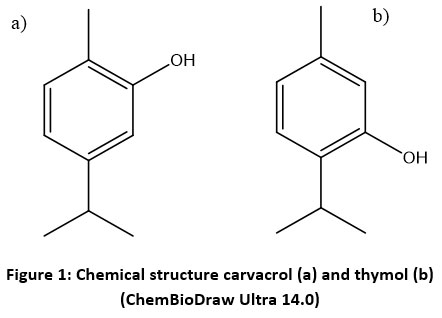 |
Figure 1: Chemical structure carvacrol (a) and thymol (b) (ChemBioDraw Ultra 14.0). |
Melaleuca alternifolia (tea tree) is a plant native to Australia and is highly valued for its medicinal and cosmetic uses, owing to its potent antiseptic, antimicrobial, antimycotic, and antiphlogistic properties9. The active components include various natural organic compounds terpenes, monoterpenes, and sesquiterpenes, with terpinen-4-ol and α-terpineol being central to its antimicrobial effects. The hydroxyl group (-OH) in their structure (Figure 2) is primarily responsible for their biological activity, as it enables interactions with microbial cell membranes, neutralizes free radicals, and modulates inflammatory pathways.
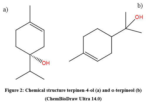 |
Figure 2: Chemical structure terpinen-4-ol (a) and α-terpineol (b) (ChemBioDraw Ultra 14.0). |
Tea tree oil has been found to initiate apoptosis in tumor cells, with terpinen-4-ol being particularly effective in activating the apoptosis pathway. Studies have demonstrated that this bioactive substance inhibits the cancer cell proliferation by increasing oxidative stress and promoting cell death10. Previous study showed that tea tree oil (TTO) from M. alternifolia reduces melanoma cell viability by triggering apoptosis11. Ireland et al. (2012) showed that topically applied 10% M. alternifolia oil in DMSO decrease cell viability in subcutaneous tumor-bearing mice12.
Cinnamomum camphora (camphor tree) of the Lauraceae family, is native to Asia, particularly China, Japan, and Taiwan13. The key compounds of its volatile oil are 1,8-cineole, limonene, alpha-pinene (Figure 3), which demonstrate a broad spectrum of biological effects, including microbe neutralization, inflammation modulation, and tumor suppression growth. The biological activity of 1,8-cineole is primarily attributed to its epoxide functional group. The aromatic compound limonene owes its activity to the conjugated double bonds in its cyclohexene ring, enhancing its antioxidant and anti-cancer potential. Alpha-pinene exhibits antimicrobial and anti-inflammatory effects due to its reactive bicyclic structure and conjugated double bonds, which interact with biological membranes and enzymes. Extracts and individual bioactive compounds from C. camphora have significant cytotoxic effects on various tumor cell types, induced by apoptosis, inhibition of cell proliferation, and modulation of signaling pathways involved in tumor growth. Previous research has indicated that certain compounds of C. camphora essential oil (Camphor EO) possess inhibitory and anti-mutagenic properties against several human cancer cell types14.
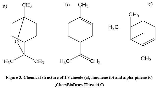 |
Figure 3: Chemical structure of 1,8 cineole (a), limonene (b) and alpha-pinene (c) (ChemBioDraw Ultra 14.0) |
This research intends to assess the cellular toxicity of O. compactum, M. alternifolia, and C. camphora essential oils on human lung carcinoma (H460 cells), cervical adenocarcinoma (HeLa cells), and colorectal carcinoma (HCT116 cells) by evaluation their impact on cell proliferation. There is evident lack of studies focused on the tumor-suppressive potential of these essential oils in the aforementioned cultured cells. The findings will contribute to understanding the efficacy of these plant extracts as natural anticancer agents. Therefore, this study may contribute valuable insights for future research.
Experimental
Essential oils
This study employed the following essential oils:
compactum EO (Pranarom International, Belgium)
Tea tree oil (BIOETERICA, Zagreb, Croatia)
Camphora EO (Volimo prirodno, Mostar, Bosnia and Herzegovina)
Essential oils were prepared by dissolving them in Dulbecco’s Modified Eagle Medium (DMEM) at concentrations of 0.01 μL/mL, 0.05 μL/mL, 0.1 μL/mL, 0.5 μL/mL, and 1 μL/mL. These stock solutions were used for subsequent experimental applications. The MTT test was performed using a microplate reader (Multiskan EX, Thermo Labsystems, Austria), and the standard chemotherapeutic agents used in the control of the MTT test were doxorubicin and etoposide (Sigma Aldrich).
Cell cultures
In this study, we utilized human malignant cell lines commonly employed in toxicological assessments: H460 epithelial lung carcinoma cells, HeLa epithelial cells derived from uterine and cervical adenocarcinoma, and HTC116 epithelial cells from colorectal cancer. These cell lines were selected for their relevance in evaluating the cytotoxic effects of the tested compounds.
Since the cell lines used in this study are adherent, they were cultured in Dulbecco’s modified Eagle medium (DMEM), enriched with 2 mM L-glutamine, 10% fetal bovine serum (FBS), 100 μg/ml streptomycin, and 100 U/ml penicillin, at 37°C with 5% CO2. Upon reaching 80-90% confluence, cells were detached using 0.25% trypsin and resuspended in fresh medium. All procedures were carried out under sterile conditions to avoid contamination.
MTT-based cell viability assay
On the initial day, three study-defined cell lines were plated separately into 96-well microtiter plates at a density of 1.5×10⁴ cells/mL. Essential oils were introduced into the wells at five defined concentrations. Following a 72-hour incubation, cell proliferation was assessed using the MTT (3-[4,5-dimethylthiazol-2-yl]-2,5 diphenyl tetrazolium bromide) assay, which assess the metabolic activity via dehydrogenase in viable cells. This colorimetric assay is based on the measurement of the reduction of yellow tetrazolium salts by mitochondrial enzymes in metabolically active cells, leading to the formation of crystalline purple formazan15. After removing the basal medium, 40 μL of MTT reagent (0.5 μg/μL) was transferred to each well. Following a four-hour incubation, the formed precipitates were solubilized in 160 μL of dimethyl sulfoxide (DMSO), and absorbance readings were taken at 570 nm using a microplate reader (Multiskan EX, Thermo Labsystems, Austria), with the obtained absorbance value being directly proportional to the viability of cancer cells. The percentage of cell growth (PG) for each treatment group was calculated using one of the formulas given bellow.
In case of (At – A0) ≥ 0 the calculation was performed by using the expression:
In case of (At – A0) < 0 the calculation was performed by using the expression:
where:
A0 represents the mean absorbance before the exposure,
At represents the mean absorbance after 72 hours of exposure, and
Ac represents the mean absorbance of untreated cells after 72 hours.
Results were presented as concentration-response curves, with negative values indicating cytotoxic effects. A -100% value corresponds to the total loss of cell viability at the given concentration. Additionally, the growth inhibition (GI50) was also calculated, reflecting the concentration at which 50% of cell growth was inhibited.
Statistical analysis
The normality in data distribution was assessed with the Shapiro-Wilk statistical test, while further verification was carried out via Skewness and Kurtosis analysis, as well as graphical methods, including the Q-Q plot and histogram. Given that the data did not follow a normal distribution, non-parametric statistical approach was employed for subsequent analyses. Specifically, the Kruskal-Wallis test was utilized to evaluate differences among groups, while Spearman’s rank correlation analysis was applied to evaluate the associations between variables.
A p-value of less than 0.05 was considered significant. Statistical analyses were carried out using software provided by IBM SPSS, V21.0.
Results and Discussion
The concentration-dependent variation of PG (%) for O. compactum EO, TTO, and Camphor EO on H460, HeLa, and HCT116 cell cultures are given below.
Citotoxicity of Origanum compactum essential oil
To evaluate the cytotoxic activity of O. compactum EO MTT assay was performed. Cell viability was evaluated in three cell lines H460, HeLa, and HCT116 cells following exposure to the O. compactum EO at five different concentration values (0.1–1 µL/mL), allowing for a dose-dependent assessment of cytotoxic effects. According to the Kruskal-Wallis test, a statistically significant difference in cell viability distribution across concentration groups (p = 0.014) was found indicating that increasing concentrations of O. compactum essential oil significantly affect cell survival.
The treatment of H460, HeLa, and HCT116 cells with increasing concentrations of O. compactum essential oil led to a pronounced, dose-dependent decline in cell viability (Figure 4). Notably, at the highest concentration (1 µL/mL), fewer than 30% of H460 and HeLa cells remained viable, whereas HCT116 cells exhibited greater resistance, maintaining approximately 60% viability (Table 1). The increase in HCT116 cells resistance to O. compactum essential oil compared to H460 and HeLa cells can be attributed to several factors. These include complex mechanisms of drug resistance in colon cancer cells with enhanced antioxidant defense and increased expression of p-glycoprotein transporter. Additionally, HCT116 cells harbor TP53 mutations, which can impair apoptosis, and may have slower proliferation rates or metabolic adaptations that reduce susceptibility to cytotoxic agents16,17.
Table 1: Dose-response profile for O. compactum essential oil tested in vitro on H460, HeLa and HCT116 cell lines
| Concentration (µL/mL) | H460 | HeLa | HCT116 |
| 0.01 | 101.4 | 107.7 | 85.4 |
| 0.05 | 87.2 | 79.6 | 62.6 |
| 0.1 | 62.6 | 53.4 | 28.5 |
| 0.5 | -82.0 | -43.0 | -42.2 |
| 1.0 | -74.8 | -72.8 | -38.1 |
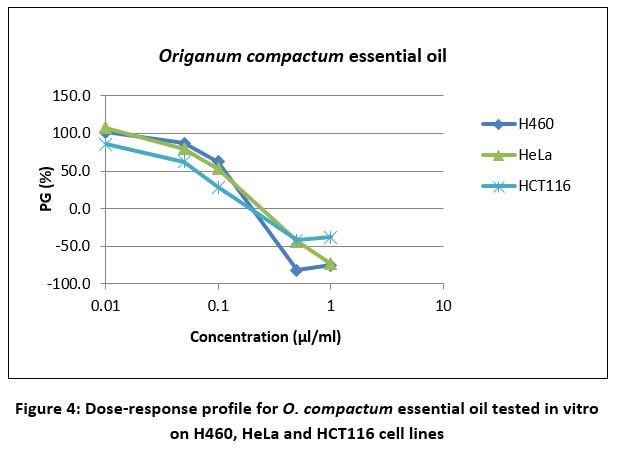 |
Figure 4: Dose-response profile for O. compactum essential oil tested in vitro on H460, HeLa and HCT116 cell lines |
Although HCT116 cells appeared more resistant to O. compactum essential oil as a potential cytotoxic agent, statistical analysis did not confirm this observation. No statistically significant difference in cell viability among the three examined cell lines (p = 0.993) was found by the Kruskal-Wallis test indicating that the distribution of percentage of cell growth (%) was consistent across all groups. Furthermore, Spearman’s rank correlation analysis demonstrated a strong negative correlation (ρ = -0.912, p = 1), suggesting an inverse association between the tested variables. These findings indicate that despite variations in cell lines, no significant difference in cytotoxicity was found, while the observed correlation reflects a strong negative trend without statistical significance (Figure 5).
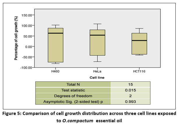 |
Figure 5: Comparison of cell growth distribution across three cell lines exposed to O.compactum essential oil. |
Citotoxicity of Melaleuca alternifolia essential oil
Recent studies have explored the potential anticancer effects of M. alternifolia essential oil. Topically applied, it demonstrates direct cytotoxicity against subcutaneous tumors in mice, suggesting its potential as a topical anticancer treatment12. In vitro studies showed that M. alternifolia essential oil exhibites strong cytotoxicity against human melanoma and squamous carcinoma cell lines, inducing apoptosis and cell cycle arrest18. Furthermore, TTO which contains terpinen-4-ol, synergistically enhance the effectiveness of targeted melanoma therapies by activating apoptosis11. These observations highlight the utility of TTO as an anticancer agent, particularly for skin cancers. However, there is evident lack of research focusing on other carcinoma cell lines.
In the current study, MTT assay of H460, HeLa, and HCT116cell lines following 72-hour incubation with five different concentrations of M. alternifolia essential oil in a microtiter plate demonstrated a dose-dependent response. A significant increase in cytotoxicity was observed with increasing oil concentration, with the most pronounced effect occurring when the concentration levels were adjusted from 0.5 to 1 μL/mL (Figure 6). According to the Kruskal-Wallis test, a notable difference was detected in cell viability across the concentration groups (p = 0.017), indicating that increasing concentrations of tea tree oil markedly influence cell survival.
The in vitro cytotoxicity results of M. alternifolia essential oil on H460, HeLa, and HCT116 cancer cells are outlined in Table 2. At concentrations between 0.01 and 0.1 μL/mL, the essential oil had no significant effect on cell viability. However, exposure to 0.5 μL/mLinduced a cytotoxic response in all three cell lines, with HCT116 exhibiting the highest sensitivity, while H460 was the least sensitive, respectively. After incubation with 0.5 μL/mL of M. alternifolia essential oil, the percentage of cell growth in H460, HeLa, and HCT116 cells was 78.4%, 56.3%, and 42.8%, respectively. Cell viability declined further with increasing concentrations, with the highest concentration (1 μL/mL) exhibiting the strongest cytotoxic effect across all three cell lines.
Table 2: Dose-response profile for M. alternifolia essential oil tested in vitro on H460, HeLa and HCT116 cell lines
| Concentration (µL/mL) | H460 | HeLa | HCT116 |
| 0.01 | 101.1 | 101.5 | 99.1 |
| 0.05 | 95.5 | 100.6 | 87.6 |
| 0.1 | 92.0 | 100.9 | 84.9 |
| 0.5 | 78.4 | 56.3 | 42.8 |
| 1.0 | -36.7 | -34.3 | -44.5 |
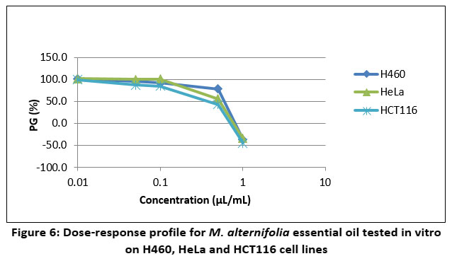 |
Figure 6: Dose-response profile for M. alternifolia essential oil tested in vitro on H460, HeLa and HCT116 cell lines |
The three cell lines demonstrated similar sensitivity to M. alternifolia essential oil, as supported by data interpretation. The Kruskal-Wallis test suggested no considerable difference in cell viability across the cell lines (p = 0.482), implying that the distribution of cell growth percentages was uniform among the groups. Additionally, Spearman’s rank correlation analysis showed a very weak negative correlation (ρ = -0.187, p = 1), suggesting a slight inverse relationship between the variables, though the correlation was not statistically significant (Figure 5).
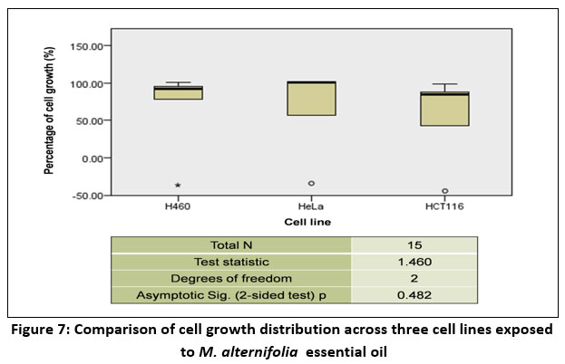 |
Figure 7: Comparison of cell growth distribution across three cell lines exposed to M. alternifolia essential oil. |
Citotoxicity of Cinnamomum camphora essential oil
Cell proliferation was evaluated with the MTT colorimetric protocol, where the reduction of MTT to formazan reflects viable cell metabolism following exposure to C. Camphora essential oil. The results of the in vitro MTT assay on H460, HeLa, and HCT116 cancer cell lines are shown in Table 3 and Figure 8. The essential oil reduced cell viability in a dose-dependent manner. The percentage of cell growth was similar for HeLa and HCT116 cells, while it differed for H460 cells. Cytotoxicity of C. Camphora essential oil was evaluated at five different concentrations. At lower concentrations (0.01-0.1 μL/mL), all three cell lines exhibited similar sensitivity, but concentrations above 0.5 μL/mL induced significantly higher cytotoxic effects in H460 cells compared to HeLa and HCT116 cells. Researchers have shown that essential oils demonstrate cytotoxic effects on various cancer cell lines, with different cell types showing varying sensitivities19,20,21. H460 cells, in particular, exhibit increased sensitivity due to the complex interplay of cellular defense mechanisms, oxidative stress, and apoptosis. The findings from Wu et al. (2010), Lu et al. (2011) further highlight the intricate mechanisms that contribute to the H460 cells’ heightened sensitivity to cytotoxic agents, including essential oils22,23.As shown in Table 3, more than 70% of H460 cells did not survive the 72-hour incubation with 1 μL/mL C. Camphora essential oil, while the percentage of cell growth for HeLa and HCT116 cells was -3.9% and 2%, respectively.
Table 3: Dose-response profile for C. camphora essential oil tested in vitro on H460, HeLa and HCT116 cell lines
| Concentration (µL/mL) | H460 | HeLa | HCT116 |
| 0.01 | 98.3 | 100.0 | 100.6 |
| 0.05 | 82.2 | 96.6 | 98.4 |
| 0.1 | 73.0 | 88.9 | 85.1 |
| 0.5 | 31.7 | 78.2 | 67.4 |
| 1.0 | -72.0 | -3.9 | 2.0 |
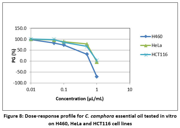 |
Figure 8: Dose-response profile for C. camphora essential oil tested in vitro on H460, HeLa and HCT116 cell lines. |
As shown in Fig. 9, although the cytotoxicity of the essential oil was observed different for H460 cells, there was no statistical difference within the tree cell lines (p=0.543). The three cell lines exhibited similar sensitivity to C. camphora essential oil, as confirmed by statistical analysis. As shown in Figure 9, while the cytotoxicity of the essential oil appeared different for H460 cells, the difference between the three cell lines was not statistically significant (p = 0.543). Furthermore, Spearman’s rank correlation analysis indicated a weak positive correlation (ρ = 0.265), suggesting a slight direct relationship between the variables, though this correlation was not statistically significant (p = 0.341).
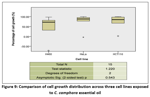 |
Figure 9: Comparison of cell growth distribution across three cell lines exposed to C. camphora essential oil. |
Growth inhibition effects of essential oils on H460, HeLa and HCT116
The GI50 values were determined for M. alternifolia, O. compactum, and C. camphora essential oils across H460, HeLa, and HCT116 cell lines. The findings revealed variations in the sensitivity of the cell lines to the essential oils (Table 4).
Table 4: GI50 values for essential oils
| Cell lines | O. compactumGI50 (nL/mL) | M.alternifoliaGI50 (nL/mL) | C. camphoraGI50 (nL/mL) |
| Lung carcinoma (H460) | 109±34 | 646±114 | 373±131 |
| Cervical adenocarcinoma (HeLa) | 73±23 | 432±43 | 760±210 |
| Colorectal carcinoma (HCT116) | 154±76 | 539±16 | 750±163 |
For the H460 cells, the GI50 values were 646 ± 114 nL/mLfor M. alternifolia, 109 ± 34 nL/mLfor O. compactum, and 373 ± 131 nL/mLfor C. camphora. The O. compactum essential oil exhibited the lowest GI50, suggesting that these cells were most sensitive to its cytotoxic effects, followed by C. camphora and M. alternifolia.
In the HeLa cells, the GI50 values were 432 ± 43 nL/mLfor M. alternifolia, 73 ± 23 nL/mLfor O. compactum, and 760 ± 210 nL/mLfor C. camphora. Notably, O. compactum again showed the lowest GI50, indicating a higher potency compared to the other two essential oils. In contrast, C. camphora was the least effective in reducing HeLa proliferation, requiring the highest concentration for a 50% inhibition of cell growth.
For the HCT116 colorectal carcinoma cells, the GI50 values were 539 ± 16 nL/mL for M. alternifolia, 154 ± 76 nL/mL for O. compactum, and 750 ± 163 nL/mL for C. camphora. Here, M. alternifolia showed moderate cytotoxicity, while O. compactum was more potent than C. camphora, which again showed the weakest inhibition at higher concentrations.
These results clearly show that the essential oils demonstrated varying degrees of growth inhibition across different cancer cell lines. The highest growth inhibition (lowest GI50) was observed in HeLa cells treated with O. compactum (GI50 = 73 ± 23 nL/mL), while the lowest growth inhibition showed C. camphora (GI50 = 760 ± 210 nL/mL). In general, O. compactum exhibited the strongest cytotoxic effects across multiple cell lines, while M. alternifolia and C. camphora showed variable effectiveness depending on the cancer type.
Table 5: GI50 for standard chemotherapeutics (doxorubicin and etoposide)
| Standard chemoterapeutic agent | H460 (ng/mL) | HCT116 (ng/mL) | HeLa (ng/mL) |
| Doxorubicin | 5.18 | 22.03 | 1.74 |
| Etoposide | 324.70 | 1294.76 | 82.40 |
Although doxorubicin and etoposide were used as controls in the MTT assay, they can also serve for comparing the results of essential oils. Doxorubicin is the most effective of all the tested compounds in all cell lines (with the lowest GI50 values), confirming its high effectiveness as a chemotherapy drug. Among all the essential oils, O. compactum stands out as particularly effective, especially in H460 and HeLa cells, with GI50 values comparable to or even lower than those of doxorubicin.
Essential oils are typically used in low concentrations; however, they are often combined with other compounds that share similar mechanisms of action in pharmaceuticals, cosmetics, and food products, potentially increasing human exposure and health risks. Substances targeting the same cellular pathways may exhibit additive, synergistic, potentiating, or even antagonistic effects, emphasizing the need for an integrative approach in cytotoxicity assessment24,25. Future research should investigate interactions between active phytochemicals in essential oils and conventional anticancer drugs to optimize combination therapies while minimizing toxicity26. Additionally, environmental factors such as oxidative stress, inflammation, and microbiome composition may influence the bioactivity and metabolism of these compounds, underscoring the importance of comprehensive studies to ensure both therapeutic efficacy and safety.
Conclusion
This study demonstrated that the cytotoxic effects of O. compactum, M. alternifolia, and C. camphora essential oils vary across different cancer cell lines, highlighting their potential as natural anticancer agents. O. compactum exhibited the strongest inhibitory effects, particularly in cervical (HeLa) and lung (H460) cancer cells, suggesting its potential as a promising candidate for further research. It is evident that aromatic compounds like carvacrol and thymol exert a strong influence and are of considerable significance.
M. alternifolia showed moderate cytotoxicity, indicating potential therapeutic applications, although with lower efficacy compared to O. compactum. Conversely, C. camphora demonstrated the weakest cytotoxic effects, which may limit its use as a standalone treatment but suggests potential as an adjuvant in combination therapies.
To fully assess the clinical potential of these essential oils, further toxicological studies are necessary to establish safe and effective dosing regimens while minimizing risks of irritation and systemic side effects, especially for topical, rectal, or vaginal applications. Additionally, future research should explore the possibility of combining these essential oils with conventional anticancer treatments to enhance therapeutic efficacy through potential synergistic interactions.
Acknowledgment
Our research was funded by the Ministry of Education and Science of Tuzla Canton, Bosnia and Herzegovina (no.10/1-11-21343-5/23). The authors would like to thank the Ruđer Bošković Institute from Zagreb (Croatia) for their help with the experimental part of the work.
Conflicts of Interest
The mentioned authors have no conflicts of interest regarding the publication of this paper.
References
- de Sousa, D.P.; Damasceno, R.O.S.; Amorati, R.; Elshabrawy, H.A.; de Castro, R.D.; Bezerra, D.P.; Nunes, V.R.V.; Gomes, R.C.; Lima, T.C. Essential oils: Chemistry and pharmacological activities. Biomolecules. 2023, 13(7),1144.
CrossRef - Vostinaru, O.; Heghes, S.C.; Filip, L. Safety profile of essential oils – Essential Oils-Bioactive Compounds. New Persp Appl. 2020, 27, 1-3.
CrossRef - Bouyahya, A.; Guaouguaou, F.E.; Dakka, N.; Bakri, J. Pharmacological activities and medicinal properties of endemic Moroccan medicinal plant Origanum compactum (Benth) and their main compounds. Asian Pac. J.Trop. Dis. 2017, 7, 628-40.
- Bouyahya, A.; Jamal, A.; Edaoudi, F.; Et-Touys, A.; Bakri, Y.; Dakka, N. Origanum compactum Benth: A review on Phytochemistry and Pharmacological Properties. Med Aromat Plants. 2016, 5, 4.
CrossRef - Ibišević, M.; Husejnagić, D.; Kazanović, R.; Arsić, I. Antibacterial Activity of Origanum Compactum Essential Oil Tested on Vaginal and Cervical Clinical Bacterial Strains. Acta Fac Med Naiss. 2019, 36(3), 219-228.
CrossRef - Bouyahya, A.; Zengin, G.; Belmehdi, O.; Bourais, I.; Chamkhi, I.; Taha, D.; Bakri, Y. Origanium compactum Benth., from tradicional use to biotechnological applications. J.Food Biochem. 2020; 44(8), 13251.
CrossRef - Wahid, C.; Bouchra, M.; Mohamed, H. Antiproliferative activity of Origanum compactum extract on lung cancer and hepatoma cells. Arab.J.Med.Aromat.Plants. 2015, 1(1), 1-13.
- 8.El Finou, H.; Bammou, M.; Mohti, H.; Abdelhamid, Z.; El Rhaffari, L. Antiproliferative, anti-inflammatory, and antioxidant activity of aqueous extracts from Origanum compactum benth aerial part. Vegetos. 2024, 37, 1257–1263.
CrossRef
- Kairey, L.; Agnew, T.; Bowles, E.Y; Barkla, B.J.; Wardle, J.; Lauche, R. Efficacy and safety of Melaleuca alternifolia (tea tree) oil for human health: A systematic review of randomized controlled trials. Front Pharmacol. 2023, 14, 1116077.
CrossRef - Greay, S. J.; Ireland, D. J.; Kissick, H. T; Levy, A.; Beilharz, M.W.; Riley, T.V.; Carson, C.F. Induction of necrosis and cell cycle arrest in murine cancer cell lines by Melaleuca alternifolia (tea tree) oil and terpinen-4-ol. Cancer Chemother Pharmacol. 2010, 65(5), 877–888.
CrossRef - Di Martile, M.; Garzoli, S.; Sabatino, M.; Valentini, E.; D`Aguanno, S.; Ragno, R.; Del Bufalo, D. Antitumor effect of Melaleuca alternifolia essential oil and its main component terpinen-4-ol in combination with target therapy in melanoma models. Cell death dis. 2021, 7(1), 127.
CrossRef - Ireland, D.J; Greay, S.J.; Hooper, C.M.; Kissick, H.T.; Filion, P.; Riley, T.V.; Beilharz, M.W. Topically applied Melaleuca alternifolia (tea tree) oil causes direct anti-cancer cytotoxicity in subcutaneous tumour bearing mice. J. Dermatol. Sci. 2012, 67(2), 120-9.
CrossRef - Lee, S.H.; Kim, D.S.; Park, S.H.; Park, H. Phytochemistry and Applications of Cinnamomum camphoraEssential Oils. Molecules. 2022, 27(9), 2695.
CrossRef - Hamidpour, R.; Hamidpour, S.; Hamidpour, M.; Shahlari, M. Camphor (Cinnamomum camphora), a traditional remedywith the history of treating several diseases. Int. J. Case Rep. Images. 2013, 4(2), 86-90.
CrossRef - Horozić, E.; Ademović,; Dautović, E.; Kolarević, L,; Cilović Kozarević, E.; Karić, E.; Ibišević, M.; Huseinović, E.; Husejnagić, D. Evaluation of antioxidant, antibacterial and cytotoxic effects of pink pepper fruit essential oil. Glas. hem. tehnol. Bosne Herceg. 2024, 62,1-6.
- Adamsen, B.L.; Kravik, K.L.; Clausen, O.P.; De Angelis, P.M. Apoptosis, cell cycle progression and gene expression in TP53-depleted HCT116 colon cancer cells in response to short-term 5-fluorouracil treatment. Int. J. Oncol. 2007, 31(6), 1491-500.
CrossRef - Kadioglu, O.; Saeed, M.; Mahmoud, N.; Azawi, S.; Mrasek, K.; Liehr, T.; Efferth, T. Identification of potential novel drug resistance mechanisms by genomic and transcriptomic profiling of colon cancer cells with p53 deletion. Arch. Toxicol. 2021, 95(3), 959-74.
CrossRef - Ramadan, M.A.; Shawkey, A.E.; Rabeh, M.A.; Abdellatif, A.O. Expression of P53, BAX, and BCL-2 in human malignant melanoma and squamous cell carcinoma cells after tea tree oil treatment in vitro. Cytotechnology. 2019, 71, 461-73.
CrossRef - Özkan, A.; Erdoğan, A. Evaluation of cytotoxic, membrane, and DNA damaging effects of Thymus revolutus Célak essential oil on different cancer cells. Turk. J. Med. Sci. 2017, 47(2), 702-14.
CrossRef - Benedetti, S.; Nasoni, M.G.; Luchetti, F.; Palma, F. New insights into the cytotoxic effects of Thymus vulgaris essential oil on the human triple-negative breast cancer cell line MDA-MB-231. Toxicol. in Vitro. 2023, 93, 105705.
CrossRef - Trang, D.T.; Hoang ,T.K.; Nguyen, T.T.; Van Cuong, P.; Dang, N.H.; Dang, H.D.; Nguyen Quang, T.; Dat, N.T. Essential oils of lemongrass (Cymbopogon citratus Stapf) induces apoptosis and cell cycle arrest in A549 lung cancer cells. BioMed. Res. Int. 2020, 1, 5924856.
CrossRef - Wu, S.H.; Hang, L.W.; Yang, J.S.; Chen, H.Y.; Lin ,H.Y.; Chiang, J.H.; Lu, C.C.; Yang, J.L.; Lai, T.Y.; Ko, Y.C.; Chung, J.G. Curcumin induces apoptosis in human non-small cell lung cancer NCI-H460 cells through ER stress and caspase cascade-and mitochondria-dependent pathways. Anticancer Res. 2010, 30(6), 2125-33.
CrossRef - Lu, H.F.; Chie, Y.J.; Yang, M.S.; Lu, K.W.; Fu, J.J.; Yang, J.S; Chen, H.Y.; Hsia, T.C.; Ma, C.Y.; Ip ,S.W.; Chung, J.G. Apigenin induces apoptosis in human lung cancer H460 cells through caspase-and mitochondria-dependent pathways. Hum. Exp. Toxicol. 2011, 30(8), 1053-61.
CrossRef - Dautović, E.; Šljivić Husejnović, M.; Bergant, M.; Sabitović, D.; Srabović, N.; Smajlović, A.; Begić, L.; Softić, A. Lead and cadmium induced cytotoxic and genotoxic effects on HL-60 and Jurkat cell lines. Genet. Appl. 2019, 3(1), 57-64.
CrossRef - Šljivić Husejnović, M.; Bergant, M.; Janković, S.; Žižek, S.; Smajlović, A.; Softić, A.; Musić O.; Antonijević, B. Assessment of Pb, Cd and Hg soil contamination and its potential to cause cytotoxic and genotoxic effects in human cell lines (CaCo-2 and HaCaT). Environ. Geochem. Health. 2018, 40, 1557-72.
CrossRef - Šljivić Husejnović, M.; Dautović, E.; Softić, A.; Džambić, A.; Halilčević, D.; Karić, E. Evaluation of the synergistic cytotoxic effect of prednisone and acetaminophen against HeLa cells. World J. Pharm. Pharm. Sci. 2022, 11(11), 84-95.
Accepted on: 08 Apr 2025


ISSN Online: 2231-5039

![]()


