Determination of Free Cysteine Levels and Measurement of Certain Biochemical Markers in Blood Sera from Women with Polycystic Ovary Syndrome (A Pilot Study Supporting the PMOS Hypothesis).
 , Noor Abdul-Khalq Abas1 and Nashwan Hussein Ali2
1Department of Biotechnology, College of Applied Science, University of Samarra, Samarra, Iraq
2Department of Applied Chemistry, College of Applied Sciences, University of Samarra, Samarra, Iraq
Corresponding Author E-mail: Aumrthaarjoad@uosamarra.edu.iq
Download this article as:
ABSTRACT:Polycystic ovary syndrome is a multifaceted endocrine and metabolic disorder in women of reproductive age, associated with menstrual irregularities, infertility, hyperandrogenism, oxidative stress, type 2 diabetes, and insulin resistance. In this study, we examined the relationship between the oxidative stress axis and the metabolic axis to evaluate the PMOS hypothesis. We collected 120 venous blood samples and assigned them to two groups: 60 from affected participants and 60 from unaffected participants. Clinical assessments were performed using a variety of ELISA kits and a spectrophotometer. The results demonstrated statistically significant differences in the study variables between the affected and healthy groups, as well as statistically significant positive correlations between cysteine and fasting blood glucose levels, and between cysteine and insulin resistance. Overall, these findings indicate that the syndrome’s complications are not confined to the ovaries, but also encompass stress- and metabolism-related disorders and complications, alongside other systemic conditions.
KEYWORDS:Cysteine; Glutathione; Oxidative stress; PCOS
Polycystic ovary syndrome
Is a medical condition that affects many women and targets the ovaries; it is a common disorder affecting women of reproductive age. The effects of this disorder are not limited to the ovaries alone, but also affect reproductive and mental health, and it also causes metabolic disorders as well as affecting the blood vessels and the heart.1 This syndrome is characterised by clear symptoms such as acne, hirsutism, obesity, as well as other symptoms such as increased In androgen secretion, insulin resistance, and changes in the shape of the ovaries due to the appearance of cysts ovulation disorder metabolic syndrome and others. This diversity of disorders and symptoms has presented clinicians with significant challenges in determining the appropriate treatment, as symptoms vary from one patient to another, in addition to individual differences in general health. As for clinical diagnosis, there are numerous diagnostic methods, such as measuring certain serum and urine markers, in addition to some studies that have identified the syndrome at the genomic level.2 Although there has been notable progress in clinical diagnosis for many cases, there are still many cases suffering from diagnostic gaps that prolong the time to diagnosis and consequently affect the duration of treatment.3 This syndrome involves elevated levels of visceral fat, which leads to insulin resistance even in the absence of obesity, resulting in hormonal imbalances in the ovaries, the pituitary gland and the hypothalamus.4 Recently, the term PCOS has come to be scientifically inaccurate, as some affected women do not develop cysts on their ovaries, whilst the disorder encompasses several manifestations, including an imbalance in the hormones of the ovaries, the endocrine system and the hypothalamus. Metabolism and psychological disorders.5 The term PCOS has been changed to Polyendocrine Metabolic Ovarian Syndrome(PMOS) by international consensus, with the participation of 56 academic and clinical organisations, as well as specialists and researchers, which will contribute in future to increased early diagnosis, improved healthcare and enhanced treatment response.6
Biomarkers overviews
Cysteine
Cysteine-L (Cys-L) is a semi-essential amino acid and is therefore one of the building blocks required for protein synthesis. This amino acid contains sulphur in the form of a thiol group (-SH) at the end of its side chain.7,8 The (-SH) group is responsible for the high reactivity of this amino acid; consequently, cysteine is responsible for many of its biological functions in the human body. This amino acid is characterised by its ability to form disulphide bridges, a type of covalent bond. It plays a pivotal role in protein folding and the stability of its tertiary structure, thereby supporting its biological activity. Cysteine is considered a potential contributing factor in the pathophysiology of polycystic ovary syndrome.9 In this context, cysteine plays a vital role; it is a monothiol reducing agent and a biomarker of oxidative stress,10 as well as being the building block of glutathione. Oxidative stress is considered one of the main causes of polycystic ovary syndrome, and here cysteine plays a role in protecting cells from oxidative stress through the synthesis of glutathione, as well as protecting oocytes and granulosa cells. Granulosa cells and oocytes require sufficient levels of glutathione, synthesised from cysteine, to counteract the toxic effects of reactive oxygen species (ROS) consequently, any disruption to these pathways leads to poor oocyte quality and impaired follicle development.11
Blood sugar
Glucose is the primary form of carbohydrate in the body and its main source of energy; it is transported via the blood to the cells under the regulation of the hormone insulin, which is secreted by the pancreas. Therefore, any disorder leading to insufficient insulin secretion or a reduced cellular response to it (insulin resistance) will cause glucose to accumulate in the bloodstream and prevent it from entering the cells properly, resulting in elevated blood sugar levels (hyperglycaemia).12 This metabolic imbalance and insulin resistance constitute one of the most common pathophysiological hallmarks associated with polycystic ovary syndrome (PCOS). Polycystic ovary syndrome (PCOS) is classified as a metabolic and reproductive disorder, and is closely associated with an increased risk of type 2 diabetes and impaired glucose tolerance compared with women who do not have the condition. The link between PCOS and type 2 diabetes stems from the presence of both insulin resistance and hyperinsulinaemia, which make the syndrome a precursor to diabetes.13 This condition progresses to full-blown diabetes when there is a total or relative deficiency in insulin levels as a result of a slowing or decline in the compensatory response of the pancreatic beta cells, and their inability to cope with persistent cellular resistance.14
Triglycerides
Triglyceride are hydrophobic (non-polar) lipid molecules, structurally composed of a single glycerol molecule bound to three fatty acids. These molecules are the primary reservoir for energy and fat storage in the human body.Triglycerides are produced mainly via the glycerol phosphate pathway and are stored in the adipose tissue of certain organs, such as the muscles and the liver; the amount stored is influenced by an individual’s nutritional status. Although this biological mechanism is essential for life, any imbalance may lead to serious metabolic disorders, such as obesity, steatosis and hypertriglyceridaemia.15 Similarly, with regard to polycystic ovary syndrome, many women with the condition typically suffer from abnormal lipid levels, such as elevated levels of triglycerides (TG) and low-density lipoprotein (LDL), and reduced levels of high-density lipoprotein (HDL). These abnormalities increase the risk of cardiovascular disease and diabetes by contributing to the development of metabolic syndrome.16
Insulin resistance
Insulin resistance is defined as the reduced ability or inability of tissues containing insulin receptors to be stimulated by insulin. All tissues containing insulin receptors can develop insulin resistance, but the tissues primarily responsible for this resistance are the liver, skeletal muscle and adipose tissue. Insulin resistance impairs glucose utilisation, leading to a compensatory increase in insulin production by beta cells and hyperinsulinaemia.17 Insulin resistance is a common feature among many women with polycystic ovary syndrome. The interactions between hyperandrogenism and hyperinsulinaemia lead to a vicious circle that exacerbates the symptoms of polycystic ovary syndrome, the severity of which varies from patient to patient.It is now well established that polycystic ovary syndrome (PCOS) is usually accompanied by severe insulin resistance and glucose intolerance, as well as obesity. In at least 50 % of women with polycystic ovary syndrome18, insulin resistance is linked to the serine phosphorylation of insulin receptors. This process, mediated by an external factor acting on insulin receptors most likely a serine/threonine kinase is a key mechanism of insulin resistance in humans and is linked to genetic variants that affect insulin receptor signalling. The activity of the P450c17 enzyme, a key regulator of androgen synthesis, is influenced by serine phosphorylation. Consequently, polycystic ovary syndrome in adolescence is considered a model condition for the development of glucose metabolism disorders and insulin resistance.19,20
Materials and Methods
The study samples were collected from Tikrit Teaching Hospital and the Specialist Hospital in Samarra between 15 /11/ 2025 to 17 /2/ 2026. The study comprised 120 blood samples, with 5 ml of blood drawn from each sample. Venous blood was drawn using a single-use medical syringe and collected in a sealed gel tube. The sample was then centrifuged at 3,600 revolutions per minute for 30 minutes. The serum was then extracted and stored in a freezer at –20 °C until the various tests were carried out. Data on these women were collected using a pre-prepared questionnaire; the serum was then drawn using a micro-pipette, divided into several portions, and distributed into Eppendorf tubes. It was stored at –20 °C until biochemical analysis was carried out. Free cysteine was measured using an ELISA kit supplied by Biorbyt Ltd and fasting blood glucose and triglycerides were measured using an analysis kit for a spectrophotometer supplied by BIOLABO SAS, while insulin resistance was assessed using the TYGindex formula, which requires measurement of fasting blood glucose and triglycerides as in the following equation:
“TyG index = Ln [fasting triglyceride (mg/dl) × fasting glucose (mg/dl) /2]”.21
Statical analysis
The present study was statistically analysed using the statistical software SPSS (Statistical Package for the Social Sciences), version (27) , to calculate the mean, standard deviation and p-value (p < 0.001). Correlation analysis was used to determine the strength of the relationship between the study variables.
Results and Discussion
We compared levels of free cysteine, fasting blood glucose and insulin resistance in the blood sera of women with polycystic ovary syndrome. The results of our current study revealed statistically significant differences in all three variables across the study groups, as shown in table 1. The results of our study revealed positive correlations between free cysteine levels and fasting blood glucose levels, as well as a positive correlation between cysteine levels and insulin resistance, as shown in Figure 1 and 2.
Table 1: Mean standard deviation of the study variables for the patient and control groups
|
Group/ |
Mean ± SD Patients |
Mean ± SD control |
P. Value |
|
Cysteine |
9.150±2.241 | 6.547±0.900 |
< 0.001** |
|
Fasting blood sugar |
90.270±12.460 | 79.980±4.478 |
< 0.001** |
|
Triglycerides |
112.950±32.675 | 84.900±8.094 |
< 0.001** |
|
Insulin resistance |
8.486±0.408 | 8.124±0.126 |
< 0.001** |
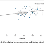 |
Figure 1: Correlation between cysteine and fasting blood glucose Click here to View Figure |
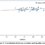 |
Figure 2: Correlation between cysteine and insulin resistance Click here to View Figure |
Table (1) shows a statistically significant increase in cysteine levels. The results of our study are consistent with those of the study conducted by(Ye et al., 2022)22.Which demonstrated a statistically significant increase in cysteine levels in women with polycystic ovary syndrome (PCOS). That study examined 50 amino acids, during which the researchers observed a disturbance in amino acid metabolism and found that levels of most amino acids tended to be elevated, including cysteine. Cysteine is the amino acid that determines the rate of glutathione production; glutathione is considered one of the body’s most powerful antioxidants. Therefore, elevated cysteine levels reflect the cells’ attempt to compensate for a deficiency and combat the increased oxidative stress associated with polycystic ovary syndrome.
Figure 1.shows the study demonstrates a statistically significant positive correlation between cysteine levels and blood glucose levels; the findings of our study are consistent with those reported by( Stabler,. 2020).23 It is believed that this effect results from a deficiency or absence of insulin in the presence of elevated blood glucose levels, coupled with the activation of proteolytic pathways in peripheral tissues, leading to the release of excess sulphur-containing amino acids, including cysteine, into the bloodstream. At the same time, the chronic oxidative stress associated with the hyperglycaemia experienced by women with polycystic ovary syndrome leads to damage to the cell membrane and disruption of amino acid transporter function. This results in impaired cellular uptake of cysteine from the bloodstream, and consequently an increased accumulation of cysteine in the serum.
The findings of our study are also consistent with (Wijekoon et al., 2007)24. who found elevated levels of cysteine and sulphur-containing amino acids; this positive correlation is attributed to the metabolic disturbances associated with diabetes, as growth-promoting hormones, including insulin, play a direct regulatory role in the activation of enzymes involved in the metabolism of sulphur-containing amino acids. In cases of insulin dysregulation, such as in diabetes, the activity of the transsulphurylation pathway increases as a result of the direct hormonal effect that stimulates key enzymes such as cysteathionine beta-synthase (CBS) and betaine homocysteine methyltransferase (BHMT). This excessive enzymatic activity leads to accelerated consumption of homocysteine and its chemical conversion within cells, resulting in the accumulation of cysteine levels in the blood.
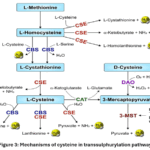 |
Figure 3: Mechanisms of cysteine in transsulphurylation pathway.25 Click here to View Figure |
Whilst our study differs(Akl & Ahmed, 2025)26. That study, the sulphur transport pathway relies primarily on energy (ATP) to activate and convert the amino acid methionine into cysteine However, in the case of hyperglycaemia, this rise leads to an increase in free fatty aacids which in turn leads to increased oxidative stress. This stress disrupts the electron transport chain within the mitochondria, leading to what is known as ‘mitochondrial suffocation’, which results in a reduction in ATP production. Due to the energy deficit caused by diabetes, the activity of the enzymes regulating this pathway is disrupted, most notably cystathionine β-synthase (CBS) and cystathionine γ-lyase (CGL). As a result of the inhibition of these enzymes and the failure of the transsulphurylation pathway to fulfil its function, the cells are unable to synthesise cysteine locally, and consequently cysteine levels decrease.
Table 1 shows that there is a statistically significant increase in triglyceride levels among women with PCOS. The results of our study are consistent with those of (Ulloque-Badaracco et al.,2025)27 and (Zheng et al., 2022)28. who observed a marked increase in triglyceride and cholesterol levels in female patients with polycystic ovary syndrome (PCOS) compared with control groups, suggesting a high prevalence of dyslipidaemia. These patients show a significant increase not only in individual lipid levels, but also in composite metabolic indices and ratios such as the triglyceride-to-glucose index (TyG), the total cholesterol-to-high-density lipoprotein cholesterol ratio (TC/HDL-C), and the triglycerides-to-high-density lipoprotein cholesterol ratio (TG/HDL-C). Physiologically, this dyslipidaemia is attributed to the interaction between insulin resistance and hyperandrogenaemia; insulin resistance leads to increased lipolysis in peripheral tissues and increased production of low-density lipoproteins in the liver, which also explains why these findings are associated with a high body mass index (BMI) and low levels of physical activity in the majority of women with polycystic ovary syndrome.
Table 1. shows a statistically significant increase in the level of insulin resistance among women with PCOS, and the results of our study are consistent with those of (Chen et al., 2025; Purwar and Nagpure, 2022; Szkodziak et al., 2025) 29 , 30,31. The study illustrates how peripheral tissues (such as muscles and the liver) become insulin-resistance in the glucose metabolism pathway which explains the blood glucose readings in many affected women, whilst the ovarian axis remains sensitive to the stimulatory effect of insulin. The resulting compensatory hyperinsulinaemia leads to excessive production of androgens (such as testosterone and DHEAS), which in turn adversely affects insulin sensitivity and secretion, trapping the patient in a vicious cycle associated with an increased lifetime risk of diabetes. The effects of this metabolic imbalance and elevated androgen levels extend to directly affect the central nervous system functions essential for ovulation; Research and histological analyses confirm that insulin resistance physiologically leads to the production of smaller eggs and a marked delay in their production and maturation, a combination of factors that also affects FSH levels and causes menstrual cycle disorders in patients.
Figure 2. This shows a statistically significant positive correlation, with the results of our current study being consistent with those reported by (Dong et al., 2020)32.It has been shown that high levels of cysteine can increase metabolic processes within the body, leading to an increase in hydrogen sulphide (H₂S). This increase acts as a negative regulator that inhibits insulin secretion from pancreatic cells, thereby directly contributing to the development of insulin resistance a prominent feature in the tissues of women with PCOS. Consequently, elevated levels of sulphur-containing amino acids, such as cysteine, disrupt intracellular signalling pathways. Excessive cysteine metabolism is also associated with impaired glutathione (GSH) levels and increased oxidative stress; this imbalance leads to increased production of free radicals and reactive oxygen species within the mitochondria, causing cellular damage that paves the way for metabolic disorders and insulin resistance.
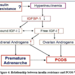 |
Figure 4: Relationship between insulin resistance and PCOS.33 Click here to View Figure |
Conclusion
We conclude that there are statistically significant differences between the study variables; furthermore, free cysteine is an effective marker for assessing oxidative stress in women with PCOS, and the positive correlations between it and metabolic markers (fasting blood glucose, insulin resistance) support the PMOS hypothesis.
Funding Sources
The author(s) received no financial support for the research, authorship, and/or publication of this article.
Conflict of Interest
The author(s) do not have any conflict of interest.
Data Availability Statement
This statement does not apply to this article.
Ethics Statement
This research did not involve human participants, animal subjects, or any material that requires ethical approval.
References
- Dokras A. Polycystic ovary syndrome in 2025—insights and innovations. Fertil Steril. 2025 Nov;124(5):907-9.
CrossRef - Teede HJ, Tay CT, Laven JJE, Dokras A, Moran LJ, Piltonen TT, et al. Recommendations from the 2023 international evidence-based guideline for the assessment and management of polycystic ovary syndrome. J Clin Endocrinol Metab 2023;108:2447–69.
CrossRef - Sung N, Amir J, Alwahab UA, Falcone T. Polycystic ovary syndrome: An update on diagnosis and management. Cleveland Clin J Med. 2026 Mar;93(3):176-183.
CrossRef - Yang Y, Cheng J, Liu C, Zhang X, Ma N, Zhou Z, et al. Gut microbiota in women with polycystic ovary syndrome: an individual based analysis of publicly available data. eClinicalMedicine. 2024 Nov;77:102884.
CrossRef - Teede HJ, Bahri Khomami M, Morman R, Laven JSE, Joham AE, Costello MF, et al. Polyendocrine metabolic ovarian syndrome, the new name for polycystic ovary syndrome: a multistep global consensus process. 2026 Jun 6;407(10545):2329-39.
- Tewari S, Biswas R, Feroz F, Chowdhury N, et al. Polyendocrine Metabolic Ovarian Syndrome (PMOS): A new paradigm in women’s endocrine health. The Pharma Innovation. 2026 Jan;15(5):58-64.
CrossRef - OtasevicV,KoracB.AminoAcids:Metabolism.In:Encyclopedia of Food and Health. Elsevier; 2016. P. 149-155.
CrossRef - Ali V, Behera S, Nawaz A,EqubalA,PandeyK.ChapterThree– Unique thiol metabolism in trypanosomatids: Redox homeostasis and drug resistance. Advances in Parasitology. 2022;117:75-155.
CrossRef - Ponzar N, Pozzi N. Probing the conformational dynamics of thiol-isomerases using non-canonical amino acids and single-molecule FRET. Methods. 2023 Jun;214:8-17.
CrossRef - Liu Y, Yu Z, Zhao S, Cheng L, Man Y, Gao X, et al. Oxidative stress markers in the follicular fluid of patients with polycystic ovary syndrome correlate with a decrease in embryo quality. J Assist Reprod Genet. 2020 Nov;38(2):471-7.
CrossRef - Voros C, Chatzinikolaou F, Papadimas G, Polykalas S, Mavrogianni D, Koulakmanidis AM, et al. Oxidative Stress and SIRT1-Nrf2 Anti-Ferroptotic Pathways in Granulosa Cells: A Molecular Key to Follicular Atresia and Ovarian Aging. IJMS. 2026 Jan 18.
CrossRef - Antar SA, Ashour NA, Sharaky M, Khattab M, Ashour NA, Zaid RT, et al. Diabetes mellitus: Classification, mediators, and complications; A gate to identify potential targets for the development of new effective treatments. Biomed Pharmacother. 2023 Dec;168:115734.
CrossRef - Livadas S, Anagnostis P, Bosdou JK, Bantouna D, Paparodis R. Polycystic ovary syndrome and type 2 diabetes mellitus: A state-of-the-art review. World J Diabetes. 2022 Jan 15;13(1):5-26.
CrossRef - Skelin Klemen M, Kopecky J, Dolenšek J, Stožer A. Human Beta Cell Functional Adaptation and Dysfunction in Insulin Resistance and Its Reversibility. Nephron. 2023 Oct 26.
CrossRef - Viecili PRN, da Silva B, Hirsch GE, Porto FG, Parisi MM, Castanho AR, et al. Chapter One – Triglycerides Revisited to the Serial. Advances in Clinical Chemistry. 2017.
- Mishra N, Mishra P, Kapoor V, Kishun J, Kumar A, Singh U. Lipid variations in different polycystic ovary syndrome phenotypes: A systematic review & meta-analysis. Indian J Med Res. 2025 Jun 30;161(5):491–501.
CrossRef - Freeman AM, Acevedo LA, Pennings N. Insulin Resistance. StatPearls. 2023 Aug 17.
- Zhao H, Zhang J, Cheng X, Nie X, He B. Insulin resistance in polycystic ovary syndrome across various tissues: an updated review of pathogenesis, evaluation, and treatment. Journal of Ovarian Research. 2023;16:9.
CrossRef - Dunaif A. Insulin Resistance and the Polycystic Ovary Syndrome: Mechanism and Implications for Pathogenesis. Endocrine Reviews. 1997 Dec 1;18(6):774-800.
CrossRef - Sun Y, Ji H, Sun W, An X, Lian F. Triglyceride glucose (TyG) index: A promising biomarker for diagnosis and treatment of different diseases. European Journal of Internal Medicine. 2025 Jan;131:3–14.
CrossRef - Bremer AA, Miller WL. The serine phosphorylation hypothesis of polycystic ovary syndrome: a unifying mechanism for hyperandrogenemia and insulin resistance. Fertility and Sterility. 2008 May;89(5):1039-48.
CrossRef - Ye W, Xie T, Di X, Zhou Z, Zhao X, Li Z, et al. Amino acid signatures in relation to polycystic ovary syndrome and increased risk of different metabolic disturbances. Reproductive BioMedicine Online. 2022;45(3):570-579.
CrossRef - Stabler SP. Alterations in Sulfur Amino Acids as Biomarkers of Disease. J Nutr. 2020 Oct;150(Suppl 1):2532S-2537S.
CrossRef - Wijekoon EP, Brosnan ME, Brosnan JT. Homocysteine metabolism in diabetes. Biochem Soc Trans. 2007 Oct;35(5):1175-1179.
CrossRef - Rahman MA, Glasgow JN, Nadeem S, Reddy VP, Saini V, Steyn AJ, et al. The Role of Host-Generated H2S in Microbial Pathogenesis: New Perspectives on Tuberculosis. Front Cell Infect Microbiol. 2020 Nov 27;10:586923.
CrossRef - Akl MM, Ahmed A. Disruption of the transsulfuration pathway as a sulfur-driven etiology of insulin resistance: proinsulin misfolding, disulfide bond deformation, and PDI dysregulation. Explor Endocr Metab Dis. 2025;2:101444.
CrossRef - Ulloque-Badaracco JR, Alarcon-Braga EA, Hernandez-Bustamante EA, Cabrera-Guzmán JC, Dotto-Vasquez G, Maldonado-Basurto GF, et al. Triglyceride–glucose index and lipid ratios in women with and without polycystic ovary syndrome: a systematic review and meta-analysis. Ther Adv Endocrinol Metab. 2025 Mar 31;16:20420188251328840.
CrossRef - Zheng Y, Yin G, Chen F, Lin L, Chen Y. Evaluation of Triglyceride Glucose Index and Homeostasis Model of Insulin Resistance in Patients with Polycystic Ovary Syndrome. International Journal of Women’s Health. 2022 Dec 23;14:1821-1829.
CrossRef - Chen X, Wan Y, Xie L. Insulin resistance in polycystic ovary syndrome: pathophysiological mechanisms of menstrual dysfunction and evidence-based treatment strategies. Biol Reprod. 2025 Dec 16;113(6):1340-1354.
CrossRef - Purwar A, Nagpure S. Insulin Resistance in Polycystic Ovarian Syndrome. Cureus. 2022 Oct 16;14(10):e30351.
CrossRef - Szkodziak P, Szkodziak F, Paszkowski T, et al. Insulin resistance in polycystic ovary syndrome phenotypes and the vicious cycle model in its etiology. Scientific Reports. 2025 Nov 28;15:42649.
CrossRef - Dong Z, Gao X, Chinchilli VM, Sinha R, Muscat J, Winkels RM, Richie JP Jr. Association of sulfur amino acid consumption with cardiometabolic risk factors: Cross-sectional findings from NHANES III. EclinicalMedicine. 2020 Feb 1;19:100248.
CrossRef - Wang HS, Wang TH. Polycystic ovary syndrome (PCOS), insulin resistance and insulin-like growth factors (IGFs)/IGF-binding proteins (IGFBPs). Chang Gung Med J. 2003 Aug 1;26(8).
Accepted on: 19 Sep 2026
Second Review by: Dr. Junaid Ahmed
Final Approval by: Dr. Fozia Z. Haque


ISSN Online: 2231-5039