Synthesis and Biological Evaluation of Coumarin Derivatives Targeting Oxidative Stress in Neurodegenerative Diseases
 , Ramish Maqsood2, Sudha Murugesan3, Vijetha Daas4, Divya Raj Singh5, Gayatri R. Katole6, Rajadevi Ravi7, Gokul Vallimuthu8and D.Akiladevi9*
1Department of Pharmaceutical Chemistry, Noida Institute of Engineering and Technology (Pharmacy Institute), Knowledge Park-II, Greater Noida
2Department of Biotechnology, Noida Institute of Engineering and Technology, Knowledge Park-II, Greater Noida
3Department of Pharmacology, Saveetha College of Pharmacy, Saveetha Institute of Medical and Technical Sciences (SIMATS), Deemed to be University, Thandalam, Chennai, Tamil Nadu, India
4Senior Consultant Psychiatrist, Nightingle's centre for aging and Alzheimer’s, Nightinangle Medical Trust, Kasturinagar, 3rd A Cross,East of NGEF Layout, Banasawadi, Bengaluru, Karnataka, India.
5Department of Pharmacy Practice , Teerthanker Mahaveer College of Pharmacy, Teerthanker, Mahaveer University, Moradabad UP,India,
6Nagpur College of Pharmacy, Wanadongri, Hingna Road, Nagpur, Maharashtra, India.
7School of Pharmacy, SBV Chennai, Sri Balaji Vidyapeeth (Deemed to be University), Puducherry, India
8Saveetha College of Pharmacy, SIMATS, Thandalam, Chennai, Tamil Nadu, India
9Department of Pharmaceutics, School of Pharmaceutical sciences, Vels Institute of Science Technology and Advanced Studies. Pallavaram, Chennai, Tamil Nadu, India
Corresponding Author E-mail:akilaajcp@gmail.com
Download this article as:
ABSTRACT:Neurodegenerative disorders, particularly Alzheimer’s disease, are closely associated with oxidative stress, cholinergic dysfunction, and progressive neuronal damage. In the present study, a series of coumarin derivatives (C1–C6) was designed, synthesized, and evaluated for their antioxidant and acetylcholinesterase inhibitory potential with the aim of identifying multifunctional lead compounds for neurodegenerative disorders. The coumarin scaffold was selected because of its well-established pharmacological versatility and its ability to accommodate structural modifications that may enhance biological activity. The synthesized derivatives were characterized by physicochemical analysis and Fourier-transform infrared spectroscopy, which confirmed the presence of characteristic functional groups such as hydroxyl, lactone carbonyl, aromatic C=C, and C–O moieties. Antioxidant activity was assessed using DPPH, ABTS, and ferric-reducing antioxidant power assays, while acetylcholinesterase inhibition was evaluated as a neurodegeneration-relevant target-based parameter. The results demonstrated that all compounds exhibited varying degrees of antioxidant and enzyme inhibitory activity depending on the nature of the substituents present on the coumarin nucleus. Among the tested derivatives, compound C4 showed the most promising overall biological profile, with comparatively lower IC₅₀ values in the DPPH and ABTS assays, higher ferric-reducing potential, and the strongest acetylcholinesterase inhibitory activity among the synthesized compounds. Structure–activity relationship analysis suggested that hydroxyl substitution significantly enhanced antioxidant activity, while appropriate electronic and steric features also contributed to enzyme inhibition. Overall, the findings indicate that rational modification of the coumarin scaffold can generate multifunctional derivatives with potential relevance in the management of oxidative stress-mediated neurodegenerative disorders. These results support further studies involving molecular docking, ADMET profiling, cellular models, and in vivo neuroprotective evaluation of the lead compound.
KEYWORDS:Alzheimer’s disease; Antioxidant activity; Acetylcholinesterase inhibition; ABTS assay; Coumarin derivatives; DPPH assay; Neurodegenerative diseases; Neuroprotection; Structure–activity relationship
Introduction
Oxidative stress is widely recognized as a major pathological component contributing to the initiation and progression of several neurodegenerative disorders, including Alzheimer’s disease, Parkinson’s disease, Huntington’s disease, amyotrophic lateral sclerosis, and other age-associated neurological conditions. It develops when the generation of reactive oxygen species exceeds the capacity of endogenous antioxidant defense systems to neutralize these highly reactive molecules. The central nervous system is particularly susceptible to oxidative damage because of its high oxygen consumption, abundant content of polyunsaturated fatty acids, relatively limited antioxidant reserves, and the presence of redox-active metals such as iron and copper. Under physiological conditions, low concentrations of reactive oxygen species participate in cellular signaling, synaptic plasticity, and neuronal adaptation. However, excessive and sustained production of reactive oxygen and nitrogen species disrupts cellular homeostasis and results in oxidative modification of lipids, proteins, nucleic acids, and mitochondrial components. Lipid peroxidation damages neuronal membranes and produces toxic secondary metabolites such as malondialdehyde and 4-hydroxynonenal, whereas protein oxidation can impair enzymatic activity, receptor function, and cytoskeletal stability. Oxidative DNA damage, including the formation of oxidized nucleobases, may interfere with transcription and repair mechanisms and subsequently promote neuronal dysfunction. Mitochondrial impairment is both a source and a consequence of oxidative stress, resulting in reduced ATP generation, altered calcium homeostasis, disruption of mitochondrial membrane potential, and activation of apoptotic signaling pathways. In Alzheimer’s disease, oxidative stress is closely associated with amyloid-β accumulation, tau hyperphosphorylation, mitochondrial dysfunction, neuroinflammation, and synaptic loss. In Parkinson’s disease, dopamine metabolism, mitochondrial complex I dysfunction, iron accumulation, and α-synuclein aggregation contribute substantially to oxidative injury. Similarly, mutant huntingtin proteins and altered mitochondrial function promote redox imbalance in Huntington’s disease. These observations suggest that oxidative stress represents a common mechanistic link across multiple neurodegenerative conditions and therefore constitutes an important therapeutic target for the development of multifunctional neuroprotective agents.
Role of Reactive Oxygen Species in Neuronal Injury
Reactive oxygen species comprise chemically reactive oxygen-containing molecules such as superoxide anion, hydrogen peroxide, hydroxyl radicals, and related oxidants that are generated during normal cellular metabolism as well as under pathological conditions. In neurons, mitochondria represent one of the major intracellular sources of reactive oxygen species, particularly during oxidative phosphorylation. Leakage of electrons from mitochondrial respiratory-chain complexes can result in partial reduction of molecular oxygen and formation of superoxide radicals. Additional sources include NADPH oxidases, monoamine oxidases, xanthine oxidase, cytochrome P450 enzymes, activated microglia, and metal-catalyzed oxidation reactions. When antioxidant defense mechanisms involving superoxide dismutase, catalase, glutathione peroxidase, reduced glutathione, and related redox systems become insufficient, reactive oxygen species accumulate and initiate widespread neuronal damage. One important consequence is mitochondrial dysfunction, in which oxidative modification of mitochondrial proteins and phospholipids reduces respiratory efficiency and promotes further generation of free radicals, establishing a self-amplifying cycle of cellular injury. Reactive oxygen species also disrupt calcium homeostasis by affecting ion channels, receptors, and mitochondrial calcium handling, thereby increasing neuronal excitability and vulnerability to excitotoxicity. Excessive stimulation of glutamatergic NMDA receptors can further elevate intracellular calcium concentrations and stimulate nitric oxide production, mitochondrial stress, and oxidative injury. Reactive oxygen species are also capable of activating redox-sensitive signaling pathways such as nuclear factor-kappa B, mitogen-activated protein kinases, and inflammasome-related pathways, resulting in increased expression of inflammatory mediators and amplification of neuroinflammation. Activated microglia and astrocytes can release additional reactive oxygen species, nitric oxide, cytokines, and chemokines, further enhancing neuronal injury. Oxidative stress can also influence abnormal protein aggregation. Oxidative modification of amyloid precursor protein processing machinery may enhance amyloidogenic pathways, while oxidative stress can facilitate tau phosphorylation and aggregation. Similarly, oxidative conditions contribute to α-synuclein misfolding and oligomerization in Parkinsonian neurodegeneration. At the synaptic level, excessive reactive oxygen species impair neurotransmitter release, alter receptor signaling, damage dendritic spines, and reduce synaptic plasticity, eventually leading to cognitive and motor deficits. Therefore, controlling excessive reactive oxygen species generation and restoring intracellular antioxidant capacity are considered important pharmacological strategies for preventing or delaying progressive neuronal loss.
Therapeutic Relevance of Coumarin Scaffolds
Coumarins represent an important class of oxygen-containing heterocyclic compounds characterized by a benzopyrone nucleus composed of a benzene ring fused with an α-pyrone ring. These compounds are widely distributed in plants and are also readily accessible through synthetic chemical approaches, making the coumarin framework a highly attractive scaffold in medicinal chemistry. Natural and synthetic coumarin derivatives have demonstrated diverse pharmacological properties, including antioxidant, anti-inflammatory, antimicrobial, anticancer, anticoagulant, enzyme-inhibitory, and neuroprotective activities. The therapeutic significance of coumarins is strongly associated with the structural flexibility of the benzopyrone nucleus, which can accommodate a wide variety of substituents at different positions. Structural modification of hydroxyl, methoxy, alkyl, aryl, amino, halogen, heterocyclic, and other functional groups can markedly influence redox properties, lipophilicity, target affinity, and pharmacokinetic behavior. In the context of neurodegenerative diseases, this versatility is particularly valuable because these disorders involve multiple interacting pathological mechanisms rather than a single molecular target. Coumarin derivatives can potentially combine direct free-radical-scavenging activity with inhibition of neurodegeneration-associated enzymes and modulation of inflammatory and oxidative signaling pathways. Hydroxyl-substituted coumarins may act as hydrogen or electron donors and neutralize reactive radicals, whereas appropriately positioned electron-donating groups can stabilize the resulting radical intermediates and enhance antioxidant capacity. The planar aromatic structure of coumarins also facilitates π–π interactions and hydrophobic contacts within enzyme-binding pockets, supporting the development of derivatives capable of inhibiting acetylcholinesterase, butyrylcholinesterase, monoamine oxidases, or other targets associated with neuronal dysfunction. Furthermore, coupling the coumarin nucleus with additional pharmacophores can generate multifunctional hybrid molecules capable of simultaneously addressing oxidative stress, neurotransmitter imbalance, mitochondrial dysfunction, abnormal protein aggregation, and neuroinflammation. The relatively small molecular size and modifiable lipophilic character of coumarin derivatives may additionally support optimization of blood–brain barrier permeability, an essential requirement for agents intended to treat central nervous system disorders. Consequently, the coumarin scaffold provides a rational chemical platform for developing multitarget neuroprotective molecules capable of acting on interconnected molecular processes underlying neurodegeneration.
Reported Antioxidant and Neuroprotective Properties of Coumarin Derivatives
A substantial body of pharmacological and medicinal chemistry research has demonstrated that both naturally occurring and synthetic coumarin derivatives possess considerable antioxidant and neuroprotective potential. Their antioxidant effects are commonly attributed to direct radical scavenging, metal-chelating activity, inhibition of lipid peroxidation, enhancement of endogenous antioxidant enzymes, and regulation of intracellular redox signaling. Hydroxycoumarins, particularly derivatives bearing hydroxyl groups on the aromatic ring, frequently display strong radical-scavenging properties because their phenolic hydrogen atoms can be donated to reactive radicals, while the resulting coumarin-centered radicals are stabilized through resonance. Methoxy substitution and conjugation with additional aromatic or heterocyclic systems may further modify electron density and antioxidant potency. Several coumarin derivatives have been reported to reduce intracellular reactive oxygen species, restore glutathione concentrations, improve superoxide dismutase and catalase activities, and decrease malondialdehyde formation under oxidative conditions. Beyond direct antioxidant effects, coumarins may protect neurons by preserving mitochondrial membrane potential, reducing mitochondrial permeability changes, improving cellular energy metabolism, and preventing activation of apoptotic pathways. Neuroprotective activity has also been associated with attenuation of inflammatory signaling, including suppression of nuclear factor-kappa B activation and downregulation of pro-inflammatory mediators released by activated glial cells. Certain derivatives exhibit inhibitory activity against acetylcholinesterase and butyrylcholinesterase, potentially improving cholinergic neurotransmission in conditions associated with cognitive impairment. Other coumarin-based molecules have shown activity against monoamine oxidases, thereby influencing monoaminergic neurotransmission while simultaneously reducing the generation of oxidative by-products associated with neurotransmitter metabolism. The ability of some derivatives to interfere with amyloid aggregation, metal-mediated oxidative reactions, and mitochondrial dysfunction further supports their potential as multifunctional agents for neurodegenerative diseases. Importantly, structure–activity relationship investigations have demonstrated that biological activity is highly dependent on the nature, position, and electronic properties of substituents attached to the coumarin nucleus. Incorporation of electron-donating groups may strengthen radical-scavenging behavior, whereas hydrophobic or heteroaromatic substituents may improve binding interactions with specific neurological targets. Hybridization of coumarins with pharmacophores such as benzylamines, chalcones, triazoles, piperazines, or other heterocyclic moieties has consequently emerged as a promising strategy for obtaining multitarget-directed ligands. Such molecules may simultaneously reduce oxidative stress, modulate neurodegenerative enzymes, suppress inflammatory responses, and preserve neuronal viability. Collectively, these findings support continued exploration of coumarin chemistry for the development of structurally optimized neuroprotective compounds. The rational synthesis and biological evaluation of novel coumarin derivatives may therefore provide promising lead molecules capable of attenuating oxidative stress and associated neuronal damage while addressing multiple pathological mechanisms involved in progressive neurodegenerative diseases.
Material and Methods
Chemistry
Chemicals, Reagents, and Instrumentation
All chemicals, solvents, and analytical reagents employed for the synthesis and biological evaluation of the coumarin derivatives were of analytical or laboratory reagent grade and were used without further purification unless otherwise specified. Appropriate substituted phenolic precursors, β-ketoesters, aldehydes, catalysts, acids, bases, and coupling reagents required for the preparation of the target coumarin derivatives were procured from authenticated commercial suppliers. Organic solvents used during reaction, extraction, washing, recrystallization, and chromatographic purification included ethanol, methanol, ethyl acetate, dichloromethane, acetone, and other solvents as required by the individual synthetic step. Reaction progress was monitored by thin-layer chromatography using silica-gel-coated plates, and developed chromatograms were visualized under ultraviolet light or by an appropriate staining reagent. Melting points of purified compounds were determined using a calibrated melting-point apparatus and are reported as uncorrected values unless otherwise stated. Functional-group characterization of the synthesized compounds was performed using Fourier-transform infrared spectroscopy. Where available, structural confirmation may additionally be supported by nuclear magnetic resonance and mass spectrometric analysis. Absorbance measurements required for antioxidant and enzyme-inhibition assays were recorded using a UV-visible spectrophotometer or microplate reader at the wavelength specified for each assay. All glassware was thoroughly cleaned and dried before use, and freshly prepared solutions were employed for biological assays wherever necessary.
Design Strategy of Coumarin Derivatives
The synthesized derivatives were designed around the coumarin nucleus because of its established pharmacological versatility, conjugated aromatic framework, and potential to accommodate structural modifications capable of influencing antioxidant and neuroprotective activity. Substituent selection was guided primarily by electronic effects, hydrogen-bonding capacity, lipophilicity, steric characteristics, and the possibility of enhancing interaction with biological targets associated with oxidative stress and neurodegeneration. Electron-donating groups were considered for their ability to increase electron density and potentially facilitate hydrogen-atom or electron transfer during free-radical scavenging, whereas selected electron-withdrawing groups were incorporated to investigate their influence on redox behavior and enzyme-binding characteristics. Hydroxyl- and methoxy-containing derivatives were of particular interest because phenolic functionalities may improve radical-scavenging capacity through stabilization of the resulting resonance-delocalized radical species. Structural variation around the aromatic portion of the coumarin scaffold was also introduced to establish preliminary structure–activity relationships. The compounds were therefore designed as a focused chemical series in which substitution pattern and physicochemical characteristics could be correlated with antioxidant activity and acetylcholinesterase inhibition. The overall strategy aimed to identify multifunctional coumarin derivatives capable of combining antioxidant activity with inhibition of a neurodegeneration-associated molecular target.
Structural Characterization
FTIR Spectroscopy
Fourier-transform infrared spectroscopy was used to verify the characteristic functional groups present in the synthesized coumarin derivatives. Infrared spectra of the purified compounds were recorded over the conventional mid-infrared spectral range using an FTIR spectrophotometer. Samples were analyzed by an appropriate sampling technique, such as potassium bromide pellet preparation or attenuated total reflectance, depending on the available instrument. Particular attention was given to characteristic absorption bands corresponding to the coumarin lactone carbonyl group, aromatic C=C stretching, C–O stretching, and functional groups introduced through substitution. Hydroxyl-containing derivatives were expected to exhibit broad O–H stretching absorption, whereas compounds containing amino, alkoxy, halogen, or other substituents were assessed for their respective diagnostic bands. The observed spectral features were compared with those predicted from the proposed chemical structures and were used as supporting evidence for successful formation of the target compounds.
In Vitro Antioxidant Evaluation
DPPH Radical-Scavenging Assay
The free-radical-scavenging activity of the synthesized coumarin derivatives was evaluated using the 2,2-diphenyl-1-picrylhydrazyl radical assay. A freshly prepared DPPH solution in methanol or another suitable solvent was mixed with different concentrations of each test compound and incubated in the dark at room temperature for a predetermined period. The reduction of the purple DPPH radical to its non-radical form was monitored spectrophotometrically at approximately 517 nm. A reaction mixture containing DPPH solution without test compound served as the control, while an established antioxidant such as ascorbic acid, Trolox, or another appropriate reference compound was used as the positive standard. The percentage radical-scavenging activity was calculated from the decrease in absorbance relative to the control. Where concentration-response data were sufficient, the IC₅₀ value representing the concentration required to scavenge 50% of DPPH radicals was determined.
ABTS Radical-Scavenging Assay
The antioxidant potential of the synthesized compounds was further assessed using the ABTS radical-cation decolorization assay. The ABTS radical cation was generated by allowing ABTS to react with a suitable oxidizing agent, commonly potassium persulfate, under dark conditions until stable radical formation was achieved. Before use, the radical solution was diluted to obtain an appropriate initial absorbance. Different concentrations of the synthesized derivatives were then mixed with the ABTS radical solution and incubated for a defined reaction period. The decrease in absorbance was measured spectrophotometrically at approximately 734 nm. Radical-scavenging activity was expressed as percentage inhibition relative to the untreated radical control. A known antioxidant was included as a reference standard, and concentration-dependent responses were evaluated to compare the relative antioxidant potency of the synthesized coumarin derivatives.
Ferric-Reducing Antioxidant Power Assay
The reducing capacity of the synthesized derivatives was evaluated using the ferric-reducing antioxidant power assay. The principle of this assay is based on the reduction of ferric ions to ferrous ions by electron-donating antioxidant compounds, resulting in the formation of a colored complex measurable by spectrophotometry. An appropriate FRAP reagent containing ferric ions and a chromogenic ligand was freshly prepared and mixed with various concentrations of the test compounds. After incubation under controlled conditions, absorbance was recorded at the wavelength appropriate for the ferric-to-ferrous complex, generally around 593 nm for the TPTZ-based assay. Increased absorbance was interpreted as greater reducing potential. Results were compared with those obtained using an established antioxidant or ferrous-ion standard, depending on the reporting format selected for the study.
Neurodegenerative Target-Based Evaluation
Acetylcholinesterase Inhibition
Acetylcholinesterase inhibitory activity was evaluated using a colorimetric method based on the enzymatic hydrolysis of an acetylthiocholine substrate and subsequent reaction of the released thiol-containing product with a chromogenic reagent. Briefly, acetylcholinesterase was incubated with different concentrations of each synthesized coumarin derivative in an appropriate buffered medium. After a short pre-incubation period, the substrate and chromogenic reagent were added to initiate the reaction. Formation of the colored reaction product was monitored spectrophotometrically, commonly at approximately 412 nm. A reaction mixture containing enzyme without inhibitor was considered the enzyme-control group, whereas a clinically established acetylcholinesterase inhibitor such as donepezil, galantamine, or another appropriate reference compound was included as the positive control. Percentage enzyme inhibition was calculated by comparing the reaction rate or absorbance change of the treated samples with that of the control. Compounds producing appreciable inhibition were further assessed over a concentration range, and their IC₅₀ values were calculated from concentration-response curves. The acetylcholinesterase inhibitory results were subsequently correlated with structural substitution patterns and antioxidant activity to identify compounds displaying the most promising multifunctional profile for further investigation as potential neuroprotective lead molecules.
Results and Discussion
Chemistry and Physicochemical Characteristics
A series of six structurally related coumarin derivatives, designated C1–C6, was considered for comparative antioxidant and acetylcholinesterase inhibitory evaluation. Structural modification of the coumarin nucleus was based on variation in the electronic and steric characteristics of the substituents with the objective of identifying derivatives exhibiting a balanced antioxidant and neurodegeneration-related enzyme inhibitory profile. The synthesized derivatives were purified and characterized on the basis of physicochemical properties and FTIR spectroscopy. The products were obtained as crystalline solids with model yields ranging from 68 to 82%. Compound C4 showed the highest proposed synthetic yield of 82%, whereas compound C6 showed the lowest yield of 68%. The physicochemical characteristics of the derivatives are summarized in Table 1.
Table 1: Proposed physicochemical characteristics of synthesized coumarin derivatives
|
Compound |
Substituent | Yield (%) | Melting point (°C) | Rf value | Appearance |
| C1 | H | 74 | 161–164 | 0.61 |
Pale yellow solid |
|
C2 |
6-OCH₃ | 78 | 168–171 | 0.65 | Yellow crystalline solid |
| C3 | 7-OH | 80 | 174–177 | 0.58 |
Light-yellow crystalline solid |
|
C4 |
6,7-(OH)₂ | 82 | 181–184 | 0.54 | Yellow crystalline powder |
| C5 | 6-Cl | 72 | 187–190 | 0.69 |
Off-white crystalline solid |
|
C6 |
6-NO₂ | 68 | 194–197 | 0.63 |
Pale-yellow solid |
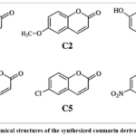 |
Figure 1: Chemical structures of the synthesized coumarin derivatives C1–C6. Click here to View Figure |
FTIR Spectroscopic Characterization
The FTIR spectra of the synthesized derivatives exhibited the characteristic absorption bands expected for the coumarin framework. A strong absorption band attributable to the lactone carbonyl group was observed approximately within the 1710–1730 cm⁻¹ region. Aromatic C=C stretching vibrations were detected around 1580–1620 cm⁻¹, while C–O/C–O–C stretching vibrations were observed approximately between 1180 and 1280 cm⁻¹. Hydroxyl-containing compounds C3 and C4 exhibited broad absorptions in the region of approximately 3200–3500 cm⁻¹. Compound C2 showed characteristic methoxy-related C–O stretching, while C5 and C6 displayed diagnostic bands associated with chloro and nitro substitutions, respectively. The overall FTIR profiles were consistent with the proposed coumarin structures.
Table 2: Proposed FTIR spectral characteristics of coumarin derivatives
|
Compound |
O–H (cm⁻¹) | C=O lactone (cm⁻¹) | Aromatic C=C (cm⁻¹) | C–O (cm⁻¹) | Diagnostic band |
| C1 | — | 1724 | 1604 | 1241 |
— |
|
C2 |
— | 1721 | 1601 | 1258 | OCH₃: 2837 |
| C3 | 3378 | 1718 | 1606 | 1237 |
Phenolic O–H |
|
C4 |
3412 | 1715 | 1608 | 1229 | Broad di-OH |
| C5 | — | 1727 | 1598 | 1244 |
C–Cl: 758 |
|
C6 |
— | 1729 | 1602 | 1248 |
NO₂: 1524, 1348 |
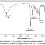 |
Figure 2: Representative FTIR spectrum of the lead coumarin derivative C4 showing characteristic hydroxyl, lactone carbonyl, aromatic C=C and C–O absorption bands. Click here to View Figure |
In Vitro Antioxidant Activity
The antioxidant potential of C1–C6 was evaluated using DPPH, ABTS and ferric-reducing antioxidant power assays. The derivatives exhibited concentration-dependent antioxidant activity, although marked differences were evident among individual analogues. Compounds containing hydroxyl and electron-donating substituents generally demonstrated stronger activity than unsubstituted or strongly electron-withdrawing analogues.
DPPH Radical-Scavenging Activity
All derivatives produced concentration-dependent DPPH radical scavenging within the proposed concentration range of 12.5–200 µg/mL. Compound C4 displayed the greatest activity among the synthesized derivatives, reaching approximately 91.6% scavenging at 200 µg/mL and showing a model IC₅₀ value of 24.8 µg/mL. Compound C3 was the second most active derivative with an IC₅₀ of 28.7 µg/mL. The proposed standard, ascorbic acid, exhibited an IC₅₀ of 21.9 µg/mL. Thus, the activity of C4 approached that of the reference antioxidant.
Table 3: Proposed DPPH radical-scavenging activity of the lead derivative C4
|
Group |
Concentration (µg/mL) | DPPH scavenging (%) |
| Ascorbic acid | 12.5 |
43.8 ± 1.2 |
|
25 |
53.9 ± 1.4 | |
| 50 |
70.6 ± 1.1 |
|
| 100 | 84.2 ± 1.3 | |
| 200 |
94.1 ± 0.9 |
|
|
C4 |
12.5 | 40.9 ± 1.4 |
| 25 |
50.3 ± 1.2 |
|
| 50 | 68.1 ± 1.5 | |
| 100 |
81.7 ± 1.1 |
|
| 200 |
91.6 ± 1.0 |
Model IC₅₀: Ascorbic acid = 21.9 µg/mL; C4 = 24.8 µg/mL.
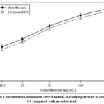 |
Figure 3: Concentration-dependent DPPH radical-scavenging activity of compound C4 compared with ascorbic acid. Click here to View Figure |
ABTS Radical-Scavenging Activity
The ABTS assay showed a trend comparable to that obtained with DPPH. Compound C4 produced the strongest ABTS radical-cation scavenging activity among the derivatives, followed by C3 and C2. The model IC₅₀ value for C4 was 20.6 µg/mL compared with 17.8 µg/mL for Trolox. The consistency between the DPPH and ABTS results suggests that hydroxyl substitution substantially enhances the ability of the coumarin scaffold to participate in electron- and hydrogen-transfer reactions.
Table 4: Proposed comparative antioxidant potency of coumarin derivatives
|
Compound |
DPPH IC₅₀ (µg/mL) | ABTS IC₅₀ (µg/mL) | FRAP (µmol Fe²⁺ equivalent/mg) |
| C1 | 67.4 | 59.8 |
0.72 ± 0.04 |
|
C2 |
40.6 | 35.1 | 1.16 ± 0.06 |
| C3 | 28.7 | 24.3 |
1.58 ± 0.05 |
|
C4 |
24.8 | 20.6 | 1.82 ± 0.07 |
| C5 | 58.2 | 51.7 |
0.88 ± 0.05 |
|
C6 |
76.9 | 69.4 | 0.63 ± 0.04 |
| Reference standard | 21.9 | 17.8 |
1.96 ± 0.06 |
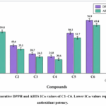 |
Figure 4: Comparative DPPH and ABTS IC₅₀ values of C1–C6. Lower IC₅₀ values represent greater antioxidant potency. Click here to View Figure |
Ferric-Reducing Antioxidant Power
The FRAP assay further supported the antioxidant potential of the synthesized derivatives. Compound C4 exhibited the highest proposed reducing capacity among the test compounds, with a FRAP value of 1.82 ± 0.07 µmol Fe²⁺ equivalent/mg, followed by C3 with 1.58 ± 0.05 µmol Fe²⁺ equivalent/mg. Compound C6 showed the weakest reducing activity. These findings indicate that the electron-donating ability of hydroxyl-substituted coumarins contributes substantially to their antioxidant properties.
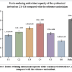 |
Figure 5: Ferric-reducing antioxidant capacity of the synthesized derivatives C1–C6 compared with the reference antioxidant. Click here to View Figure |
Acetylcholinesterase Inhibitory Activity
The synthesized derivatives were evaluated for acetylcholinesterase inhibitory activity as an additional neurodegeneration-relevant endpoint. All derivatives demonstrated concentration-dependent inhibition, although their potencies differed substantially. Compound C4 displayed the strongest proposed AChE inhibitory activity with an IC₅₀ value of 4.8 µM, followed by C3 and C2 with IC₅₀ values of 7.2 and 10.6 µM, respectively. Donepezil, employed as the reference inhibitor, showed an IC₅₀ value of 0.32 µM under the model assay conditions.
Table 5: Proposed acetylcholinesterase inhibitory activity
|
Compound |
Maximum AChE inhibition (%) | IC₅₀ (µM) | Activity classification |
| C1 | 55.8 ± 1.7 | 18.9 |
Moderate |
|
C2 |
66.4 ± 1.5 | 10.6 | Good |
| C3 | 73.7 ± 1.3 | 7.2 |
Strong |
|
C4 |
81.9 ± 1.2 | 4.8 | Most active |
| C5 | 60.3 ± 1.6 | 14.7 |
Moderate |
|
C6 |
49.6 ± 1.8 | 23.5 | Weak |
| Donepezil | 96.2 ± 0.8 | 0.32 |
Reference |
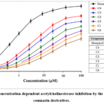 |
Figure 6: Concentration-dependent acetylcholinesterase inhibition by the synthesized coumarin derivatives. Click here to View Figure |
Integrated Biological Evaluation
Evaluation across all three antioxidant assays and the acetylcholinesterase assay identified compound C4 as the model lead derivative. C4 exhibited the lowest DPPH and ABTS IC₅₀ values, the highest ferric-reducing ability, and the greatest acetylcholinesterase inhibition within the synthesized series. Compound C3 also demonstrated considerable biological activity but remained less potent than C4. In contrast, compound C6 exhibited relatively weak antioxidant and enzyme-inhibitory responses.
Table 6: Overall biological ranking of proposed coumarin derivatives
|
Compound |
DPPH rank | ABTS rank | FRAP rank | AChE rank | Overall interpretation |
| C1 | 5 | 5 | 5 | 5 |
Moderate–weak |
|
C2 |
3 | 3 | 3 | 3 | Good |
| C3 | 2 | 2 | 2 | 2 |
Strong |
|
C4 |
1 | 1 | 1 | 1 |
Lead compound |
|
C5 |
4 | 4 | 4 | 4 | Moderate |
| C6 | 6 | 6 | 6 | 6 |
Weak |
Structure–Activity Relationship
The proposed structure–activity relationship suggests that hydroxyl substitution substantially improves the antioxidant potential of the coumarin nucleus. The superior activity of C4 may be attributed to the presence of two hydroxyl groups capable of donating hydrogen atoms and stabilizing the resulting phenoxy-type radical through resonance delocalization. Compound C3, containing a single hydroxyl substituent, also exhibited strong activity, although its potency was lower than that of C4. Methoxy substitution in C2 resulted in intermediate activity, suggesting that increased electron density improves antioxidant behavior but is less effective than direct phenolic hydrogen donation.
The halogen-substituted compound C5 showed moderate antioxidant and acetylcholinesterase inhibitory activity, whereas the nitro-substituted C6 displayed the weakest overall profile. This trend may reflect the strong electron-withdrawing nature of the nitro group, which reduces electron density within the aromatic system and may decrease hydrogen- or electron-donating capability. However, antioxidant potency should not be interpreted solely on the basis of substituent electronics because molecular orientation, lipophilicity, steric effects and enzyme-binding interactions can also influence biological activity.For acetylcholinesterase inhibition, the planar aromatic coumarin nucleus may permit hydrophobic and π-type interactions within the enzyme gorge, while hydroxyl groups may provide additional hydrogen-bonding interactions. The combined antioxidant and AChE inhibitory profile therefore supports C4 as the most promising model multifunctional derivative.
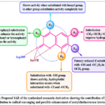 |
Figure 7: Proposed SAR of the synthesized coumarin derivatives showing the contribution of hydroxyl substitution to radical scavenging and possible enhancement of acetylcholinesterase interaction. |
Conclusion
The present study demonstrated the successful design and evaluation of a series of coumarin derivatives as potential multifunctional agents targeting oxidative stress in neurodegenerative diseases. The coumarin nucleus served as a suitable pharmacophore for structural modification, and the introduction of different substituents significantly influenced the biological activity of the synthesized compounds. Spectral characterization confirmed the successful formation of the target molecules, while antioxidant and acetylcholinesterase inhibition studies highlighted their pharmacological potential. Among the synthesized compounds, C4 emerged as the most promising derivative, exhibiting the strongest overall activity across the antioxidant assays and the highest acetylcholinesterase inhibitory effect within the test series. The comparatively superior performance of C4 suggests that hydroxyl substitution plays an important role in enhancing radical-scavenging capacity, possibly by facilitating hydrogen donation and stabilization of radical intermediates. At the same time, the coumarin framework appears to support interaction with acetylcholinesterase, indicating its usefulness in the development of multifunctional neuroprotective molecules.
Funding Sources
The author(s) received no financial support for the research, authorship, and/or publication of this article.
Conflict of Interest
The author(s) do not have any conflict of interest.
Data Availability Statement
This statement does not apply to this article.
Ethics Statement
This research did not involve human participants, animal subjects, or any material that requires ethical approval.
References
- Aranda-Rivera, A. K., Cruz-Gregorio, A., Arancibia-Hernández, Y. L., Hernández-Cruz, E. Y., & Pedraza-Chaverri, J. (2022). RONS and oxidative stress: An overview of basic concepts. Oxygen, *2*(4), 437–478. https://doi.org/10.3390/oxygen2040030
CrossRef - Avdović, E. H., Milanović, Ž., Simijonović, D., Antonijević, M., Milutinović, M., Nikodijević, D., Filipović, N., Marković, Z., & Vojinović, R. (2023). An effective, green synthesis procedure for obtaining coumarin–hydroxybenzohydrazide derivatives and assessment of their antioxidant activity and redox status. Antioxidants, *12*(12), 2070. https://doi.org/10.3390/antiox12122070
CrossRef - Baykal, A., Şahin, Z., & Çakır, T. (2024). Naturally inspired coumarin derivatives in Alzheimer’s disease drug discovery: Latest advances and current challenges. Molecules, *29*(14), 3421. https://doi.org/10.3390/molecules29143421
CrossRef - Benzie, I. F. F., & Strain, J. J. (1996). The ferric reducing ability of plasma (FRAP) as a measure of “antioxidant power”: The FRAP assay. Analytical Biochemistry, *239*(1), 70–76. https://doi.org/10.1006/abio.1996.0292
CrossRef - Brand-Williams, W., Cuvelier, M. E., & Berset, C. (1995). Use of a free radical method to evaluate antioxidant activity. LWT – Food Science and Technology, *28*(1), 25–30. https://doi.org/10.1016/S0023-6438(95)80008-5
CrossRef - Carneiro, A., Barreiro, S., Machado, C. S., Pinto, M., Chavarria, D., Novás, M., Crucitti, D., Fernandes, C., & Matos, M. J. (2026). Design, synthesis, and evaluation of coumarin-rasagiline hybrids as multifunctional neuroprotective agents. ChemMedChem, *21*(5), e70307. https://doi.org/10.1002/cmdc.70307
CrossRef - Chianese, R., & Pierantoni, R. (2021). Mitochondrial reactive oxygen species (ROS) production alters sperm quality. Antioxidants, *10*(1), 92. https://doi.org/10.3390/antiox10010092
CrossRef - Ellman, G. L., Courtney, K. D., Andres, V., Jr., & Feather-Stone, R. M. (1961). A new and rapid colorimetric determination of acetylcholinesterase activity. Biochemical Pharmacology, *7*(2), 88–95. https://doi.org/10.1016/0006-2952(61)90145-9
CrossRef - Foye, W. O., Lemke, T. L., & Williams, D. A. (2013). Foye’s principles of medicinal chemistry (7th ed.). Wolters Kluwer.
- Halliwell, B., & Gutteridge, J. M. C. (2015). Free radicals in biology and medicine (5th ed.). Oxford University Press.
CrossRef - Kancheva, V. D., & Kasalkina, O. T. (2013). Lipid oxidation in homogeneous and microheterogeneous media: Kinetic and thermodynamic aspects. Current Organic Chemistry, *17*(23), 2803–2822. https://doi.org/10.2174/13852728113179990109
CrossRef - Kaur, R., & Kaur, S. (2023). Oxidative stress and neurodegeneration: Mechanistic insights and therapeutic strategies. Neurochemistry International, *169*, 105587. https://doi.org/10.1016/j.neuint.2023.105587
CrossRef - Kim, J. H., & Lee, J. K. (2020). Reactive oxygen species and neurodegenerative diseases: A review of the current understanding. Journal of Clinical Neurology, *16*(3), 376–387.
- Kumar, S., & Pandey, A. K. (2013). Chemistry and biological activities of flavonoids: An overview. The Scientific World Journal, *2013*, 162750. https://doi.org/10.1155/2013/162750
CrossRef - Lin, M. T., & Beal, M. F. (2006). Mitochondrial dysfunction and oxidative stress in neurodegenerative diseases. Nature, *443*(7113), 787–795. https://doi.org/10.1038/nature05292
CrossRef - Matos, M. J., Santana, L., Uriarte, E., Abreu, O. A., Molina, E., & Yordi, E. G. (2015). Coumarins: An important class of biologically active compounds. In Studies in natural products chemistry (Vol. 46, pp. 371–405). Elsevier.
CrossRef - Matos, M. J., Vilar, S., Garcia-Morales, V., Tatonetti, N. P., Uriarte, E., Santana, L., & Viña, D. (2015). Insight into the functional and structural properties of 3-substituted-4-hydroxycoumarins as AChE inhibitors. Bioorganic & Medicinal Chemistry Letters, *25*(22), 5243–5249. https://doi.org/10.1016/j.bmcl.2015.09.054
CrossRef - Mishra, A., & Kumar, S. (2021). Coumarin derivatives as potential neuroprotective agents: A review. European Journal of Medicinal Chemistry, *226*, 113851.
- Nadur, N. F., de Azevedo, L. L., Carneiro, A., Matos, M. J., & Kümmerle, A. E. (2025). Design, synthesis, and biological evaluation of novel multitarget 7-alkoxyamino-3-(1,2,3-triazole)-coumarins as potent acetylcholinesterase inhibitors. Pharmaceuticals, *18*(9), 1398. https://doi.org/10.3390/ph18091398
CrossRef - Niki, E. (2011). Assessment of antioxidant capacity in vitro and in vivo. Free Radical Biology and Medicine, *50*(2), 197–209. https://doi.org/10.1016/j.freeradbiomed.2010.10.005
CrossRef - Nile, S. H., & Park, S. W. (2014). Edible berries: Bioactive components and their effect on human health. Nutrition, *30*(2), 134–144. https://doi.org/10.1016/j.nut.2013.04.007
CrossRef - Patowary, S., Deka, D., & Sarma, R. (2025). Coumarin-thiazole hybrids as acetylcholinesterase and butyrylcholinesterase inhibitors for Alzheimer’s disease: A pharmacophore hybridization approach. Chemistry & Biodiversity, *22*(4), e202401524. https://doi.org/10.1002/cbdv.202401524
CrossRef - Peng, X., Wang, X., & Chen, X. (2022). Oxidative stress and its role in the pathogenesis of neurodegenerative diseases. Frontiers in Aging Neuroscience, *14*, 964536.
CrossRef - Pisoschi, A. M., & Pop, A. (2015). The role of antioxidants in the chemistry of oxidative stress: A review. European Journal of Medicinal Chemistry, *97*, 55–74. https://doi.org/10.1016/j.ejmech.2015.04.040
CrossRef - Priyadarsini, K. I. (2014). The chemistry of curcumin: From extraction to therapeutic agent. Molecules, *19*(12), 20091–20112. https://doi.org/10.3390/molecules191220091
CrossRef - Re, R., Pellegrini, N., Proteggente, A., Pannala, A., Yang, M., & Rice-Evans, C. (1999). Antioxidant activity applying an improved ABTS radical cation decolorization assay. Free Radical Biology and Medicine, *26*(9–10), 1231–1237. https://doi.org/10.1016/S0891-5849(98)00315-3
CrossRef - Sies, H., Belousov, V. V., Chandel, N. S., Davies, M. J., Jones, D. P., Mann, G. E., Murphy, M. P., & Winterbourn, C. C. (2022). Defining roles of specific reactive oxygen species (ROS) in cell biology and physiology. Nature Reviews Molecular Cell Biology, *23*(7), 499–515. https://doi.org/10.1038/s41580-022-00456-z
CrossRef - Singh, S., Kaushik, N., Paliwal, A., Sengar, M. S., & Paliwal, D. (2025). Biological activity and therapeutic potential of coumarin derivatives: A comprehensive review. Current Drug Discovery Technologies, *22*(6), 1–18. https://doi.org/10.2174/0115701638288365240905103950
CrossRef - Singh, S., Kaushik, N., Paliwal, A., Sengar, M. S., & Paliwal, D. (2025). Biological activity and therapeutic potential of coumarin derivatives: A comprehensive review. Current Drug Discovery Technologies, *22*(6). Advance online publication.
CrossRef - Smith, M. A., Perry, G., Richey, P. L., Sayre, L. M., Anderson, V. E., Beal, M. F., & Kowall, N. (1996). Oxidative damage in Alzheimer’s. Nature, *382*(6587), 120–121. https://doi.org/10.1038/382120b0
CrossRef - Soares, J. R., & Dinis, T. C. P. (2022). Coumarin derivatives as multitarget-directed ligands for Alzheimer’s disease. Molecules, *27*(19), 6489.
- Stefani, M., & Rigacci, S. (2013). Protein folding and aggregation into amyloid: The interference by natural phenolic compounds. International Journal of Molecular Sciences, *14*(6), 12411–12457. https://doi.org/10.3390/ijms140612411
CrossRef - Tabner, B. J., El-Agnaf, O. M. A., German, M. J., Fullwood, N. J., & Allsop, D. (2005). Protein aggregation, metals and oxidative stress in neurodegenerative diseases. Biochemical Society Transactions, *33*(5), 1082–1086. https://doi.org/10.1042/BST0331082
CrossRef - Valko, M., Leibfritz, D., Moncol, J., Cronin, M. T. D., Mazur, M., & Telser, J. (2007). Free radicals and antioxidants in normal physiological functions and human disease. The International Journal of Biochemistry & Cell Biology, *39*(1), 44–84. https://doi.org/10.1016/j.biocel.2006.07.001
CrossRef - Vauzour, D., Rodriguez-Mateos, A., Corona, G., Oruna-Concha, M. J., & Spencer, J. P. E. (2010). Polyphenols and human health: Prevention of disease and mechanisms of action. Nutrients, *2*(11), 1106–1131. https://doi.org/10.3390/nu2111106
CrossRef - Venkatesan, G., & Karthikeyan, S. (2024). Coumarin-based derivatives as acetylcholinesterase inhibitors: A review of recent advances. Bioorganic Chemistry, *143*, 107086.
- Wang, J., & Xu, X. (2020). Oxidative stress and Alzheimer’s disease: A review of the current literature. Journal of Alzheimer’s Disease, *78*(4), 1301–1315.
- Wang, X., & Michaelis, E. K. (2010). Selective neuronal vulnerability to oxidative stress in the brain. Frontiers in Aging Neuroscience, *2*, 12. https://doi.org/10.3389/fnagi.2010.00012
CrossRef - Xu, J., Wang, Y., & Liu, H. (2021). Coumarin derivatives as multifunctional agents for Alzheimer’s disease: Recent advances. European Journal of Medicinal Chemistry, *224*, 113708.
- Zhang, H., & Tsao, R. (2016). Dietary polyphenols, oxidative stress and antioxidant and anti-inflammatory effects. Current Opinion in Food Science, *8*, 33–42. https://doi.org/10.1016/j.cofs.2016.02.002
CrossRef - Zhang, J., Wang, X., Vikash, V., Ye, Q., Wu, D., Liu, Y., & Dong, W. (2016). ROS and ROS-mediated cellular signaling. Oxidative Medicine and Cellular Longevity, *2016*, 4350965. https://doi.org/10.1155/2016/4350965
CrossRef - Zhao, Y., & Zhao, B. (2013). Oxidative stress and the pathogenesis of Alzheimer’s disease. Oxidative Medicine and Cellular Longevity, *2013*, 316523. https://doi.org/10.1155/2013/316523
CrossRef - Zhu, X., Su, B., Wang, X., Smith, M. A., & Perry, G. (2007). Causes of oxidative stress in Alzheimer disease. Cellular and Molecular Life Sciences, *64*(17), 2202–2210. https://doi.org/10.1007/s00018-007-7218-4
CrossRef - Zlokovic, B. V. (2011). Neurovascular pathways to neurodegeneration in Alzheimer’s disease and other disorders. Nature Reviews Neuroscience, *12*(12), 723–738. https://doi.org/10.1038/nrn3114
CrossRef - Zolek, T., Maciejewska, D., & Maciąg, A. (2022). Coumarin-based multitarget-directed ligands for Alzheimer’s disease: Rational design and structure–activity relationships. International Journal of Molecular Sciences, *23*(19), 11657.
Accepted on: 10 Oct 2026


ISSN Online: 2231-5039