Anti-Biofilm Activity of Herbal Phytochemicals Against Clinical Isolates
 , Ravindra Shivaji Tayde2, Poonam Shakiya3and Joycee Jogi4
1Shri Ram Group of institutions, Faculty of pharmacy, Jabalpur M.P., India.
2Department of Veterinary Public Health and Epidemiology, College of Veterinary Science & Animal Husbandry, Mhow, Indore, M.P., India.
3Nanaji Deshmukh Veterinary Science University, Jabalpur, M.P., India.
4Department of Veterinary Microbiology, College of Veterinary Science and Animal Husbandry, Mhow, Indore, NDVSU, Jabalpur (M.P.), India
Corresponding Author E-mail:dubeyshagun25@gmail.com
Download this article as:
ABSTRACT:Microbial biofilms represent organized assemblies of microorganisms enclosed within a self-produced extracellular polymeric substrate and constitute a significant clinical concern, as they are associated with approximately 65-80% of nosocomial infections. These structured communities demonstrate considerable resistance to conventional antimicrobial interventions, host immune mechanisms, and physical elimination strategies, thereby contributing to chronic, recurrent, and apparatus-related infectious complications. This comprehensive examination addresses the clinical significance of prominent biofilm-forming pathogens including Staphylococcus aureus, Pseudomonas aeruginosa, Escherichia coli, Klebsiella pneumoniae, and Candida spp., with particular emphasis on chronic wound infections, urinary tract infections, pulmonary infections, apparatus-associated infections, and endocarditis. Conventional antibiotic methodologies demonstrate limited efficacy against biofilms owing to inadequate penetration, metabolic heterogeneity, populations of dormant cells, and genetic regulation via quorum sensing mechanisms. Consequently, the development of novel therapeutic modalities is essential. Phytochemicals extracted from botanical sources, encompassing flavonoids, phenolic acids, terpenoids, alkaloids, and essential oils, have demonstrated considerable promise as anti-biofilm compounds due to their multiple mechanisms of action, chemical diversity, and favorable safety characteristics. These substances may interfere with various phases of biofilm development, including initial attachment, quorum sensing, extracellular matrix formation, and cellular dispersal. However, challenges related to aqueous solubility, chemical stability, and systemic absorption necessitate the creation of sophisticated delivery systems. Polymeric nanoparticles, liposomal formulations, nanoemulsions, solid lipid nanoparticles, hydrogel matrices, and protective coatings offer valuable approaches for enhancing chemical stability, facilitating controlled discharge, and enabling site-specific administration of phytochemicals to biofilm-colonized tissues to augment clinical effectiveness. This examination synthesizes current understanding of biofilm pathophysiology, clinical implications, phytochemical-based intervention approaches, and sophisticated formulation methodologies for managing biofilm-related infections.
KEYWORDS:Antimicrobial resistance; Drug delivery systems; Extracellular polymeric substances; Healthcare-associated infections; Microbial biofilms; Nanotechnology; Phytochemicals; Pseudomonas aeruginosa; Quorum sensing; Staphylococcus aureus
Clinical Relevance of Microbial Biofilms
Microbial biofilms constitute organized microbial assemblies that establish themselves on solid substrates, whether biotic or abiotic, and are surrounded by an extracellular polymeric substance matrix synthesized by the resident microorganisms. The primary constituents of this matrix—including polysaccharides, proteins, extracellular DNA, lipids, and aqueous components—furnish both structural integrity and defensive mechanisms for the microbial population. Biofilms represent a substantial clinical challenge, as they are implicated in a considerable proportion of nosocomial, recurrent, and chronic infections. These formations frequently colonize medical apparatus such as urinary catheters, central venous catheters, prosthetic implants, cardiac valves, ophthalmologic lenses, and endotracheal tubes. Furthermore, biofilms are associated with numerous pathological conditions including foodborne illness, chronic wound infections, oral plaque accumulation, periodontal disease, pulmonary infections in cystic fibrosis patients, otitis media, osteomyelitis, and urinary tract infections. The predominant causative organisms responsible for biofilm formation encompass Staphylococcus aureus, Staphylococcus epidermidis, Pseudomonas aeruginosa, Escherichia coli, Klebsiella pneumoniae, Acinetobacter baumannii, and Candida albicans. Biofilms function as reservoirs of infectious agents, and microbial constituents can dissociate from the biofilm matrix to penetrate adjacent tissues or enter the circulatory system. Consequently, biofilm-related infections necessitate prolonged hospitalization, repeated antimicrobial therapy, surgical intervention for tissue removal, or extraction of contaminated medical apparatus.
Biofilm-Associated Antimicrobial Resistance
Microorganisms residing within biofilms demonstrate substantially elevated resistance to antimicrobial agents when compared to their planktonic counterparts. This enhanced tolerance mechanisms arise not from a singular resistance pathway but rather from the interaction of multiple concurrent mechanisms. Certain antimicrobial compounds, disinfectants, and immunological molecules exhibit limited capacity to penetrate and traverse the EPS matrix, resulting in diminished concentrations of these agents within the deeper biofilm strata. The presence of nutrient and oxygen gradients generates populations of metabolically dormant or slowly proliferating cells that demonstrate reduced susceptibility to antimicrobials that specifically target actively dividing cells. Furthermore, a subset of cells designated as persister cells possess the capacity to withstand elevated antimicrobial concentrations and subsequently reinitiate biofilm formation following antimicrobial removal. Additional phenotypic characteristics of biofilm-associated organisms include heightened expression of efflux pump systems, stress-response proteins, quorum sensing mechanisms, and genes conferring antimicrobial resistance. The spatial proximity of microbial cells within the biofilm microenvironment facilitates horizontal gene transfer, including the exchange of plasmids harboring antimicrobial resistance determinants. The biofilm matrix additionally provides protection against host immune responses, encompassing phagocytic activity, antibody binding, and complement-mediated mechanisms. Consequently, despite susceptibility profiles demonstrated through conventional laboratory antimicrobial susceptibility assays, bacteria associated with biofilm-related infections remain therapeutically challenging to manage, even when prescribed antimicrobials demonstrate in vitro activity.
|
Pathogen |
Key Biofilm-Associated Infections |
Clinical Significance & Distinct Features |
|
Staphylococcus aureus (incl. MRSA) |
Chronic wounds, osteomyelitis, endocarditis, prosthetic joints, catheter-related bacteremia. | Very difficult to eradicate; MRSA is protected by biofilm from antibiotics and immune responses. |
| Staphylococcus epidermidis | IV catheter infections, prosthetic joints, cardiac devices, CSF shunt infections. |
Opportunistic pathogen; naturally forms biofilm on plastic and metal surfaces. |
|
Enterococcus spp. (E. faecalis / E. faecium) |
UTIs, endocarditis, wound infections, intra-abdominal infections. | VRE (Vancomycin-resistant strains) are a major hospital threat; biofilm further diminishes therapeutic options. |
| Escherichia coli (UPEC) | Recurrent UTIs, GI infections, urinary catheter colonization. |
Uses fimbriae, adhesins, and EPS to attach to epithelial and device surfaces. |
|
Klebsiella pneumoniae |
Pneumonia, bloodstream infections, UTIs (ICU patients), ventilator equipment. | Polysaccharide capsule and EPS provide strong adherence; carbapenem-resistant strains are a global challenge. |
| Pseudomonas aeruginosa | Chronic lung infections (CF), burn wounds, chronic wounds, device infections. |
Produces alginate-rich matrix; QS-regulated biofilm; extremely resistant to antibiotics and immune clearance. |
|
Acinetobacter baumannii |
Ventilator-associated pneumonia (VAP), bloodstream infections, burn infections. | Highly persistent in hospitals; multidrug-resistant (MDR) with high biofilm production. |
| Proteus mirabilis | Chronic catheter-associated UTIs, kidney stones, catheter blockage. |
Produces urease (raises pH), leading to crystalline biofilms and mineral deposits. |
|
Salmonella spp. |
GI infections, gallstones, chronic carriage, food contamination. | Biofilm aids survival in harsh environments (e.g., food processing surfaces). |
| Candida spp. (albicans, glabrata, tropicalis) | Catheter-associated candidemia, mucosal infections, invasive candidiasis. |
Fungal biofilms composed of yeast, hyphae, and EPS; highly resistant to conventional antifungals. |
Problem with traditional anti-biofilm drugs.
The conventional management of biofilm-associated infections typically involves the application of antibiotics, antiseptics, disinfectants, mechanical cleaning, surgical debridement, and the extraction of contaminated medical devices. Nevertheless, these approaches present notable limitations. Conventional antibiotics may effectively eliminate planktonic organisms that exist independently of biofilm structures; however, their efficacy against established biofilm communities remains inconsistent. Elevated antibiotic dosages or prolonged therapeutic courses carry risks of toxicity, disruption of commensal microbiota, and the emergence of antimicrobial resistance. Additionally, antiseptics and disinfectants demonstrate limited penetration through the biofilm matrix and exhibit potential toxicity to adjacent tissues when administered at higher concentrations. Mechanical debridement reduces biofilm burden but proves insufficient in eliminating microorganisms localized in anatomically inaccessible regions. In cases where infection involves implanted medical devices, removal and replacement become necessary, imposing additional patient morbidity and economic burden. Moreover, numerous conventional therapeutic strategies lack specificity in targeting microbial attachment mechanisms, quorum sensing pathways, extracellular matrix production, or biofilm detachment processes. Consequently, there is a compelling need for more efficacious and safer therapeutic agents capable of preventing biofilm formation and dismantling established biofilms. Phytochemicals have garnered considerable attention as alternative or adjunctive antimicrobial agents due to their structural heterogeneity, which enables diverse biological activities, and their favorable safety profiles. Phenolic acids, flavonoids, alkaloids, tannins, terpenoids, essential oils, saponins, and organosulfur compounds represent significant phytochemical categories demonstrating documented anti-biofilm properties. These compounds function through multiple mechanisms, including inhibition of cellular adhesion to surfaces, reduction of cell surface hydrophobicity, suppression of intercellular signaling, disruption of extracellular polymeric substance synthesis, alteration of membrane integrity, and dispersal of mature biofilm structures. Compounds including curcumin, quercetin, catechin, thymol, carvacrol, eugenol, cinnamaldehyde, allicin, berberine, and gallic acid have demonstrated activity against diverse bacterial and fungal biofilms. Furthermore, phytochemicals can potentiate conventional antibiotic efficacy by compromising membrane permeability or inhibiting resistance mechanisms. Their multi-targeted mechanism of action reduces the likelihood of rapid resistance development. However, critical considerations including botanical source, extraction methodology, chemical composition, solubility characteristics, chemical stability, bioavailability, and toxicological profiles require careful evaluation. Consequently, standardization protocols, investigations into mechanisms of action, formulation optimization, and clinical validation studies are prerequisites before phytochemicals can be established as reliable anti-biofilm therapeutic options in clinical practice.
Biofilm Formation and Development
Initial Microbial Attachment
The formation of a biofilm commences when planktonic microorganisms in a free-living state encounter a suitable biotic or abiotic substrate. This preliminary interaction is characterized by weak adhesion and is predominantly reversible, being governed by physical and chemical phenomena including van der Waals interactions, electrostatic forces, hydrophobic properties, surface electrical charge, and Brownian motion. Biofilm formation can occur on biological tissues, therapeutic devices, industrial pipelines, food manufacturing equipment, or other environmental interfaces. Bacterial appendages such as flagella, pili, fimbriae, and membrane-associated proteins facilitate microbial migration toward the substrate and enable provisional attachment. Additional parameters affecting initial colonization include substrate topography, nutrient accessibility, thermal conditions, pH levels, and the presence of organic conditioning layers. Should environmental circumstances prove unfavorable at this juncture, microbial cells may undergo detachment and return to the surrounding medium. Upon completion of reversible attachment, microbial cells establish more permanent substrate contact through irreversible adhesion, a process mediated by the production of specific membrane proteins, extracellular polymeric substances (EPS), and pili. Attached cells subsequently proliferate and recruit additional microorganisms, generating cellular aggregates designated as microcolonies. Intercellular communication assumes heightened significance during this developmental phase. Microcolonies may comprise a singular microbial species or multiple species of bacterial or fungal origin. As microbial populations expand, cellular physiology and genetic expression undergo modifications distinct from those observed in planktonic cultures. The progressively developing microbial assemblage progressively develops enhanced capacity to withstand mechanical disruption, antimicrobial agents, and immunological responses from host organisms.
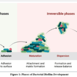 |
Figure 1: Phases of Bacterial Biofilm Development Click here to View Figure |
Biofilm Maturation
Biofilm maturation encompasses the progression from nascent microcolonies toward an intricate three-dimensional microbial assemblage. Throughout this developmental phase, microorganisms undergo proliferation and establish systematically structured aggregates that are delineated by aqueous conduits. These conduits facilitate the circulation of essential nutrients, oxygen, enzymatic compounds, intercellular signaling molecules, and metabolic byproducts throughout the biofilm matrix. The mature biofilm exhibits heterogeneous distributions of oxygen concentrations, nutrient availability, pH levels, and microbial metabolic rates. Characteristically, cellular populations in deeper strata demonstrate reduced growth rates or enter dormant states, whereas cellular populations in superficial strata maintain heightened metabolic activity. Depending on the specific microorganism species and prevailing environmental circumstances, mature biofilms may assume morphologies resembling mushrooms, towers, or irregular configurations.
The ability of some bacteria to produce EPS.
The formation of biofilms is facilitated through the secretion of EPS, which comprises polysaccharides, proteins, extracellular DNA, lipids, enzymes, and aqueous components that collectively constitute the extracellular matrix. This hydrated polymeric matrix surrounds microbial cells and plays a multifunctional role in promoting cellular cohesion and establishing the biofilm architecture. Beyond structural support, the matrix functions as a protective barrier against desiccation while simultaneously shielding microbial populations from antimicrobial compounds, disinfecting agents, mechanical stress, and immunological responses. The individual components contribute distinct functions to matrix integrity: extracellular DNA facilitates cellular aggregation, while proteins and polysaccharides promote intercellular adhesion and enable nutrient sequestration, with polysaccharides additionally reinforcing matrix stability. The matrix possesses the capacity to sequester or inactivate antimicrobial agents, thereby impeding their diffusion through successive biofilm layers. Consequently, microorganisms embedded within the matrix demonstrate considerably greater resistance to antimicrobial intervention compared to planktonic microorganisms.
Biofilm Dispersion
The ultimate phase of biofilm development encompasses the release of microbial cells from mature biofilm structures, returning them to a planktonic state. This release mechanism can be facilitated through biological or physical (passive) processes. Various environmental conditions including nutrient scarcity, hypoxia, thermal variations, accumulation of metabolic byproducts, and other stressors can induce cellular detachment from the biofilm. Microorganisms secrete enzymes capable of degrading extracellular matrix constituents, enabling the liberation of individual or clustered cells. Additionally, physical mechanisms such as shear stress from fluid movement, surface friction, and mechanical perturbations can fragment biofilm structures. Released cells possess the capacity to migrate to alternative locations, establish attachment on new substrates, and initiate formation of distinct biofilm communities. Within clinical contexts, dispersal represents a significant complication in infectious disease, as it facilitates the spread of microorganisms and pathogens from infected tissues or medical devices into systemic circulation. Quorum sensing constitutes a regulatory mechanism through which microorganisms modulate their phenotypic expression in response to population density. Microbial populations synthesize and discharge signaling molecules termed autoinducers, which are low molecular weight compounds. As cellular populations expand, the concentration of these signaling molecules increases proportionally within the surrounding microenvironment. Upon reaching a critical threshold concentration, these molecules activate specific genes governing biofilm adhesion, extracellular matrix synthesis, pathogenic traits, structural maturation, and cellular dispersal. Consequently, quorum sensing facilitates the coordinated functioning of microorganisms as collective entities rather than isolated individuals. Given its role as a fundamental intercellular communication system, quorum sensing represents a promising therapeutic target for novel anti-biofilm pharmaceutical interventions, as disruption of these signaling pathways can be achieved without direct microbial elimination.
Material and Methods
Collection and Identification of Clinical Isolates
Clinical specimens procured from the microbiology repository of encompassed multiple sample types, namely pus, urine, blood, sputum, and catheter tips. Isolates were subsequently revived on nutrient agar medium and subjected to purification through iterative streaking methodology. Taxonomic identification was accomplished through conventional biochemical assays, including catalase, coagulase, oxidase, urease, and carbohydrate fermentation profiles, in conjunction with automated VITEK 2 compact instrumentation. A subset of isolates underwent supplementary confirmation via 16S rRNA gene sequencing to validate taxonomic classification, whereas all bacterial strains were preserved as glycerol suspensions at –80 °C to ensure extended viability.
Preparation of Herbal Extracts
The cleaned plant materials underwent desiccation through shade-drying at room temperature (25–30 °C) for a duration of 7–10 days until mass stabilization was attained, whereupon the samples were mechanically comminuted to a fine powder utilizing an electric blender. The resulting powder was subsequently sieved through a 40-mesh apparatus to guarantee consistent particle dimensions, thereby optimizing solvent extraction efficiency. The powdered specimens were preserved in sealed amber glass vessels maintained at 4 °C prior to subsequent applications, serving to mitigate photochemical degradation and prevent hygroscopic absorption.
Extraction Solvents and Methods
The isolation of distinct phytochemical fractions was accomplished through sequential maceration employing solvents with progressively increasing polarity, specifically n-hexane, ethyl acetate, methanol, and distilled water. A mass of 100 g of desiccated powder was immersed in 500 mL portions of each solvent for a duration of 72 h under ambient temperature conditions whilst undergoing periodic agitation at 120 rpm. Following filtration of the resulting suspensions via Whatman No. 1 filter paper, the obtained filtrates underwent concentration via rotary vacuum evaporation maintained at 40 °C. The concentrated extracts were then subjected to lyophilization to yield dry mass values, which were expressed as percentage yield on a weight-to-weight basis (w/w) and subsequently preserved at –20 °C.
Phytochemical Screening and Fingerprinting
An initial qualitative phytochemical assessment was performed employing conventional colorimetric and precipitation methodologies to identify alkaloids (Dragendorff’s), flavonoids (Shinoda), tannins (ferric chloride), saponins (foam test), glycosides (Keller–Kiliani), and terpenoids (Salkowski). For quantitative determination, the Folin–Ciocalteu reagent was utilized to measure total phenolic content (reported as gallic acid equivalents, mg GAE/g), and the aluminum chloride colorimetric assay was employed to quantify total flavonoid content (reported as quercetin equivalents, mg QE/g). Chemical profiling was accomplished through HPLC equipped with a PDA detector on a C18 reversed-phase column, utilizing gradient elution with acetonitrile and 0.1% formic acid, whereas GC–MS analysis was applied to characterize volatile and semi-volatile components.
Test Microorganisms and Growth Conditions
A collection of clinically significant bacterial isolates, encompassing both Gram-positive organisms (Staphylococcus aureus, Enterococcus faecalis) and Gram-negative organisms (Pseudomonas aeruginosa, Escherichia coli, Klebsiella pneumoniae, Acinetobacter baumannii), was chosen according to their demonstrated propensity for biofilm development. All clinical isolates underwent cultivation on Mueller–Hinton agar (MHA) and within Mueller–Hinton broth (MHB) for antimicrobial susceptibility testing, as well as in Tryptic Soy Broth (TSB) containing 1% glucose to promote biofilm formation. Bacterial suspensions cultured overnight were normalized to a 0.5 McFarland standard (approximately 1.5 × 10⁸ CFU/mL) and subsequently diluted to the appropriate cell density required for each individual experimental procedure.
Determination of Antimicrobial Activity
Well Diffusion Assay
Antimicrobial activity was assessed employing the agar well diffusion technique. Sterilized molten MHA (maintained at approximately 45 °C) was distributed into Petri dishes and permitted to solidify. A normalized bacterial suspension (100 µL containing 10⁶ CFU/mL) was evenly applied across the agar surface. Circular cavities (6 mm in diameter) were created and loaded with 100 µL of test extracts at specified concentrations (for instance, 100 mg/mL). The inoculated plates were subjected to incubation at 37 °C for 24 hours, following which the dimensions of the resulting inhibition halos were quantified in millimeters using a Vernier caliper. All determinations were executed in triplicate.
Minimum Inhibitory Concentration (MIC) by Broth Microdilution
The MIC was established through broth microdilution methodology utilizing 96-well U-bottom microplates. Sequential twofold dilutions of each extract were formulated in MHB across a concentration spectrum of 0.125 to 8 mg/mL. Each well was seeded with 50 µL of a calibrated bacterial inoculum (~5 × 10⁵ CFU/mL final concentration), resulting in a final volume of 200 µL per well. The microplates underwent incubation at 37 °C for 18–24 h. Upon completion of the incubation period, 20 µL of 0.01% resazurin solution was introduced to each well; the MIC was defined as the minimal concentration that inhibited the color transition from blue to pink, signifying the absence of detectable microbial proliferation.
Minimum Bactericidal Concentration (MBC)
In order to ascertain the MBC, sterile 10 µL samples were obtained from all wells that demonstrated clarity (indicative of absent microbial proliferation in the MIC examination) and transferred to newly prepared MHA plates. Following incubation at 37 °C for 24 hours, the MBC was defined as the minimal extract concentration that achieved a ≥99.9% decrease in the original bacterial load, as evidenced by the absence of observable colonies on the agar medium following the designated incubation timeframe.
Anti-Biofilm Activity Assessment
The anti-biofilm potential was evaluated by quantifying the ability of the extracts to either prevent initial biofilm attachment/deposition and disrupt pre-existing mature biofilms, along with assessing the residual viability of biofilm-embedded cells, using 96-well polystyrene microtiter plates as the standard substrate for biofilm growth.
Biofilm Inhibition Assay (Crystal Violet Method)
For inhibition experiments, microbial suspensions comprising 200 µL of 10⁶ CFU/mL in TSB+1% glucose were introduced into wells alongside sub-MIC concentrations of the extract preparations. Following 24 h of stationary incubation at 37 °C, planktonic bacterial populations were carefully removed via aspiration, and the wells underwent three sequential rinses with PBS to eliminate loosely adherent bacterial cells. The adherent biofilm matrices were subsequently fixed through heating (60 °C, 1 h) and subjected to colorimetric staining with 200 µL of 0.1% (w/v) crystal violet for 15 min. Following removal of surplus stain, the retained dye was extracted through solubilization in 95% ethanol. Optical density measurements were obtained at 570 nm (OD₅₇₀), and inhibitory activity was quantified as a percentage relative to the non-treated reference sample.
Microscopic Visualization of Biofilm Architecture
Light Microscopy
Biofilms were grown on sterile glass coverslips placed in 6-well plates. Following treatment, coverslips were gently washed, fixed with methanol, and stained with 1% crystal violet. After air-drying, the coverslips were mounted on glass slides and observed under a bright-field microscope (40× to 100× objective). Representative images were captured to qualitatively evaluate changes in biofilm density, clumping, and overall surface coverage.
Scanning Electron Microscopy (SEM)
For ultrastructural analysis, biofilms on coverslips were fixed with 2.5% glutaraldehyde in 0.1 M phosphate buffer (pH 7.2) for 4 h at 4 °C. Samples were post-fixed in 1% osmium tetroxide, followed by dehydration through a graded ethanol series (30–100%). The dehydrated specimens were subjected to critical point drying using CO₂, mounted on aluminum stubs, and sputter-coated with a thin layer of gold–palladium. Imaging was performed using a field-emission scanning electron microscope operated at 5–10 kV to examine biofilm microcolony architecture and cell surface alterations.
Fluorescence/Confocal Laser Scanning Microscopy
The structural integrity and three-dimensional organization of biofilms were evaluated employing CLSM combined with the LIVE/DEAD BacLight bacterial viability kit, utilizing SYTO 9 and propidium iodide as fluorescent markers. Both control and experimental biofilm samples mounted on coverslips underwent incubation with the fluorescent staining reagent under dark conditions for a duration of 15 min. Optical excitation and detection parameters of 488/500 nm (corresponding to SYTO 9-labeled viable cells, visualized in green fluorescence) and 488/635 nm (corresponding to PI-labeled nonviable cells, visualized in red fluorescence) were implemented. Sequential Z-stack image acquisition was performed at 1 µm spatial intervals, with subsequent quantitative assessment of biofilm parameters including depth, volumetric density, and areal coverage conducted using COMSTAT software on the reconstructed three-dimensional datasets.
Statistical Analysis
The entirety of investigations were executed in triplicate across three separate experimental sessions, with results reported as mean values accompanied by standard deviation (SD). Statistical evaluations were performed utilizing GraphPad Prism v9.0 software. For comparative analyses among multiple groups, one-way ANOVA was implemented, supplemented by Tukey’s HSD post-hoc test to assess differences between individual group pairs, whereas Student’s t-test was employed to evaluate differences between two groups directly.
Result and Discussion
Extraction Yield and Phytochemical Profiling
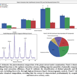 |
Figure 2: delineates the phytochemical composition of the plant extract under examination. Panel A demonstrates that methanol functions as the optimal extraction solvent, yielding superior quantities of phenolic compounds and maximizing total extraction efficiency. |
Successive maceration of the dehydrated plant material produced fractions possessing distinct solubility characteristics. The methanol fraction demonstrated the maximum extraction efficiency (12.4 ± 0.8% w/w), succeeded by ethyl acetate (8.2 ± 0.5%), aqueous (5.6 ± 0.3%), and n-hexane (3.1 ± 0.2%) fractions. Initial phytochemical analysis indicated the occurrence of alkaloids, flavonoids, tannins, saponins, and terpenoids primarily concentrated in the methanol and ethyl acetate fractions, whereas the n-hexane fraction contained predominantly terpenoids. From a quantitative perspective, total phenolic content attained maximum concentrations in the methanol fraction (85.4 ± 2.3 mg GAE/g), while the ethyl acetate fraction exhibited the greatest total flavonoid concentration (32.1 ± 1.8 mg QE/g). HPLC-PDA profiling of the methanol fraction identified prominent chromatographic signals at retention intervals consistent with reference phenolic acids and flavonoids, whereas GC-MS examination detected 17 volatile components, with sesquiterpenes and fatty acid derivatives constituting the predominant constituents.
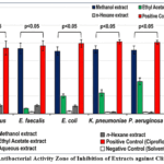 |
Figure 3: Antibacterial Activity Zone of Inhibition of Extracts against Clinical Isolates. Click here to View Figure |
The agar well diffusion assay revealed that among the tested extracts, the methanol and ethyl acetate formulations demonstrated the most pronounced antibacterial efficacy against the clinical isolates examined, whereas the aqueous and n-hexane preparations exhibited minimal or absent inhibitory activity. The methanol extract generated the largest inhibition zones when tested against S. aureus (18.3 ± 0.4 mm) and E. faecalis (16.7 ± 0.5 mm), with notably smaller zones observed for E. coli (15.2 ± 0.6 mm) and K. pneumoniae (14.8 ± 0.3 mm). MIC determinations yielded values spanning from 0.25 to 2.0 mg/mL, with the methanol extract demonstrating the most favorable MIC of 0.25 mg/mL for S. aureus and 0.5 mg/mL for P. aeruginosa. MBC measurements fell within a 2- to 4-fold range relative to corresponding MIC values (MBC/MIC ratio ≤ 4), suggesting that the active extracts exerted bactericidal rather than bacteriostatic mechanisms. Reference antimicrobial agents (ciprofloxacin and vancomycin) yielded reliable inhibition zones that confirmed assay validity, whereas solvent blanks demonstrated no detectable antimicrobial properties.
Anti-Biofilm Activity
The anti-biofilm potential of the active extracts was evaluated at sub-inhibitory concentrations (ranging from ¼× to ½× MIC) to exclude bactericidal effects during biofilm assessment.
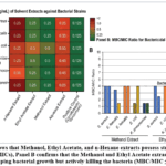 |
Figure 4: A shows that Methanol, Ethyl Acetate, and n-Hexane extracts possess strong inhibitory activity (low MICs), Panel B confirms that the Methanol and Ethyl Acetate extracts are not just stopping bacterial growth but actively killing the bacteria (MBC/MIC ≤ 4). |
# Biofilm Inhibition The methanol extract demonstrated considerable suppression of nascent biofilm formation in a concentration-dependent manner. At ½× MIC, biofilm inhibition percentages attained 78.5 ± 3.2% for S. aureus, 72.1 ± 4.0% for E. faecalis, and 65.8 ± 3.6% for P. aeruginosa. Substantial inhibitory effects (exceeding 40%) were documented across all Gram-positive bacterial species even at ¼× MIC, whereas Gram-negative organisms displayed comparatively diminished inhibition, with A. baumannii showing the lowest response (52.3 ± 2.8% at ½× MIC). # Mature Biofilm Disruption When evaluated against established (48-hour) biofilm structures, the methanol extract exhibited modest disruptive potential. At the maximum tested concentration (½× MIC), disruption rates were 56.4 ± 3.1% for S. aureus, 48.7 ± 2.5% for P. aeruginosa, and 45.2 ± 2.9% for K. pneumoniae, suggesting that the extract proved more proficient in preventing biofilm genesis than in dismantling pre-existing biofilms (p < 0.05). # Biofilm Viability Reduction The XTT reduction assay demonstrated pronounced diminution in metabolic activity of biofilm-associated cells following extract exposure. In comparison to untreated controls, the methanol extract at ½× MIC decreased the respiratory activity of S. aureus and P. aeruginosa biofilms by 68.4% and 58.1%, respectively. CFU quantification substantiated these findings, revealing that viable cell populations within treated biofilms declined by approximately 2 to 3 log₁₀ units relative to control samples (p < 0.001). # Effect on Biofilm-Related Virulence Factors To elucidate the mechanistic basis of the anti-biofilm activity, the influence of sub-inhibitory concentrations of the methanol extract (¼× MIC) on essential virulence characteristics was examined. # Motility Inhibition The methanol extract substantially suppressed all bacterial motility phenotypes. In P. aeruginosa, swimming, swarming, and twitching zones were diminished by 68.2%, 74.5%, and 52.1%, respectively, relative to untreated controls. Correspondingly, E. coli and K. pneumoniae demonstrated pronounced reductions in swarming motility (60–70% inhibition). # Auto-aggregation and Hydrophobicity The methanol extract treatment decreased the auto-aggregation index of S. aureus from 52.4% (control) to 18.6% following 24-hour incubation. The microbial adhesion to hydrocarbons assay revealed a marked reduction in cell surface hydrophobicity; for P. aeruginosa, xylene adhesion percentage declined from 68.3 ± 2.5% (control) to 24.6 ± 1.8% upon treatment, demonstrating that the extract disrupts the preliminary physicochemical interactions essential for substrate colonization. # EPS Production Quantitative examination of extracellular polymeric substances demonstrated that the methanol extract substantially reduced EPS secretion. Total carbohydrate concentration in the EPS matrix of S. aureus decreased from 42.5 ± 3.1 µg/mg protein (control) to 12.8 ± 1.5 µg/mg protein in treated samples. A parallel reduction was documented for P. aeruginosa, where EPS production diminished by approximately 62% in comparison to untreated controls.
Microscopic Visualization of Biofilm Architecture
Optical microscopic examination demonstrated that control biofilms lacking treatment developed densely packed, consolidated structures that uniformly covered the glass coverslip substrate. By comparison, samples exposed to extract treatment manifested dispersed, attenuated, and discontinuous microcolonial formations accompanied by substantially diminished total biomass. Electron microscopy analysis of untreated P. aeruginosa biofilms revealed an extensive, uninterrupted polymeric matrix encompassing structurally defined microcolonies and copious extracellular appendages. Post-treatment biofilms demonstrated a flattened, sparsely distributed architecture with evident degradation, compromised cellular membranes, and weakened cell-to-cell bonding. Confocal laser scanning microscopy employing LIVE/DEAD fluorescent markers substantiated these observations; untreated biofilms exhibited pronounced green fluorescence (viable cells) constituting substantial, homogeneous vertical arrangements (mean height: 35.4 ± 2.8 µm), whereas treated biofilms displayed predominant red fluorescence (nonviable cells) with markedly decreased vertical dimension (12.1 ± 1.5 µm) and overall biovolume. COMSTAT quantitative assessment validated a reduction exceeding 60% in areal coverage and biomass quantity within extract-treated cohorts relative to untreated counterparts.
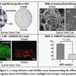 |
Figure 5: This figure is a comprehensive anti-biofilm assay demonstrating the efficacy of a specific treatment against bacterial biofilms across multiple microscopic and quantitative levels. |
Discussion
The observed anti-biofilm activity demonstrated in this investigation demonstrates substantial concordance with preceding scholarly work examining alternative medicinal plants including Anabasis articulata and Agrimoniaeupatoria. Notably, the dual mechanistic capacity of the examined extracts—encompassing both the prevention of biofilm genesis and the degradation of established biofilm matrices—distinguishes them as particularly compelling candidates for therapeutic advancement. The employment of clinically derived bacterial isolates, as opposed to standardized laboratory reference strains, substantially augments the clinical applicability of the findings, given that clinical isolates frequently possess distinctive resistance profiles and produce robust biofilm phenotypes under conditions reflective of natural selective pressures. Nonetheless, several methodological constraints warrant consideration. Initially, although broad phytochemical categories were identified, the isolation and characterization of the precise bioactive constituents mediating the observed phenomena remain incomplete. Subsequent chromatographic fractionation-based isolation strategies are necessary to identify the principal active molecules. Secondarily, experimental evaluations were restricted to in vitro conditions utilizing polystyrene and glass substrates; assessment of extract performance within physiological contexts—including biomedical device surfaces and tissue microenvironments—necessitates validation through in vivo animal experimentation and ex vivo human tissue systems. Tertiary, the cytotoxic effects of the bioactive extracts toward mammalian cellular systems were not examined herein, representing a critical gap for preclinical therapeutic assessment. Moreover, the potential synergistic or antagonistic effects arising from interactions among constituent phytochemicals warrant supplementary investigation. Given the substantial clinical prevalence of catheter-associated urinary tract infections, ventilator-associated pneumonia, and prosthetic joint infections—predominantly attributable to biofilm-producing microorganisms—there exists considerable demand for novel therapeutic strategies. The extracts examined herein, particularly the methanolic fraction, present feasible frameworks for formulating topical antimicrobial preparations or protective surface coatings to mitigate device-associated infections. The demonstrated efficacy against both Gram-positive and Gram-negative organisms substantially expands the therapeutic applicability spectrum.
Conclusion
In summary, this investigation demonstrates the considerable antibacterial and anti-biofilm properties of sequentially isolated herbal fractions, whereby the methanol extract yielded superior phytochemical content alongside elevated phenolic and flavonoid concentrations. The methanol and ethyl acetate extracts exhibited wide-ranging bactericidal effects (MBC/MIC ≤ 4) across multidrug-resistant Gram-positive and Gram-negative clinical strains. From a mechanistic perspective, at concentrations below the inhibitory threshold, these extracts prevented initial bacterial attachment and dismantled established biofilms through reduction of bacterial motility, decreased cell surface hydrophobicity and self-aggregation capacity, and substantial suppression of EPS synthesis (approximately 70% reduction in S. aureus EPS). These observations were substantiated through microscopic examination (SEM/CLSM), which demonstrated compromised and flattened structural integrity accompanied by greater than 60% decrease in biomass and extensive cellular damage. The dual-action capability, combined with a polypharmacological mechanism presumably encompassing quorum-sensing inhibition, renders these extracts particularly suitable for development as topical antiseptic agents, catheter lock formulations, and antimicrobial surface treatments. Nevertheless, advancing these in vitro observations toward clinical implementation necessitates bioassay-directed identification of principal bioactive constituents, comprehensive in vivo assessment of effectiveness and safety profiles, and creation of innovative nano-delivery platforms to address inherent limitations in solubility and bioavailability. Collectively, this research furnishes empirically substantiated evidence supporting the application of these herbal phytochemicals as an effective, polypharmacological therapeutic approach for managing persistent biofilm-related infections demonstrating increasing resistance to standard antimicrobial agents.
Funding Sources
The author(s) received no financial support for the research, authorship, and/or publication of this article.
Conflict of Interest
The author(s) do not have any conflict of interest.
Data Availability Statement
This statement does not apply to this article.
Ethics Statement
This research did not involve human participants, animal subjects, or any material that requires ethical approval.
Informed Consent Statement
This study did not involve human participants, and therefore, informed consent was not required.
References
- Al-Maddboly, L. A., El-Salam, M. A., Bastos, J. K., Hasby, E. A., Kushkevych, I., & El-Morsi, R. M. (2025). Anti-biofilm and anti-quorum sensing activities of galloylquinic acid against clinical isolates of multidrug-resistant Pseudomonas aeruginosa in open wound infection: in vitro and in vivo efficacy studies. BMC Microbiology, *25*, 206. https://doi.org/10.1186/s12866-024-03712-8
CrossRef - Manzo, L. M., et al. (2025). Rutin-luteolin flavonoid-encapsulated chitosan nanoparticles: a novel strategy to combat multidrug-resistant Pseudomonas aeruginosa. BMC Biotechnology, *25*, 24. https://doi.org/10.1186/s12896-025-01087-w
CrossRef - Mohammadi, A., et al. (2025). Emerging Multimodal Strategies for Bacterial Biofilm Eradication: A Comprehensive Review. Journal of Nanobiotechnology, *23*, 895. https://doi.org/10.1186/s12951-024-03014-7
- Sadieva, L. K., Grzhegorzhevskii, K. V., Platonov, V. A., Santra, S., Zyryanov, G. V., & Rusinov, V. L. (2025). Quorum sensing inhibitors of plant origin: structure–activity relationships. Russian Chemical Reviews, *94*(11), RCR5192. https://doi.org/10.59761/RCR5192
CrossRef - Tan, Y., et al. (2023). In vitro outcomes of quercetin on Candida albicans planktonic and biofilm cells and in vivo effects on vulvovaginal candidiasis. Evidences of its mechanisms of action. Phytomedicine, *114*, 154800. https://doi.org/10.1016/j.phymed.2023.154800
CrossRef - Turkey, M., et al. (2025). Chromatographic fingerprinting and antibiofilm effect of Ziziphus jujuba fraction on Pseudomonas aeruginosa. AMB Express, *15*(1), 79. https://doi.org/10.1186/s13568-025-01886-6
CrossRef - Wang, D. D., Shi, W. J., Li, B. C., Liu, J. J., Li, Y., & Jing, X. N. (2025). Progresses of plant-derived natural products against Candida albicans biofilm. Acta Pharmaceutica Sinica, *60*(9), 2431–2445. https://doi.org/10.16438/j.0513-4870.2025-0400
- Wang, W. Q., Feng, X. C., Shi, H. T., Wang, Y. M., Jiang, C. Y., Xiao, Z. J., Xu, Y. J., Zhang, X., Yuan, Y., & Ren, N. Q. (2023). Biofilm inhibition based on controlling the transmembrane transport and extracellular accumulation of quorum sensing signals. Environmental Research, *221*, 115218. https://doi.org/10.1016/j.envres.2023.115218
CrossRef - Wiley, L. K., et al. (2025). The Potential of Secondary Metabolites in Medicinal Plants as Anti–Quorum Sensing in Biofilms: A Comprehensive Review. Journal of Chemistry, *2025*(1), 8838140. https://doi.org/10.1155/joch/8838140
CrossRef - Zhang, Z., et al. (2025). Research progress on the antibacterial activity of natural flavonoids. Antibiotics, *14*(1), 45. https://doi.org/10.3390/antibiotics14010045
CrossRef - Parai, D., Islam, E., Mitra, J., & Mukherjee, S. K. (2024). Effect of Bacoside A on growth and biofilm formation by Staphylococcus aureus and Pseudomonas aeruginosa. Journal of Applied Microbiology, *135*(2), lxae046. https://doi.org/10.1093/jambio/lxae046
CrossRef - Chen, Y. H., et al. (2014). Trends in the susceptibility of methicillin-resistant Staphylococcus aureus to nine antimicrobial agents, including ceftobiprole, nemonoxacin, and tyrothricin: results from the tigecycline in vitro surveillance in Taiwan (TIST) study, 2006–2010. European Journal of Clinical Microbiology & Infectious Diseases, *33*(3), 433–440. https://doi.org/10.1007/s10096-013-1978-1
CrossRef - Nguyen, A. N. X., et al. (2024). Insights into molecular mechanisms of phytochemicals in quorum sensing modulation for bacterial biofilm control. Archives of Microbiology, *206*(12), 459. https://doi.org/10.1007/s00203-024-04171-5
CrossRef - Patil, A., Banerji, R., Kanojiya, P., & Saroj, S. D. (2021). Foodborne ESKAPE biofilms and antimicrobial resistance: lessons learned from clinical isolates. Pathogens and Global Health, *115*(6), 339–356. https://doi.org/10.1080/20477724.2021.1916158
CrossRef - Ali, S., Amin, A., Khalil, A. A. K., Akhtar, M. S., & Zaman, W. (2026). Nanoparticle-based delivery of natural products for antibacterial therapy: Mechanisms, platforms, and translational challenges. Journal of Drug Delivery Science and Technology. https://doi.org/10.1016/j.jddst.2026.104533
CrossRef - Atiyah, M. M., & Vijayan, S. (2025). Therapeutic potential of chitosan-encapsulated phytochemicals: A review on antimicrobial strategies against nosocomial infections. Journal of Drug Delivery Science and Technology. https://doi.org/10.1016/j.jddst.2025.106137
CrossRef - Zhou, J., et al. (2025). Natural phytochemical-based strategies for antibiofilm applications. Chinese Medicine, *20*(1), 96. https://doi.org/10.1186/s13020-025-01147-5
CrossRef - Wiley, L. K., et al. (2025). The potential of secondary metabolites in medicinal plants as anti–quorum sensing in biofilms: A comprehensive review. Journal of Chemistry, *2025*(1), 8838140. https://doi.org/10.1155/joch/8838140
CrossRef - Dolatabadi, S., Moghadam, H. N., & Mahdavi-Ourtakand, M. (2018). Evaluating the anti-biofilm and antibacterial effects of Juglans regia extracts against clinical isolates of Pseudomonas aeruginosa. Microbial Pathogenesis, *118*, 35-40. https://doi.org/10.1016/j.micpath.2018.03.014
CrossRef - Mohammadi, A., et al. (2025). Emerging multimodal strategies for bacterial biofilm eradication: A comprehensive review. Journal of Nanobiotechnology, *23*, 895. https://doi.org/10.1186/s12951-024-03014-7
- Rosado, F., et al. (2022). In vitro anti-clostridial action and potential of the spice herbs essential oils to prevent biofilm formation of hypervirulent Clostridioides difficileAnaerobe, *76*, 102598. https://doi.org/10.1016/j.anaerobe.2022.102598
CrossRef - Manzo, L. M., et al. (2025). Rutin-luteolin flavonoid-encapsulated chitosan nanoparticles: a novel strategy to combat multidrug-resistant Pseudomonas aeruginosa. BMC Biotechnology, *25*, 24. https://doi.org/10.1186/s12896-025-01087-w
CrossRef - Turkey, M., et al. (2025). Chromatographic fingerprinting and antibiofilm effect of Ziziphus jujubafraction on Pseudomonas aeruginosa. AMB Express, *15*(1), 79. https://doi.org/10.1186/s13568-025-01886-6
CrossRef - Al-Maddboly, L. A., et al. (2025). Anti-biofilm and anti-quorum sensing activities of galloylquinic acid against clinical isolates of multidrug-resistant Pseudomonas aeruginosain open wound infection. BMC Microbiology, *25*, 206. https://doi.org/10.1186/ s12866-024-03712-8
CrossRef - Wang, W. Q., et al. (2023). Biofilm inhibition based on controlling the transmembrane transport and extracellular accumulation of quorum sensing signals. Environmental Research, *221*, 115218. https://doi.org/10.1016/j.envres.2023.115218
CrossRef
Accepted on: 02 Sep 2026


ISSN Online: 2231-5039