Isolation of Bacterial Endophytes from Carica Papaya, Tamarindus Indica and Moringa Oleifera Leaves
 , Anju Nayak2, Joycee Jogi3, Ajay Rai4, R K Sharma1 and Shagun Upadhyay5*
1Nanaji Deshmukh Veterinary Science University, Jabalpur, M.P. India.
2Department of Microbiology, College of Veterinary Sciences and A.H, NDVSU, Jabalpur (M.P.), India
3Department of Microbiology, College of Veterinary Sciences and A.H., MHOW, Indore, NDVSU, Jabalpur (M.P.), India.
4Department of Microbiology, College of Veterinary and Animal Sciences (CoVAS), Kishanganj, Patna, India.
5Shri Ram Group of Institutions, Faculty of Pharmacy, Jabalpur (M.P.), India.
Corresponding Author E-mail:dubeyshagun25@gmail.com
Download this article as:
ABSTRACT:The study was carried out for isolation and characterization of the potentially isolated strains of the bacteria endophytes from Carica papaya, Tamarindus indica and Moringa oleifera leaves. The leaves of Carica papaya, Tamarindus indica and Moringa oleifera are a source for endophytes that may be a source of new natural products useful in food and agroindustry, little is known about isolation and characterization of endophytes with bacterial origin in these plants. The leaves of the above mentioned plants were collected, surface sterilized and then inoculated in King's B Media for isolation of bacterial endophytes. Six pure isolates of bacteria were isolated and subjected to PCR and sequencing of the PCR products to molecularly characterize them as bacterial endophytes. The percentage similarity and classification of endophytic bacterial strains were done with help of NCBI BLASTDNA against the recorded sequences of the database. The bacterial endophytes of Moringa oleifera demonstrated a 99 % coverage respectively for Staphylococcus pasteuri coverage and the bacterial endophytes of Carica papaya and Tamarindus indica revealed 95 % respectively for coverage of Lysinibacillus sphaericus and Paenibacillus azotifigens.
KEYWORDS:Bacterial endophyte; Carica papaya; Moringa oleifera; Synergistic; Tamarindus indica
Introduction
Endophytic microbes are a source of many important bioactive molecules. The Endophytes are classified as microbes which grow in the plant tissues without causing any expression of disease. Are briefly pathogenic. Endophytes have a niche so they are known to produce a wide range of bioactive compounds. The need for new sources of antimicrobial metabolites has been great due to the ongoing development of pathogen resistance to current health care treatments and medicines. Therefore, investigation for endophytic microbes, which could be used for producing new and unique quorum sensing inhibitors, is very essential. Papaya (Carica papaya) is a fruit crop which is very popular in the tropical and subtropical fruit trees. There is a growing interest in the study of the papaya as it is much faster to mature than many of its tropical and subtropical counterparts. Considerable research has been done on the papaya fruit’s nutritional value and the medicinal properties of the papaya plant are known. But, the Papaya plant has not been studied in the field of plant endophyte research. As papaya contain a lot of sugar for example sucrose, rich in fructose and glucose, the endophytes associated with the papaya might be a good study of what papaya has to offer. Tamarindus indica is an African tree which is common throughout the Indian subcontinent. A versatile plant. The pulp of fruits was used as a spice in Asian cooking particularly in the southern part of India.
The research work done in the recent past focusses on the discovery of novel natural sources and the associated natural compounds with the potential of pharmaceutical application. The success results which have obtained from this area would be a great success in the battle against animal bacterial pathogens. Secondary metabolites produced by plants can be beneficial to treat diseases. Moringa oleifera is the most common species in the Moringaceae family. They grow well in tropical climate. In India and Nigeria this plant is grown as a crop, and is also referred to as Drumstick. It is used for foods and medicine, and a popular traditional medicine plant. The biological activities reported for Moringa oleifera are: antimicrobial, regulation of thyroid hormone, antidiabetic, antihyperglycemic and hypotensive. This wide range of properties of Moringa oleifera, warranted an investigation of the endophytes.
Material and Methods
Plant Sample Collection
Fresh, mature, and healthy leaves of Carica papaya, Tamarindus indica, and Moringa oleifera were collected for this study. After procurement, all samples were immediately transported to the laboratory and processed within 4-6 hours to prevent contamination and ensure the viability of endophytic bacteria.
Preparation of Culture Media
King’s B (KB) media was prepared according to the manufacturer’s instructions for the initial isolation of endophytic bacteria. The media components were mixed thoroughly using a hot plate to ensure complete dissolution. The prepared media was then sterilized by autoclaving at 121°C for 15-20 minutes at 15 lbs pressure. To verify sterility, the media was poured into 90 mm sterile Petri plates and incubated at 37°C for 24 hours. The absence of microbial growth confirmed the sterility of the prepared media.
Surface Sterilization of Plant Material
Leaf disinfection and isolation of endophytic bacteria were performed following the method described by Mahajan et al. (2014) with modifications. All procedures were conducted in a laminar air flow chamber using sterilized scalpels and forceps. The sterilization protocol involved multiple steps:
The leaves were initially rinsed with double distilled (dd) water for 2-3 minutes. Surface disinfection was achieved by treating the leaves with 0.1% sodium hypochlorite for 5 minutes, followed by another dd water wash for 2-3 minutes. The leaves were then sprayed with Bavistin at a concentration of 500 ml/ha and 0.01% disinfectant. Subsequently, the leaves were submerged in dd water for 5 minutes and treated with 0.05% streptomycin solution, followed by another dd water rinse. Finally, the leaves were treated with 70% ethanol and left in dd water for an additional 5 minutes. After completing the sterilization procedure, the leaves were air-dried in the laminar air flow chamber.
Sterility Check
To confirm complete surface sterilization, 1 ml of the sterilized distilled water from the final rinse was poured onto nutrient agar medium and incubated at 37°C for 24 hours. The absence of bacterial growth on these plates confirmed successful surface disinfection of the leaves.
Isolation of Endophytic Bacteria
The surface-sterilized leaves from each plant species were placed onto KB media plates and incubated at 37°C for 24 hours. Following incubation, bacterial colonies were characterized based on morphological observation and Gram’s staining. Single colonies were then inoculated into Brain Heart Infusion (BHI) broth and incubated for 24 hours.
Purification of Endophytic Bacteria
Endophytic bacterial purification was performed by sub-culturing a loopful of BHI broth culture onto 5% sheep blood agar plates. The plates were incubated at 37°C for 24 hours. A single colony was then transferred to fresh BHI broth and incubated at 37°C for 24 hours. Gram’s staining was performed to verify culture purity, and the purified cultures were preserved for further studies.
Identification of Endophytic Bacteria
Gram’s Staining
Bacterial identification was initiated through Gram’s staining. Heat-fixed smears were prepared on slides and placed in a staining tray. Crystal violet was applied for 1 minute, followed by rinsing with distilled water. Gram’s iodine was then added and left for 1 minute. After rinsing, the slides were decolorized with 95% ethyl alcohol for 5-10 seconds and immediately washed with distilled water. Safranin counterstain was applied for 1 minute, followed by final rinsing and air drying. The slides were examined under a microscope at 100X magnification.
DNA Extraction
Genomic DNA was extracted using the Instagene Matrix (Bio-Rad) kit. An isolated bacterial colony was grown overnight, picked, and suspended in 1 ml of autoclaved water in a microcentrifuge tube. The suspension was centrifuged at 10,000-12,000 rpm for 1 minute. The supernatant was discarded, and 200 µl of Instagene Matrix was added to the pellet. The mixture was incubated at 56°C for 30 minutes, vortexed, and placed in a boiling water bath at 100°C for 8 minutes. After vortexing for 10 seconds, the sample was centrifuged at 10,000-12,000 rpm for 2-3 minutes. The extracted DNA was stored at -20°C for future use.
Polymerase Chain Reaction
PCR was performed using universal bacterial primers 8F and 1541R as described in Table 1.
Table 1: Details of primers used for endophytic bacteria
|
Primer |
Sequence | Product Size | Reference |
|
8F |
AGAGTTTGGATCTGGCTCAG | 1533 bp | Zhou et al., 1997 |
| 1541R |
AAGGAGGTGATCCAGCCGCA |
The PCR reaction mixture (25 µl) was prepared as follows:
Table 2: Composition of PCR reaction mix for endophytic bacteria
|
Component |
Volume |
|
Taq Master mix |
12.5 µl |
| Forward primer |
1.0 µl |
|
Reverse primer |
1.0 µl |
| DNA template |
2.0 µl |
|
Nuclease-free water |
8.5 µl |
| Total |
25 µl |
The reaction mixture was gently mixed and centrifuged to collect all components at the bottom of the tube. PCR amplification was performed in a thermal cycler under the following conditions:
Table 3: PCR cycle conditions for endophytic bacteria
|
Temperature |
Time | No. of cycles |
| 95°C | 2 min |
1 |
|
95°C |
30 sec | 25 |
| 50°C | 30 sec | |
|
72°C |
2 min | |
| 72°C | 10 min |
1 |
|
4°C |
∞ |
Hold |
Agarose Gel Electrophoresis
PCR products were analyzed by agarose gel electrophoresis. A 1.0% agarose gel was prepared in 1x Tris Borate EDTA (TBE) buffer (35 ml) and melted in a microwave oven. After cooling to 55°C, ethidium bromide (0.5 µg/ml) was added. The gel was poured into an electrophoresis tray and placed in the gel electrophoresis assembly with 1x TBE buffer. Samples were prepared by mixing 1 µl of gel loading buffer with 5 µl of PCR products. The samples were loaded along with a 1 kb DNA ladder. Electrophoresis was performed at 70 volts for 10 minutes, followed by 50 volts for 2 hours. The agarose gels were visualized under a UV trans-illuminator and photographed.
DNA Sequencing
The PCR products of all six bacterial endophytes were sent to Eurofins Genomics India Pvt. Ltd, Bangalore for Sanger sequencing using the forward and reverse primers.
Phylogenetic Analysis
The sequences obtained for the six bacterial endophytes were subjected to phylogenetic analysis. BLAST (Basic Local Alignment Search Tool) was used for sequence alignment, and Clustal-W (CLUSTAL 2.1) was employed for multiple sequence alignment reports. Phylogenetic trees were constructed using Molecular Evolutionary Genetic Analysis (MEGA 11) version 11. The Neighbor-Joining method was used for tree construction with the Bootstrap resampling method for statistical support.
Table 4: Summary of bacterial endophytes isolated
|
Plant Species |
Isolate Code | Number of Isolates |
| Carica papaya | CP1, CP2 |
2 |
|
Moringa oleifera |
MO1, MO2 |
2 |
|
Tamarindus indica |
TI1, TI2 | 2 |
| Total |
6 |
Results
Isolation of Endophytic Bacteria
Endophytic bacteria were successfully isolated from surface-disinfected leaves of Carica papaya, Moringa oleifera, and Tamarindus indica using King’s B media. After incubation at 37°C for 24 hours, distinct bacterial colonies emerged on the culture plates. Based on morphological differences, colonies were purified by repeated streaking (five times) onto 5% sheep’s blood agar. Purified colonies were examined microscopically, and based on colony morphology and Gram’s staining characteristics, two colonies were selected from each plant species.
Gram’s Staining Results
Table 5: Gram’s Reaction of Bacterial Endophytes
|
S. No. |
Isolate Name | Plant Source | Gram’s Reaction |
| 1 | P1 | Carica papaya |
Gram positive rods |
|
2 |
P2 | Carica papaya | Gram positive rods |
| 3 | M1 | Moringa oleifera |
Gram positive rods |
|
4 |
M2 | Moringa oleifera | Gram negative rods |
| 5 | T1 | Tamarindus indica |
Gram positive cocci in clusters |
|
6 |
T2 | Tamarindus indica |
Gram positive rods |
The results demonstrated that the majority of isolates (5 out of 6) were Gram-positive rods, which is consistent with earlier studies reporting that approximately 90% of endophytic bacteria are Gram-positive rods (Reference 21). However, some studies have also reported the presence of endophytic Gram-positive cocci , as observed in isolate T1 (Staphylococcus pasteuri).
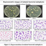 |
Figure 1: Representation of isolated bacterial endophytes Click here to View Figure |
Discussion on Isolation
Endophytic bacteria represent a highly diverse group of microorganisms. With approximately 300,000 plant species on Earth, each plant species, and often each individual plant, can harbor one or several endophytes. Many endophytes can inhabit multiple different plant hosts (Reference 23). Endophytes are potentially hyper-diverse, with estimates suggesting there could be nearly 1,000,000 endophytic bacterial taxa. This diversity likely varies geographically, from country to country, region to region, and among different climatic zones. The successful isolation of six distinct bacterial endophytes from three different plant species in this study supports the concept of endophytic diversity.
Molecular Characterization of Isolated Endophytic Bacteria
PCR Amplification of 16S rRNA Gene
All six bacterial endophyte isolates underwent genotypic characterization using the 16S rRNA gene. PCR amplification was performed using universal bacterial primers 8F and 1541R, which successfully generated amplicons of approximately 1533 bp for all isolates, in accordance with the method described by Reference 24 with minor modifications.
Table 6: Genotypic Characterization of Bacterial Endophytes
|
S. No. |
Sample | 16S rRNA Amplification | Product Size |
| 1 | P1 | Positive |
~1533 bp |
|
2 |
P2 | Positive | ~1533 bp |
| 3 | M1 | Positive |
~1533 bp |
|
4 |
M2 | Positive | ~1533 bp |
| 5 | T1 | Positive |
~1533 bp |
|
6 |
T2 | Positive |
~1533 bp |
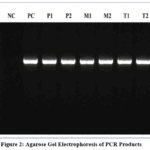 |
Figure 2: Agarose Gel Electrophoresis of PCR Products Click here to View Figure |
Phylogenetic Analysis and Species Identification
The 16S rRNA gene sequences of all six isolates were compared with deposited sequences in the NCBI GenBank database using the BLAST tool. Phylogenetic trees were constructed using the Neighbor-Joining method with Bootstrap resampling in MEGA 11.
Bacterial Endophytes of Carica papaya
Sequence Analysis: The nucleotide sequence of isolate P1 showed 98.66% similarity to Lysinibacillus sphaericus with a query cover of 95%.
Phylogenetic Placement: The phylogenetic tree (Plate 10) demonstrated that isolate P1 clustered closely with Lysinibacillus sphaericus reference strains, confirming its identification.
Discussion: Lysinibacillus sphaericus is a well-known endophytic bacterium with mosquitocidal properties and plant growth-promoting activities. Its isolation from Carica papaya leaves in this study is consistent with previous findings. Hlaing and Soe (2018) isolated Bacillus and Staphylococcus species from papaya leaves, while Kushwah (2019) identified Bacillus subtilis, Bacillus wiedmannii, and Staphylococcus pasteuri as bacterial endophytes of papaya plants. The presence of Lysinibacillus species in papaya adds to the known endophytic diversity of this plant.
Isolate P2
Sequence Analysis: The nucleotide sequence of isolate P2 showed 99.38% similarity to Paenibacillus illinoisensis with a query cover of 97%.
Phylogenetic Placement: The phylogenetic tree (Plate 11) placed isolate P2 within the Penicillus clade, closely related to P. illinoisensis reference strains.
Discussion: Paenibacillus illinoisensis is a Gram-positive, endospore-forming bacterium known for its plant growth-promoting properties, including phosphate solubilization and production of antimicrobial compounds. The high similarity (99.38%) strongly supports the identification of P2 as P. illinoisensis. This represents a notable finding, as Paenibacillus species are increasingly recognized as important plant-associated bacteria with biotechnological potential.
Bacterial Endophytes of Moringa oleifera
Sequence Analysis: The nucleotide sequence of isolate M1 showed 98.28% similarity to Paenibacillus azotifigens with a query cover of 95%.
Phylogenetic Placement: The phylogenetic tree (Plate 12) demonstrated that isolate M1 grouped with Paenibacillus azotifigens reference strains.
Discussion: Paenibacillus azotifigens is a nitrogen-fixing bacterium that contributes to plant nutrition and growth promotion. Its isolation from Moringa oleifera leaves is significant, as Moringa is known for its nutritional and medicinal properties. Previous studies have reported the isolation of Pectobacterium cypripedii and Pantoea agglomerans from the Moringa rhizosphere (Berg et al., 2009). White (2017) identified Bacillus pumilus, Citrobacter koseri, and Klebsiella pneumoniae from Moringa seeds, while Ilmi et al. (2018) reported Bacillus cereus from Moringa stems. The identification of P. azotifigens from Moringa leaves in this study expands the known endophytic community of this important medicinal plant.
Bacterial Endophytes of Cupriavidus taiwanensis
Sequence Analysis: The nucleotide sequence of isolate M2 showed 82.93% similarity to Cupriavidus taiwanensis with a query cover of 80%.
Phylogenetic Placement: The phylogenetic tree (Plate 13) placed isolate M2 within the Cupriavidus clade.
Discussion: Cupriavidus taiwanensis is a Gram-negative, rod-shaped bacterium belonging to the family Burkholderiaceae. The relatively lower similarity (82.93%) and query cover (80%) suggest that M2 may represent a novel strain or species within the Cupriavidus genus. Cupriavidus species are known for their metabolic versatility, including heavy metal resistance and degradation of environmental pollutants. The isolation of this bacterium from Moringa leaves is noteworthy and warrants further investigation for its potential biotechnological applications.
Bacterial Endophytes of Tamarindus indica
Sequence Analysis: The nucleotide sequence of isolate T1 showed 98.23% similarity to Staphylococcus pasteuri with a query cover of 99%.
Phylogenetic Placement: The phylogenetic tree (Plate 14) placed isolate T1 within the Staphylococcus clade, closely related to S. pasteuri reference strains.
Discussion: Staphylococcus pasteuri is a Gram-positive coccus belonging to the genus Staphylococcus, commonly found on human skin but also reported as a plant endophyte. Kushwah (2019) previously identified S. pasteuri as an endophyte of papaya plants. The isolation of S. pasteuri from Tamarindus indica in this study indicates that this bacterium can colonize multiple plant hosts. The presence of Gram-positive cocci as endophytes has been reported though they are less common than Gram-positive rods.
Isolate T2
Sequence Analysis: The nucleotide sequence of isolate T2 showed 97.85% similarity to Bacillus paramycoides with a query cover of 99%.
Phylogenetic Placement: The phylogenetic tree (Plate 22) placed isolate T2 within the Bacillus clade, closely related to B. paramycoides reference strains.
Discussion: Bacillus paramycoides is a recently described species within the Bacillus cereus group, known for its biocontrol potential. Chigurupati et al. (2020) previously isolated Bacillus velezensis from Tamarindus indica leaves. The identification of B. paramycoides from the same plant species in this study suggests that Tamarindus indica harbors a diverse community of Bacillus species with potential plant growth-promoting and biocontrol properties.
Conclusion
This study successfully isolated and characterized six bacterial endophytes from Carica papaya, Moringa oleifera, and Tamarindus indica leaves. The identified endophytes—Lysinibacillus sphaericus, Paenibacillus illinoisensis, Paenibacillus azotifigens, Cupriavidus taiwanensis, Staphylococcus pasteuri, and Bacillus paramycoides—represent a diverse group of bacteria with significant potential for agricultural, biotechnological, and pharmaceutical applications. The research highlights the importance of exploring endophytic microbial communities associated with medicinal plants, as these microorganisms may serve as valuable sources of novel bioactive compounds and plant growth-promoting agents. The presence of potentially novel Cupriavidus strain (M2) warrants further investigation through whole-genome sequencing and comprehensive characterization.
Funding Sources
The author(s) received no financial support for the research, authorship, and/or publication of this article.
Conflict of Interest
The author(s) do not have any conflict of interest.
Data Availability Statement
This statement does not apply to this article.
Ethics Statement
This research did not involve human participants, animal subjects, or any material that requires ethical approval.
References
- Ameen M, Mahmood A, Sahkoor A, Zia MA, Ullah MS. The role of endophytes to combat abiotic stress in plants. Plant Stress. 2024 Jun 1;12:100435.
CrossRef - Koul B, Pudhuvai B, Sharma C, Kumar A, Sharma V, Yadav D, Jin JO. Carica papaya L.: a tropical fruit with benefits beyond the tropics. Diversity. 2022 Aug 20;14(8):683.
CrossRef - Kushwaha MS, Nayak A, Rai AK, Bordoloi S, Gupta V, Jogi J, Shakya P, Himani K, Gupta B. Antibacterial Potential of Fungal Endophytes Isolated from Carica papaya. Indian Journal of Animal Research. 2023 Aug 1;1:6.
- Olubode OO, Odeyemi OM, Aiyelaagbe IO. Papaya. InTropical and subtropical fruit crops 2023 Jun 30 (pp. 617-699). Apple Academic Press.
CrossRef - Krishnan P, Bhat R, Kush A, Ravikumar P. Isolation and functional characterization of bacterial endophytes from Carica papaya fruits. J Appl Microbiol. 2012 Aug;113(2):308-17. doi: 10.1111/j.1365-2672.2012.05340.x.Epub 2012 Jun 13. PMID: 22587617.
CrossRef - Seethapathy P, Pandita D, Sankaralingam S, Pandita A. Antiviral Potential of Tamarindus indica L.: Exploring Nature’s Shield. InAntiviral Secrets of Medicinal Plants 2025 Nov 12 (pp. 201-220). CRC Press.
CrossRef - Bhattacharya E, Hazra A, Dutta M, Bose R, Dutta A, Dandapat M, Guha T, Mandal Biswas S. Novel report of Acinetobacter johnsonii as an indole-producing seed endophyte in Tamarindus indica L. Archives of Microbiology. 2024 Apr;206(4):144.
CrossRef - Abdallah EM, Alhatlani BY, de Paula Menezes R, Martins CH. Back to nature: Medicinal plants as promising sources for antibacterial drugs in the post-antibiotic era. Plants. 2023 Aug 28;12(17):3077.
CrossRef - Nassarawa SS, Nasiru MM, Magaji HA, Abdullahi FS. Moringa oleifera (Drumstick). InPhytoadaptogenic Properties of Medicinal Plants 2025 Nov 19 (pp. 77-95). CRC Press.
CrossRef - Gharsallah K, Rezig L, Rajoka MS, Mehwish HM, Ali MA, Chew SC. Moringa oleifera: Processing, phytochemical composition, and industrial applications. South African Journal of Botany. 2023 Sep 1;160:180-93.
CrossRef - Chiș A, Noubissi PA, Pop OL, Mureșan CI, Fokam Tagne MA, Kamgang R, Fodor A, Sitar-Tăut AV, Cozma A, Orășan OH, Hegheș SC. Bioactive compounds in Moringa oleifera: mechanisms of action, focus on their anti-inflammatory properties. Plants. 2023 Dec 20;13(1):20.
CrossRef - Pareek, A., Pant, M., Gupta, M.M., Kashania, P., Ratan, Y., Jain, V., Pareek, A. and Chuturgoon, A.A., 2023. Moringa oleifera: an updated comprehensive review of its pharmacological activities, ethnomedicinal, phytopharmaceutical formulation, clinical, phytochemical, and toxicological aspects. International journal of molecular sciences, 24(3), p.2098.
CrossRef - Singh AK, Sharma RK, Sharma V, Singh T, Kumar R, Kumari D. Isolation, morphological identification and in vitro antibacterial activity of endophytic bacteria isolated from Azadirachta indica (neem) leaves. Veterinary World. 2017 May 11;10(5):510.
CrossRef - Singh AK, Sharma RK, Sharma V, Singh T, Kumar R, Kumari D. Isolation, morphological identification and in vitro antibacterial activity of endophytic bacteria isolated from Morus nigra (Mulberry) leaves. Journal of Animal Research. 2017 Feb 1;7(1):155.
CrossRef - Mirjankar MR, Pattar SV, Gaddigal AT, Shivappa P, Poojari PB, Ganeshkar MP, Goder PH, Kamanavalli CM. Isolation of culturable endophytic bacteria from the roots of Aloe Vera L.(Aloe Barbadensis Miller) and their application in phytodegradation of xenobiotics. Water, Air, & Soil Pollution. 2023 Jul;234(7):451.
CrossRef - Panwar S, Duggirala KS, Yadav P, Debnath N, Yadav AK, Kumar A. Advanced diagnostic methods for identification of bacterial foodborne pathogens: contemporary and upcoming challenges. Critical Reviews in Biotechnology. 2023 Oct 3;43(7):982-1000.
CrossRef - Younas H, Nazir A, Bareen FE, Thies JE. Metabolic profile and molecular characterization of endophytic bacteria isolated from Pinus sylvestris L. with growth-promoting effect on sunflower. Environmental Science and Pollution Research. 2023 Mar;30(14):40147-61.
CrossRef - Singh R, Pandey KD, Singh M, Singh SK, Hashem A, Al-Arjani AB, Abd_Allah EF, Singh PK, Kumar A. Isolation and characterization of endophytes bacterial strains of Momordica charantia L. and their possible approach in stress management. Microorganisms. 2022 Jan 26;10(2):290.
CrossRef - Permanasaria ED, Ibadurrohmanb M, Susiloc S. Molecular identification of endophytic bacterium DBA2 isolated from the leaf of binahong (Anredera cordifolia (Ten.) Steenis) and its antagonistic activity against bacteria associated with dental caries. Asia-Pacific Journal of Molecular Biology and Biotechnology. 2024;32(1):85-92.
CrossRef - Raimi A, Adeleke R. 16S rRNA gene‐based identification and plant growth‐promoting potential of cultivable endophytic bacteria. Agronomy Journal. 2023 May;115(3):1447-62.
CrossRef - Wittmeier P, Hummel S. Agarose gel electrophoresis to assess PCR product yield: comparison with spectrophotometry, fluorometry and qPCR. Biotechniques. 2022 Apr 1;72(4):155-8.
CrossRef - Behl A, Nair A, Mohagaonkar S, Yadav P, Gambhir K, Tyagi N, Sharma RK, Butola BS, Sharma N. Threat, challenges, and preparedness for future pandemics: A descriptive review of phylogenetic analysis based predictions. Infection, Genetics and Evolution. 2022 Mar 1;98:105217.
CrossRef - Singh R, Pandey KD, Singh M, Singh SK, Hashem A, Al-Arjani AB, Abd_Allah EF, Singh PK, Kumar A. Isolation and characterization of endophytes bacterial strains of Momordica charantia L. and their possible approach in stress management. Microorganisms. 2022 Jan 26;10(2):290.
CrossRef - Chavda VR, Bhatt SB, Umaretiya VR, Gajera HP, Padhiyar SM, Kandoliya UK, Parakhia MV. Characterization and metabolomic profiling of endophytic bacteria isolated from Moringa oleifera and Piper betel leaves. Scientific Reports. 2025 Jan 3;15(1):632.
CrossRef
Accepted on: 15 Jul 2026
Second Review by: Dr. Jennicav Jove
Final Approval by: Dr. Naeem Uddin Siddiqui


ISSN Online: 2231-5039