Integrated Molecular Docking and Density Functional Theory Studies of a Polyhydroxylated Chromen-4-one flavonoid (5H3M1P1C4O) Against Multiple Protein Targets
 , P. Venkatesh2and Rajakumari Subramaniyan3
1Department of Chemistry, Velammal Engineering College, Chennai, Tamil Nadu, India
2Department of Chemistry, Pachaiyappa's college, Chennai, Tamil Nadu, India
3Department of Chemistry, Vel Tech Multi Tech Dr. Rangarajan Dr. Sakunthala Engineering College, Chennai, Tamil Nadu, India
Corresponding Author E-mail : niruban.t@gmail.com
Download this article as:
ABSTRACT:5H3M1P1C4O, a polyhydroxylated chromen-4-one flavonoid (2-3,4-dihydroxy-5,7-dihydroxy- 3,4,5-trihydroxy-6-hydroxymethoxy-chromen-4-one), was studied in relation to four protein targets: 3ERT, 6J90, 7RPU, and 8TSK. The combined computational approach utilized molecular docking and density functional theory (DFT) calculations. All targets showed energetically favorable ligand-protein complex formation in the molecular docking study, with negative binding energy values; comparative examination showed that different proteins had different binding affinities and contact distances. The best binding profile, indicating better ligand accommodation and improved non-covalent stabilization, was seen in 7RPU, out of all the systems that were investigated. 5H3M1P1C4O exhibited excellent chemical stability and appropriate reactivity for molecular recognition, as revealed by DFT calculations conducted at the B3LYP/6-311++G(d,p) level, which supplemented the docking results by shedding light on the electronic structure, charge distribution, and frontier molecular orbital features of the compound. This compound has great promise as a scaffold for future research into the structure-activity connection as well as experimental validation, thanks to the mechanistic insights provided by the combined docking and DFT results regarding the structure-interaction relationship of 5H3M1P1C4O across several protein targets.
Introduction
Cancer remains a major global health burden, with breast, lung, colorectal, pancreatic, and prostate accounting for a substantial proportion of worldwide cancer incidence and mortality.1,2 These malignancies are characterized by pronounced molecular heterogeneity, complex signaling dysregulation, and high adaptive potential, which collectively contribute to tumor progression, metastasis, and therapeutic resistance.3-5 Despite significant advances in cancer diagnosis and treatment, lung and pancreatic cancers continue to exhibit poor clinical outcomes, while breast, colorectal, and prostate cancers frequently develop resistance during prolonged therapeutic regimens. Moreover, the limited selectivity and systemic toxicity associated with many currently used anticancer agents restrict their long-term efficacy and patient compliance. Accordingly, the development of novel anticancer compounds with improved potency, selectivity, and safety profiles remains a central challenge in modern medicinal chemistry.6,7
In response to the limitations of conventional anticancer drug discovery, modern medicinal chemistry increasingly relies on computational approaches that enable the rational investigation of bioactive compounds at the molecular level. Among these, structure-based molecular docking and density functional theory (DFT) calculations represent complementary and widely accepted tools for elucidating ligand–target interactionsand intrinsic electronic properties relevant to biological activity.8-10 Molecular docking provides structural insight into binding modes, key intermolecular interactions, and relative binding affinities of small molecules toward cancer-associated protein targets, thereby supporting lead identification and optimization. In parallel, DFT calculations offer a detailed understanding of electronic structure, molecular reactivity, and stability, which are critical determinants of target recognition and binding behaviour.10-12 For the purpose of accurately assessing the global reactivity descriptors and frontier molecular orbitals of biologically active chemicals, the B3LYP functional in conjunction with the 6-311++G(d,p) basis set has seen considerable application. To rationally explore possible anticancer drugs before experimental validation, a computational approach that combines molecular docking with high-level DFT simulations utilizing Autodock Tools 1.5.7 is a strong and cost-effective option.8,9,13
Materials and Methods
Identification of the target protein
The three-dimensional crystal structures of cancer-associated proteins were retrieved from the Protein Data Bank (PDB) using the PDB IDs 3ERT, 6J90, 7RPU, and 8TSK were employed for the molecular docking analysis. A version of Autodock Tools, 1.5.7, was used for the docking analysis. All ions, water molecules, and co-crystallized ligands were removed before docking. Then, Kollman charges were applied, and polar hydrogen atoms were introduced. The PDBQT format was used to preserve the generated protein structure for future molecular docking investigations.
Ligand Retrieval and Optimization
The molecular structure of 5H3M1P1C4O was retrieved from a public chemical database and used for computational studies. The compound was procured commercially. The ligand geometry was optimized to obtain a minimum-energy conformation prior to docking analysis. Autodock tool was used to assign Gasteiger charges, define rotatable bonds, and save the ligand in PDBQT format.
Molecular docking protocol
Molecular docking simulations were carried out using the Autodock suite implemented with the Lamarckian genetic algorithm. The docking grid box was defined to encompass the active binding sites of 3ERT, 6J90, 7RPU, and 8TSK with dimensions sufficient to allow full ligand flexibility within the binding pocket. Default Autodock parameters were employed unless otherwise specified. Multiple docking runs were performed to generate diverse binding conformations. The resulting docked poses were ranked based on predicted binding energy, and the most energetically favourable conformation was selected for detailed interaction analysis using molecular visualization tools.
Density functional theory calculations
The electronic structure and reactivity characteristics of 5H3M1P1C4O were investigated by density functional theory (DFT) computations. Using the 6-311++G(d,p) basis set and the B3LYP exchange-correlation functional, geometry optimization was performed. The gas phase was used for all computations, and no symmetry restrictions were used. Calculations were made for the energies of the frontier molecular orbitals, which include the HOMO and LUMO, and the HOMO- LUMO energy gap was assessed. In order to better understand the molecular docking data, global reactivity descriptors were generated to provide light on the chemical stability and interaction potential of 5H3M1P1C4O.
Table 1: Theoretical values of bond lengths and angles for the title compound 5H3M1P1C4O. (Å: angstrom unit, °:degree.)
|
Bond Length (Å) |
DFTB3LY P/6–311++ G
(d,p) |
Bond Length (Å) | DFTB3LY P/6–311++ G
(d,p) |
Bond Angle (Å) | DFTB3LY P/6–311++ G
(d,p) |
Bond Angle (Å) | DFTB3L YP/6–311
++G (d,p) |
Bond Angle (Å) | DFTB3LY P/6–311++ G
(d,p) |
Bond Angle (Å) |
DFTB3L YP/6–311 ++G (d,p) |
| O1-C16 | 1.437 | C16-C18 | 1.522 | C16-O1-C17 | 113 | O6-C18-H40 | 106.8 | C13-C15-C17 | 109.2 | C 3 0 – C 2 6 – | 122.2 |
| O1-C17 | 1.4 | C16-H37 | 1.101 | O1-C16-O4 | 111.6 | C23-O7-C20 | 121.8 | C13-C15-H36 | 109.3 | C 2 6 – C 3 0 – | 121.9 |
| O2-C17 | 1.424 | C17-H38 | 1.097 | O1-C16-C18 | 105.4 | O7-C23-C22 | 120.4 | C16-C14-H35 | 108.2 | H 4 6 – C 2 7 – | 118.3 |
| O2-C19 | 1.37 | C18-H39 | 1.095 | O1-C16-H37 | 108.5 | O7-C23-C26 | 116.9 | C14-C16-C18 | 114.8 | C 2 7 – C 3 1 – | 119.6 |
| O3-C13 | 1.425 | C18-H40 | 1.092 | O1-C17-O2 | 107.1 | O7-C20-C19 | 121 | C14-C16-H37 | 108 | H 4 7 – C 2 8 – | 119.6 |
| O3-H41 | 0.965 | C19-C21 | 1.462 | O1-C17-C15 | 108.8 | O7-C20-C24 | 111.5 | C17-C15-H36 | 107.6 | C 2 8 – C 3 2 – | 120.4 |
| O4-C14 | 1.42 | C19-C20 | 1.367 | O1-C17-H38 | 111.4 | O8-C21-C19 | 122.2 | C15-C17-H38 | 111.2 | C 2 8 – C 3 2 – | 119.8 |
| O4-H42 | 0.966 | C20-C24 | 1.472 | C17-O2-C19 | 119.1 | O8-C21-C22 | 122.4 | C18-C16-H37 | 108.3 | C 3 0 – C 2 9 – | 120.2 |
| O5-C15 | 1.425 | C21-C22 | 1.448 | O2-C17-C15 | 108.2 | C21-O8-H50 | 100.2 | C16-C18-H39 | 109 | C 3 1 – C 3 3 – | 120.1 |
| O5-H43 | 0.965 | C22-C23 | 1.4 | O2-C17-H38 | 110 | H50-O9-C25 | 107.3 | C16-C18-H40 | 109.8 | C 3 3 – C 3 2 – | 119.8 |
| O6-C18 | 1.42 | C22-C25 | 1.427 | O2-C19-C21 | 120.2 | O9-H50-O8 | 148 | H39-C18-H40 | 108.5 | ||
| O6-H44 | 0.966 | C23-C26 | 1.389 | O2-C19-C20 | 118.6 | O9-C25-C22 | 120.5 | C21-C19-C20 | 121 | ||
| O7-C23 | 1.364 | C24-C28 | 1.404 | C13-O3-H41 | 107.5 | O9-C25-C29 | 119.4 | C19-C21-C22 | 115.4 | ||
| O7-C20 | 1.364 | C24-C27 | 1.406 | O3-C13-C14 | 107.6 | C30-O10-H51 | 110.2 | C19-C20-C24 | 127.5 | ||
| O8-C21 | 1.246 | C25-C29 | 1.388 | O3-C13-C15 | 111.4 | O10-C30-C26 | 121.7 | C20-C24-C28 | 122.2 | ||
| O9-H50 | 0.99 | C26-C30 | 1.397 | O3-C13-H34 | 109.7 | O10-C30-C29 | 116.4 | C20-C24-C27 | 118.8 | ||
| O9-C25 | 1.338 | C26-H45 | 1.083 | C13-O3-H42 | 81.9 | C31-O11-H52 | 108.8 | C21-C22-C23 | 120.4 | ||
| O10-C30 | 1.36 | C27-H46 | 1.081 | H41-O3-H42 | 142.2 | O11-C31-C27 | 119.6 | C21-C22-C25 | 121.6 | ||
| O10-H51 | 0.963 | C27-C31 | 1.385 | C14-O4-H42 | 106.8 | O11-C31-C33 | 120.8 | C23-C22-C25 | 118 | ||
| O11-C31 | 1.36 | C28-H47 | 1.079 | O4-C14-C13 | 111 | O11-H52-O12 | 112.5 | C22-C23-C26 | 122.7 | ||
| O11-H52 | 0.966 | C28-C32 | 1.393 | O4-C14-C16 | 109.9 | C33-O12-H53 | 111 | C22-C25-C29 | 120.1 | ||
| O12-C33 | 1.371 | C29-C30 | 1.398 | O4-C14-H35 | 110 | O12-C33-C31 | 115.2 | C23-C26-C30 | 117.7 | ||
| O12-H53 | 0.963 | C29-H48 | 1.082 | O4-H42-O3 | 111.2 | O12-C33-C32 | 124.7 | C23-C26-H45 | 120.1 | ||
| C13-C14 | 1.534 | C31-C33 | 1.404 | C15-O5-H43 | 108.6 | C33-O12-H52 | 82.7 | C28-C24-C27 | 119 | ||
| C13-C15 | 1.527 | C32-C33 | 1.389 | O5-C15-C13 | 106.8 | H53-O12-H52 | 166.3 | C24-C28-H47 | 120.3 | ||
| C13-H34 | 1.099 | C32-H49 | 1.086 | O5-C15-C17 | 112.8 | C14-C13-C15 | 111 | C24-C28-C32 | 120.1 | ||
| C14-C16 | 1.532 | O3-H42 | 2.231 | O5-C15-H36 | 111 | C14-C13-H34 | 108.8 | C24-C27-H46 | 120.9 | ||
| C14-H35 | 1.1 | O8-H50 | 1.718 | C18-O6-H44 | 107.3 | C13-C14-C16 | 109.3 | C24-C27-C31 | 120.8 | ||
| C15-C17 | 1.532 | O12-H52 | 2.161 | O6-C18-C16 | 111 | C13-C14-H35 | 108.3 | C25-C29-C30 | 119.7 | ||
| C15-H36 | 1.095 | O6-C18-H39 | 111.6 | C15-C13-H34 | 108.3 | C25-C29-H48 | 120.2 |
Results and discussion
Molecular geometric parameters
The optimized molecular geometry of 5H3M1P1C4O was obtained using density functional theory at the B3LYP/6-311++G(d,p) level. The calculated bond lengths and bond angles are presented in Fig. 1 and Table 1. The molecule consists of carbon, oxygen and hydrogen atoms forming a stable network of C – C, C – O, and C – H bonds. The C – C bond lengths of 5H3M1P1C4O range from 1.367 to 1.534 Å. Longer bonds such as C13 – C14 (1.534 Å), C14 – C16 (1.532 Å), and C15 – C17 (1.532 Å) correspond to single-bond character within the molecular skeleton, while shorter bonds including C19 – C20 (1.367 Å) and C23 – C26 (1.389 Å) indicate partial double-bond character and conjugation, contributing to molecular rigidity. The C – O bond distances vary between 1.246 and 1.437 Å. The shortest bond, O8 – C21 (1.246 Å), suggests strong bonding interaction, whereas longer bonds such as O1 – C16 (1.437 Å) and O3 – C13 (1.425 Å) are characteristic of single C – O linkages. O – H bond lengths lie in the range of 0.963–0.990 Å, confirming the presence of hydroxyl groups capable of hydrogen bonding. Extended O – H distances observed for O3 – H42 (2.231 Å) and O12 – H52 (2.161 Å) indicate intramolecular hydrogen-bond formation. C – H bond lengths were found between 1.079 and 1.101 Å, with the shortest values corresponding to C28 – H47 (1.079 Å) and C27 – H46 (1.081 Å), supporting the structural stability of the optimized geometry. Bond angle analysis shows values consistent with ideal tetrahedral and trigonal planar geometries. The largest bond angle, H53 – O12 – H52 (166.3°) and O9 – H50 – O8 (148.0°) further confirm strong hydrogen-bond interactions. The smallest angle, C13 – O3 – H42 (81.9°), reflects angular strain around the hydroxyl group. Aromatic ring angles such as C24 – C28 – C32 (120.1°), C28 – C32 – C33 (120.4°) and C26 – C30 – C29 (121.9°) remain close to the ideal 120°, confirming planarity and ring stability. Overall, the optimized geometric parameters indicate that 5H3M1P1C4O possesses a stable and well-defined molecular structure, supported by favourable bond lengths, bond angles, and intramolecular hydrogen bonding, which are crucial for its electronic behaviour and potential biological interactions.14-17
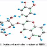 |
Figure 1: Optimized molecular structure of 5H3M1P1C4O Click here to View Figure |
Table 2: Vibrational frequency analysis of 5H3M1P1C4O: Experimental and computational comparison
|
Vibrational Modes |
Experimental Frequency (cm-1) | Frequency (cm-1) | Infrared intensity | Raman intensity | PED
Contribution (%) |
||||
| FT-IR | FT- RAMAN | Unscaled | Scaled | Relative | Absolute | Relative |
Absolute |
||
|
153 |
3350 | 3425 | 3843.71 | 3693.81 | 138.629 | 23.781 | 234.481 | 22.3105 | ν OH 100 |
| 152 | 3325 | 3380 | 3830.4 | 3681.01 | 90.7565 | 15.5688 | 150.541 | 14.3237 |
ν OH 100 |
|
151 |
3300 | 3320 | 3806.92 | 3658.45 | 47.02 | 8.06605 | 53.7717 | 5.11629 | ν OH 100 |
| 150 | 3250 | 3210 | 3796.8 | 3648.72 | 126.595 | 21.7168 | 167.975 | 15.9825 |
ν OH 98 |
|
149 |
3200 | 3082 | 3792.45 | 3644.54 | 63.9077 | 10.9631 | 70.032 | 6.66343 | ν OH 99 |
| 148 | 3085 | 2935 | 3791.26 | 3643.4 | 169.354 | 29.0518 | 129.138 | 12.2873 |
ν OH 98 |
|
147 |
3055 | 2865 | 3789.89 | 3642.08 | 57.1405 | 9.80217 | 54.9198 | 5.22553 | ν OH 98 |
| 146 | 3020 | 1662 | 3309.7 | 3180.62 | 461.023 | 79.0862 | 150.878 | 14.3558 |
ν OH 99 |
|
145 |
2950 | 1655 | 3233.97 | 3107.85 | 1.9755 | 0.33889 | 33.4572 | 3.1834 | ν CH 99 |
| 144 | 2935 | 1610 | 3216.32 | 3090.88 | 1.244 | 0.2134 | 44.0889 | 4.19498 |
ν CH 100 |
|
143 |
2925 | 1582 | 3215.09 | 3089.7 | 0.1517 | 0.02602 | 177.795 | 16.9169 | ν CH 99 |
| 142 | 2875 | 1462 | 3190.75 | 3066.31 | 1.6626 | 0.28521 | 79.7887 | 7.59176 |
ν CH 100 |
|
141 |
2860 | 1270 | 3161.32 | 3038.03 | 12.6209 | 2.16505 | 125.892 | 11.9784 | ν CH 99 |
| 140 | 2850 | 1220 | 3092.82 | 2972.2 | 27.7932 | 4.76779 | 106.962 | 10.1773 |
ν CH 79 |
|
139 |
1660 | 1172 | 3048.06 | 2929.19 | 26.5176 | 4.54896 | 13.3714 | 1.27227 | ν CH 88 |
| 138 | 1645 | 1115 | 3038.46 | 2919.96 | 30.6548 | 5.25868 | 106.142 | 10.0992 |
ν CH 88 |
|
137 |
1630 | 1088 | 3019.81 | 2902.04 | 18.5548 | 3.18298 | 88.8552 | 8.45442 | ν CH 89 |
| 136 | 1580 | 1058 | 2998.57 | 2881.63 | 53.6082 | 9.19622 | 167.686 | 15.955 |
ν CH 46 |
|
135 |
1490 | 1012 | 2983.36 | 2867.01 | 22.0902 | 3.78947 | 60.1473 | 5.72291 | ν CH 46 |
| 134 | 1465 | 855 | 2975.91 | 2859.85 | 3.1301 | 0.53695 | 26.4099 | 2.51286 |
ν CH 46 |
|
133 |
1455 | 835 | 1687.26 | 1621.46 | 582.937 | 100 | 284.171 | 27.0384 | ν CC 22 + βHOC12 |
| 132 | 1445 | 778 | 1653.64 | 1589.15 | 6.1532 | 1.05555 | 432.381 | 41.1403 |
ν CC 36 |
|
131 |
1260 | 765 | 1637.04 | 1573.2 | 181.563 | 31.1462 | 279.56 | 26.5997 | ν CC 18 |
| 130 | 1210 | 702 | 1634.71 | 1570.96 | 371.065 | 63.6544 | 43.2958 | 4.11952 |
ν CC 18 + νOC53 |
|
129 |
1175 | 615 | 1624.05 | 1560.71 | 187.909 | 32.2348 | 129.582 | 12.3295 | ν CC 14 |
| 128 | 1160 | 590 | 1607.37 | 1544.68 | 76.6864 | 13.1552 | 1050.99 | 100 |
ν CC 40 |
|
127 |
1150 | 545 | 1545.95 | 1485.66 | 160.713 | 27.5695 | 26.194 | 2.49231 | βHCC19 |
| 126 | 1120 | 520 | 1524.56 | 1465.1 | 239.1 | 41.0165 | 160.648 | 15.2854 |
ν CC 12 + βHCC14 |
|
125 |
1110 | 495 | 1501.15 | 1442.61 | 1.7228 | 0.29554 | 6.9368 | 0.66002 | ν CC 12 + βHCH91 |
| 124 | 1100 | 470 | 1490.24 | 1432.12 | 178.735 | 30.6611 | 14.3844 | 1.36865 |
ν OC 14 + βHCC12 + βCCO11 |
|
123 |
1090 | 445 | 1482.18 | 1424.37 | 1.1875 | 0.20371 | 3.7953 | 0.36112 | ν OC 14 |
| 122 | 1080 | 420 | 1451.1 | 1394.51 | 77.5271 | 13.2994 | 122.839 | 11.6879 |
ν CC 13 |
|
121 |
1070 | 395 | 1437.79 | 1381.72 | 8.3109 | 1.42569 | 12.026 | 1.14425 | ν CC 13 + τHCCC13 |
| 120 | 1050 | 370 | 1431.67 | 1375.83 | 60.7296 | 10.4179 | 47.7764 | 4.54584 |
ν CC 13 |
|
119 |
1015 | 345 | 1425.91 | 1370.3 | 6.2727 | 1.07605 | 36.3617 | 3.45975 | βHCO18 |
| 118 | 1008 | 320 | 1424.31 | 1368.76 | 83.1424 | 14.2627 | 292.676 | 27.8476 |
βHCO18 |
|
117 |
1000 | 295 | 1417.64 | 1362.35 | 26.1296 | 4.4824 | 14.16 | 1.3473 | βHCO18 |
| 116 | 860 | 270 | 1402.96 | 1348.24 | 18.9522 | 3.25116 | 2.5757 | 0.24507 |
βHCO13 + ωCCCH13 |
|
115 |
845 | 245 | 1401.34 | 1346.69 | 49.9321 | 8.56561 | 3.1578 | 0.30046 | βHCO42 + τHCOC10 |
| 114 | 830 | 220 | 1384.19 | 1330.21 | 212.877 | 36.518 | 229.694 | 21.855 |
νCC12 |
|
113 |
780 | 195 | 1383 | 1329.06 | 111.93 | 19.201 | 109.348 | 10.4042 | βHOC14 + τHCOC11 |
| 112 | 770 | 170 | 1369.07 | 1315.68 | 22.7689 | 3.90589 | 21.261 | 2.02295 |
βHCO13 |
|
111 |
760 | 145 | 1368.44 | 1315.07 | 5.4554 | 0.93585 | 5.008 | 0.4765 | βHCO13 |
| 110 | 705 | 120 | 1359.89 | 1306.85 | 22.2668 | 3.81976 | 4.0052 | 0.38109 |
βHCO13 |
|
109 |
690 | 95 | 1344.29 | 1291.86 | 17.6131 | 3.02144 | 59.0526 | 5.61875 | βHCO13 |
| 108 | 670 | 70 | 1342.48 | 1290.12 | 14.8853 | 2.5535 | 2.44 | 0.23216 |
βHCO13 |
|
107 |
650 | < 50 | 1337.22 | 1285.07 | 312.862 | 53.6699 | 57.5778 | 5.47843 | βHCO13 |
| 106 | 620 | 1313.31 | 1262.09 | 182.665 | 31.3352 | 141.79 | 13.491 |
βHCO13 |
|
|
105 |
600 | 1300.97 | 1250.23 | 14.4749 | 2.4831 | 2.9725 | 0.28283 | νOC17 | |
| 104 | 580 | 1288.5 | 1238.25 | 131.442 | 22.5482 | 22.7504 | 2.16466 |
νOC17 |
|
|
103 |
560 | 1283.76 | 1233.69 | 38.9795 | 6.68674 | 1.5464 | 0.14714 | νOC17 | |
| 102 | 540 | 1264.28 | 1214.97 | 51.8942 | 8.9022 | 1.9889 | 0.18924 |
βHOC23 |
|
|
101 |
< 520 | 1262.14 | 1212.92 | 1.9315 | 0.33134 | 0.7894 | 0.07511 | βHOC23 | |
| 100 | 1244.11 | 1195.59 | 93.5225 | 16.0433 | 2.6543 | 0.25255 |
βHOC23 |
||
|
99 |
1241.44 | 1193.02 | 203.329 | 34.8801 | 6.1869 | 0.58867 | βHOC23 | ||
| 98 | 1231.94 | 1183.89 | 14.6346 | 2.51049 | 4.7406 | 0.45106 |
βHOC23 |
||
|
97 |
1222.24 | 1174.57 | 338.645 | 58.093 | 45.1228 | 4.29336 | νOC14 | ||
| 96 | 1215.96 | 1168.54 | 125.082 | 21.4572 | 7.6617 | 0.729 |
νOC14 |
||
|
95 |
1209.93 | 1162.74 | 55.3913 | 9.50211 | 1.7006 | 0.16181 | νOC12 | ||
| 94 | 1197.18 | 1150.49 | 13.89 | 2.38276 | 26.287 | 2.50116 |
νOC12 |
||
|
93 |
1177.4 | 1131.48 | 25.1413 | 4.31287 | 15.1186 | 1.43851 | βHOC32 | ||
| 92 | 1172.04 | 1126.33 | 220.353 | 37.8005 | 2.6172 | 0.24902 |
βHOC32 |
||
|
91 |
1158.2 | 1113.03 | 56.5365 | 9.69856 | 3.955 | 0.37631 | βHOC32 | ||
| 90 | 1142.43 | 1097.88 | 31.103 | 5.33557 | 3.0025 | 0.28568 |
βHOC32 |
||
|
89 |
1131.16 | 1087.04 | 220.819 | 37.8805 | 12.1282 | 1.15398 | νOC16 | ||
| 88 | 1128.41 | 1084.4 | 103.982 | 17.8375 | 30.8338 | 2.93378 |
νOC16 |
||
|
87 |
1124.57 | 1080.71 | 21.1753 | 3.63252 | 3.0481 | 0.29002 | νOC16 | ||
| 86 | 1110.26 | 1066.96 | 26.8048 | 4.59823 | 20.5222 | 1.95265 |
νOC16 |
||
|
85 |
1099.05 | 1056.19 | 248.247 | 42.5855 | 4.1409 | 0.394 | νCC10 | ||
| 84 | 1098.51 | 1055.67 | 78.7664 | 13.512 | 5.9123 | 0.56255 |
νCC10 |
||
|
83 |
1082.73 | 1040.5 | 63.0776 | 10.8207 | 9.8217 | 0.93452 | βCCC10 | ||
| 82 | 1072.84 | 1031 | 165.164 | 28.333 | 6.2352 | 0.59327 |
βCCC10 |
||
|
81 |
1066.72 | 1025.12 | 90.0196 | 15.4424 | 4.108 | 0.39087 | βCCC10 | ||
| 80 | 1031.15 | 990.935 | 58.0001 | 9.94963 | 2.5077 | 0.2386 |
νOC12 |
||
|
79 |
1026.5 | 986.467 | 31.8523 | 5.46411 | 2.3388 | 0.22253 | νOC14 | ||
| 78 | 1008.9 | 969.553 | 60.4349 | 10.3673 | 22.5392 | 2.14457 |
νCC12 |
||
|
77 |
1002.08 | 962.999 | 86.1077 | 14.7714 | 8.0169 | 0.76279 | νCC12 | ||
| 76 | 957.39 | 920.052 | 25.1537 | 4.31499 | 13.5852 | 1.29261 |
τHCCC48 |
||
|
75 |
954.3 | 917.082 | 5.1818 | 0.88891 | 7.4196 | 0.70596 | τHCCC48 | ||
| 74 | 941.15 | 904.445 | 21.5137 | 3.69057 | 6.3467 | 0.60388 |
νCC16 |
||
|
73 |
902.04 | 866.86 | 19.5451 | 3.35287 | 3.7866 | 0.36029 | τHCCC74 | ||
| 72 | 889.7 | 855.002 | 24.2434 | 4.15884 | 2.7671 | 0.26328 |
τCCCO10 |
||
|
71 |
871.91 | 837.906 | 22.8617 | 3.92181 | 4.1713 | 0.39689 | τCCCO10 | ||
| 70 | 857.62 | 824.173 | 0.5679 | 0.09742 | 0.2187 | 0.02081 |
τHOCC26 + τHCCC43 |
||
|
69 |
853.51 | 820.223 | 8.1174 | 1.3925 | 16.6594 | 1.58511 | τHOCC26 + τHCCC43 | ||
| 68 | 833.91 | 801.388 | 104.517 | 17.9294 | 0.5622 | 0.05349 |
ωOCCC10 |
||
|
67 |
822.59 | 790.509 | 30.4207 | 5.21852 | 4.4014 | 0.41879 | ωOCCC10 | ||
| 66 | 796.14 | 765.091 | 44.7337 | 7.67385 | 34.236 | 3.2575 |
νCC34 |
||
|
65 |
791.65 | 760.776 | 32.525 | 5.5795 | 0.3828 | 0.03642 | ωOCCC17 | ||
| 64 | 785.67 | 755.029 | 23.6002 | 4.0485 | 1.1739 | 0.11169 |
ωOCCC13 |
||
|
63 |
782.87 | 752.338 | 8.8716 | 1.52188 | 1.3199 | 0.12559 | ωOCCC13 | ||
| 62 | 734.14 | 705.509 | 4.9567 | 0.8503 | 3.5529 | 0.33805 |
τCCCC10 |
||
|
61 |
717.85 | 689.854 | 4.6134 | 0.79141 | 0.6591 | 0.06271 | βCCO10 | ||
| 60 | 709.68 | 682.002 | 14.006 | 2.40266 | 4.0864 | 0.38881 |
ωOCCC14 |
||
|
59 |
695.16 | 668.049 | 53.5878 | 9.19272 | 1.013 | 0.09639 | τCCOC10 | ||
| 58 | 690.74 | 663.801 | 4.696 | 0.80558 | 4.0016 | 0.38075 |
βCCC12 |
||
|
57 |
675.59 | 649.242 | 1.4156 | 0.24284 | 6.6709 | 0.63472 | ωOCCC11 | ||
| 56 | 658.61 | 632.924 | 11.9842 | 2.05583 | 14.7621 | 1.40459 |
βCCC12 |
||
|
55 |
636.45 | 611.628 | 13.4297 | 2.3038 | 2.7499 | 0.26165 | βCCC12 | ||
| 54 | 617.96 | 593.86 | 4.2629 | 0.73128 | 9.9248 | 0.94433 |
βCCC12 |
||
|
53 |
615.66 | 591.649 | 1.528 | 0.26212 | 7.6639 | 0.72921 | βCCC12 | ||
| 52 | 605.48 | 581.866 | 25.5533 | 4.38354 | 11.8043 | 1.12316 |
βCCC16 |
||
|
51 |
598.4 | 575.062 | 8.7127 | 1.49462 | 0.3905 | 0.03716 | βCCC11 | ||
| 50 | 585.83 | 562.983 | 25.0641 | 4.29962 | 11.8209 | 1.12474 |
βCCC11 |
||
|
49 |
571.56 | 549.269 | 4.1685 | 0.71509 | 2.6385 | 0.25105 | βCCC11 | ||
| 48 | 561.98 | 540.063 | 7.4439 | 1.27696 | 4.6129 | 0.43891 |
βCCC11 |
||
|
47 |
529.74 | 509.08 | 0.3807 | 0.06531 | 4.7889 | 0.45566 | βOCC16 | ||
| 46 | 517 | 496.837 | 12.7913 | 2.19428 | 11.1347 | 1.05945 |
βOCC16 |
||
|
45 |
483.6 | 464.74 | 8.1116 | 1.39151 | 7.1576 | 0.68103 | βCOC10 | ||
| 44 | 469.62 | 451.305 | 17.518 | 3.00513 | 2.535 | 0.2412 |
βOCC11 |
||
|
43 |
465.69 | 447.528 | 2.2645 | 0.38846 | 1.8741 | 0.17832 | βCCC11 + βOCC12 | ||
| 42 | 455.4 | 437.639 | 2.4131 | 0.41396 | 1.9208 | 0.18276 |
τHOCC10 |
||
| 41 | 446.74 | 429.317 | 192.078 | 32.95 | 2.8012 | 0.26653 | τHOCC10 | ||
| 40 | 433.29 | 416.392 | 17.119 | 2.93668 | 3.2685 | 0.31099 | τHOCC10 | ||
| 39 | 431 | 414.191 | 58.4175 | 10.0212 | 4.5151 | 0.4296 | τHOCC13 | ||
| 38 | 423.92 | 407.387 | 50.8994 | 8.73154 | 3.2905 | 0.31309 | τHOCC29 | ||
| 37 | 416.67 | 400.42 | 34.3882 | 5.89913 | 5.7354 | 0.54571 | τHOCC90 | ||
| 36 | 411.25 | 395.211 | 81.8484 | 14.0407 | 0.9393 | 0.08937 | τHOCC90 | ||
| 35 | 403.26 | 387.533 | 116.007 | 19.9004 | 1.0516 | 0.10006 | τHOCC90 | ||
| 34 | 386.74 | 371.657 | 16.3816 | 2.81018 | 11.1087 | 1.05697 | τHOCC90 | ||
| 33 | 378.97 | 364.19 | 6.213 | 1.06581 | 1.884 | 0.17926 | ωOCCC10 | ||
| 32 | 372.73 | 358.194 | 3.8804 | 0.66566 | 2.2366 | 0.21281 | βCCC17 + βOCC18 | ||
| 31 | 348.11 | 334.534 | 65.0279 | 11.1552 | 1.9364 | 0.18425 | τHOCC60 | ||
| 30 | 346.26 | 332.756 | 40.3214 | 6.91694 | 1.7639 | 0.16783 | τHOCC60 | ||
| 29 | 336.72 | 323.588 | 7.517 | 1.2895 | 2.3298 | 0.22168 | τHOCC60 | ||
| 28 | 327.44 | 314.67 | 3.6346 | 0.6235 | 0.9718 | 0.09247 | τHOCC60 | ||
| 27 | 311.51 | 299.361 | 4.2485 | 0.72881 | 0.5066 | 0.0482 | βOCC26 | ||
| 26 | 305.06 | 293.163 | 6.3213 | 1.08439 | 0.0376 | 0.00358 | βOCC26 | ||
| 25 | 290.56 | 279.228 | 8.0025 | 1.37279 | 1.9549 | 0.18601 | ωCCCC27 | ||
| 24 | 265.84 | 255.472 | 14.3476 | 2.46126 | 1.5795 | 0.15029 | βOCC36 | ||
| 23 | 263.32 | 253.051 | 8.7875 | 1.50745 | 1.0866 | 0.10339 | βOCC36 | ||
| 22 | 258.79 | 248.697 | 2.5513 | 0.43766 | 0.5627 | 0.05354 | τCCCO13 | ||
| 21 | 248.13 | 238.453 | 24.94 | 4.27833 | 1.41 | 0.13416 | βCCC11 + τHOCC13 | ||
| 20 | 244.33 | 234.801 | 48.0298 | 8.23928 | 0.6171 | 0.05872 | νCC11 | ||
| 19 | 232.49 | 223.423 | 62.6991 | 10.7557 | 1.5399 | 0.14652 | νCC11 | ||
| 18 | 227.91 | 219.022 | 10.127 | 1.73724 | 1.3463 | 0.1281 | νCC11 | ||
| 17 | 222.7 | 214.015 | 8.4618 | 1.45158 | 2.8059 | 0.26698 | τCCCC32 | ||
| 16 | 199.72 | 191.931 | 5.5246 | 0.94772 | 5.3172 | 0.50592 | τCCCC32 | ||
| 15 | 187.01 | 179.717 | 2.1046 | 0.36103 | 0.6276 | 0.05972 | βCCO28 | ||
| 14 | 174.58 | 167.771 | 1.3182 | 0.22613 | 1.0614 | 0.10099 | βOCO11 | ||
| 13 | 140.29 | 134.819 | 2.5487 | 0.43722 | 0.8921 | 0.08488 | ωCCOC14 | ||
| 12 | 132.2 | 127.044 | 0.5884 | 0.10094 | 0.7244 | 0.06893 | βOCC10 | ||
| 11 | 119.58 | 114.916 | 0.9859 | 0.16913 | 0.2501 | 0.0238 | τOCCC13 | ||
| 10 | 117.44 | 112.86 | 0.8457 | 0.14508 | 0.1582 | 0.01505 | τCOCC11 | ||
| 9 | 102.98 | 98.9638 | 2.3414 | 0.40166 | 0.41 | 0.03901 | β OCC 12 | ||
| 8 | 87.48 | 84.0683 | 1.4036 | 0.24078 | 1.2029 | 0.11445 | β OCC 12 | ||
| 7 | 67.5 | 64.8675 | 0.4643 | 0.07965 | 1.9087 | 0.18161 | β OCC 12 | ||
| 6 | 59.37 | 57.0546 | 1.9509 | 0.33467 | 1.8421 | 0.17527 | β OCC 12 | ||
| 5 | 42.78 | 41.1116 | 0.8005 | 0.13732 | 1.7286 | 0.16447 | τ COCC 14 | ||
| 4 | 38.78 | 37.2676 | 0.9588 | 0.16448 | 3.4832 | 0.33142 | τ COCC 14 | ||
| 3 | 28.3 | 27.1963 | 0.6116 | 0.10492 | 3.0906 | 0.29407 | τ COCC 14 | ||
| 2 | 20.57 | 19.7678 | 0.3043 | 0.0522 | 2.2566 | 0.21471 | τ COCC 14 | ||
| 1 | 12.17 | 11.6954 | 0.1841 | 0.03158 | 0.819 | 0.07793 | τ OCOC 73 | ||
Vibrational assignment
A PerkinElmer FT-IR spectrophotometer was used to record the 5H3M1P1C4O FT-IR spectrum in the 4000-500 cm⁻¹ range, while a Bruker RFS Raman spectrometer was used to analyze the FT-Raman spectrum in the 3500-500 cm⁻¹.18 Density functional theory was used at the B3LYP/6-311++G(d,p) level to determine the theoretical vibrational frequencies. An improvement in concordance between the experimental and theoretical spectra was seen after scaling all calculated frequencies by a factor of 0.961 to account for anharmonicity and basis-set shortcomings.19 Nonlinear (3N-6) normal vibrational modes are generated by the N atoms in the studied molecule. In order to determine the modes, the VEDA4 algorithm conducted a potential energy distribution (PED) study.20 Table 2 summarizes the tested FT- IR and FT-Raman frequencies, their corresponding scaled theoretical values, and the PED contributions. As seen in Figure 2, the experimental and simulated spectra are correlated.21 The influences of solid states, interactions between molecules, temperature fluctuations, and instrumental resolution can cause small differences in observed and theoretical frequency. The experimental spectra show some weak irregularities, which are explained by instrumental noise.22-24
O–H and C–H vibrations
Phenolic O–H stretching vibrations are characteristically broad due to strong intra- and intermolecular hydrogen bonding, typically appearing in the region 3600–3200 cm⁻¹. In the present compound, a broad absorption envelope observed between 3350 and 3200 cm⁻¹ in the FT-IR spectrum is assigned to O–H stretching modes of hydroxyl groups attached to the chromen-4-one and glycosidic moieties. This broad nature is well reproduced in the simulated spectrum after appropriate scaling and band broadening, confirming extensive hydrogen-bonding interactions in the molecular framework.25
In general, the region of 3100-2850 cm⁻¹ is where you may see aromatic and aliphatic C-H stretching vibrations. The aromatic rings’ and sugar units’ C-H stretching modes are mapped to the area of 5H3M1P1C4O’s FT-IR spectrum where bands of weak to medium intensity are observed. The greater Raman activity of these modes causes them to show up more strongly in the FT-Raman spectrum, especially for symmetric stretching vibrations. The experimental assignments are supported by theoretical calculations that indicate these modes exist between 3080 and 2890 cm⁻¹, with PED contributions surpassing 85%.25
C=O and C–O stretching vibrations
The carbonyl (C=O) stretching vibration is a key spectral marker of chromen-4-one derivatives and typically appears between 1700 and 1650 cm⁻¹. In the FT-IR spectrum of the present compound, a strong absorption band observed in the region 1660–1630 cm⁻¹ is attributed to the C = O stretching vibration of the chromen-4-one ring. The corresponding Raman band appears with moderate intensity, consistent with partial symmetry reduction due to substitution effects. Theoretical calculations locate this vibration in the 1685–1650 cm⁻¹ range, with PED contributions of approximately 60–70%, supporting the experimental observation. C–O stretching vibrations associated with phenolic, ether and glycosidic linkages are expected in the 1300–1000 cm⁻¹ region. Experimentally, prominent FT-IR bands observed near 1260, 1210 and 1080 cm⁻¹, along with corresponding Raman bands, are assigned to these modes. Theoretical frequencies predicted between 1280 and 1050 cm⁻¹, with PED contributions ranging from 40% to 65%, further confirm these assignments.26
C–C skeletal vibrations
Within the 1600-1000 cm⁻¹ range, you may often find aromatic C-C stretching vibrations. According to the FT-IR spectra, the conjugated aromatic system’s C-C stretching modes are represented by bands at around 1580, 1490, 1380 and 1270 cm⁻¹. These vibrations stand out more in the FT-Raman spectrum because they are symmetric and have a delocalized π-electron character. Coherent C-C skeletal vibrations in the 1575-1265 cm⁻¹ range are predicted by theoretical calculations, with PED contributions ranging from 30% to 55%, suggesting moderate mode mixing with neighboring bending movements.11,12,24,26
Bending and deformation vibrations
C–H in-plane and out-of-plane bending vibrations are typically observed between 1450 and 1000 cm⁻¹. In the present compound, these modes are clearly detected in both FT-IR and FT-Raman spectra and are theoretically predicted in the region 1480–1010 cm⁻¹, with PED contributions ranging from 70% to 90%, indicating well-defined bending character.11,12
Skeletal deformation modes involving C–C–C and O–C–C bending vibrations are mainly located below 1000 cm⁻¹. Experimentally, bands observed around 900, 780, 650 and 550 cm⁻¹ are attributed to these deformation modes. Theoretical calculations place these vibrations between 950 and 300 cm⁻¹, with PED contributions of 40–80%, supporting the experimental assignments and confirming the structural rigidity of the molecular backbone.
Spectral correlation
Overall, the scaled DFT-calculated FT-IR and FT-Raman spectra show good agreement with the experimental results. Minor deviations between calculated and observed frequencies can be attributed to anharmonic effects, intermolecular hydrogen bonding, and solid-state interactions not fully accounted for in the gas-phase calculations. The successful correlation between experimental and theoretical vibrational data confirms the structural integrity of 5H3M1P1C4O and validates the reliability of the adopted computational methodology.
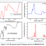 |
Figure 2: FT-IR spectra and FT-Raman spectra of 5H3M1P1C4O Click here to View Figure |
Frontier molecular orbital (FMO) analysis: (Study of HOMO–LUMO)
Frontier molecular orbitals (FMOs), namely the highest occupied molecular orbital (HOMO) and the lowest unoccupied molecular orbital (LUMO), play a crucial role in understanding the electronic structure, stability and reactivity of molecular systems. Analysis of the HOMO and LUMO energies provide valuable insight into the kinetic stability, chemical reactivity, optical polarizability, softness and hardness of a compound. These parameters are also closely associated with charge transfer processes and potential biological interactions.
Table 3: Reactivity profile and molecular stability
| Quantum Chemical Parameter | Gas
Phase |
Water | Methanol
(MeOH) |
Ethanol
(EtOH) |
DMSO |
| HOMO Energy (eV) | -6.729 | -6.254 | -6.256 | -6.258 | -6.255 |
| LUMO Energy (eV) | -2.368 | -2.281 | -2.281 | -2.281 | -2.281 |
| Ionization Energy (eV) | 6.729 | 6.254 | 6.256 | 6.258 | 6.255 |
| Electron Affinity Value (eV) | 2.368 | 2.281 | 2.281 | 2.281 | 2.281 |
| HOMO–LUMO Separation (eV) | 4.361 | 3.973 | 3.975 | 3.977 | 3.974 |
| Absolute Electronegativity (χ) | 4.549 | 4.268 | 4.269 | 4.270 | 4.268 |
| Electronic Chemical Potential (μ) | -4.549 | -4.268 | -4.269 | -4.270 | -4.268 |
| Chemical Hardness (η) | 2.181 | 1.987 | 1.988 | 1.989 | 1.987 |
| Chemical Softness (S) | 0.229 | 0.252 | 0.252 | 0.251 | 0.252 |
| Electrophilic Reactivity Index (ω) | 4.744 | 4.584 | 4.584 | 4.584 | 4.584 |
| Electronic Charge Transfer (ΔN) | 2.086 | 2.148 | 2.148 | 2.147 | 2.148 |
| Electron Donation Power (ω⁻) | 7.291 | 6.966 | 6.966 | 6.967 | 6.966 |
| Electron Acceptance Power (ω⁺) | 2.742 | 2.698 | 2.698 | 2.697 | 2.698 |
For the title compound 5H3M1P1C4O, the calculated HOMO and LUMO energies in the gas phase are −6.729 eV and −2.368 eV, respectively, resulting in an energy gap (ΔE) of 4.361 eV. This moderate HOMO–LUMO gap indicates good molecular stability along with appreciable chemical reactivity. Upon solvation in polar media such as methanol, ethanol, and DMSO, the HOMO energy shifts to −6.256 eV, −6.258 eV and −6.255 eV respectively, while the LUMO energy stabilizes at −2.281 eV in all solvent environments. Consequently, the HOMO–LUMO energy gap decreases to 3.975 eV (MeOH), 3.977 eV (EtOH) and 3.974 eV (DMSO), indicating enhanced electronic delocalization and increased reactivity in solution. The calculated frontier orbital energies and global reactivity descriptors in the gas phase and solvent media are summarized in Table 3.
The reduced HOMO–LUMO gap observed in solvent environments facilitates easier excitation of electrons from the HOMO to the LUMO, which is a desirable feature for intramolecular charge transfer and nonlinear optical (NLO) activity. Moreover, the relatively low energy gap values (< 5eV) obtained for 5H3M1P1C4O in all media suggest potential biological activity, as compounds with smaller band gaps generally exhibit improved redox behavior and enhanced ability to participate in biochemical interactions such as electron donation, electron acceptance and binding to enzyme or receptor active sites.
The solvent effect on the electronic properties of 5H3M1P1C4O was examined using the IEFPCM solvation model, considering polar solvents due to their ability to stabilize the electronic cloud of the molecule. Although slight stabilization of the frontier orbital energies is observed in solvent media, the overall changes remain marginal, indicating that the intrinsic electronic structure of the molecule is largely preserved. The spatial distributions of the HOMO and LUMO orbitals for the gas phase, methanol, ethanol and DMSO are illustrated in Fig. 3. The HOMO density is predominantly localized over electron-rich regions of the molecular framework, whereas the LUMO density is mainly distributed over electron-deficient regions, confirming effective intramolecular charge transfer. The similarity in orbital shapes across different environments highlights the solvent-independent nature of the electronic distribution.
The ionization potential decreases from 6.729 eV in the gas phase to approximately 6.25 eV in solvent media, indicating that electron removal becomes energetically more favourable in solution. Similarly, the electron affinity slightly decreases upon solvation, reflecting moderate stabilization of the LUMO level. These observations collectively confirm the enhanced charge transfer capability of 5H3M1P1C4O in polar solvents.
The electrophilicity index remains relatively high and nearly constant across all solvent environments, confirming the pronounced electrophilic character of the molecule and its strong tendency to interact with electron-rich biomolecular sites. Additionally, the decrease in chemical hardness accompanied by an increase in chemical softness in solvent media indicates improved molecular reactivity and polarizability. The low chemical potential values further suggest that the system remains thermodynamically stable and close to equilibrium. Overall, the frontier molecular orbital analysis and global reactivity descriptors presented in Table 3, together with the HOMO–LUMO visualizations shown in Fig. 3, demonstrate that 5H3M1P1C4O possesses favourable electronic characteristics, solvent-enhanced reactivity, strong electrophilic behaviour and promising optical as well as biological interaction potential.
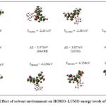 |
Figure 3: Effect of solvent environment on HOMO–LUMO energy levels of 5H3M1P1C4O Click here to View Figure |
Charge distribution (MEP) analysis
The molecular electrostatic potential (MEP) surface provides valuable insight into the charge distribution and solvation behaviour of the molecule. Although pronounced negative electrostatic potential regions are observed around the oxygen atoms of the hydroxyl and carbonyl groups, these electron-rich sites are spatially localized and do not extend over the entire molecular framework. The remaining aromatic and heterocyclic regions exhibit comparatively neutral to positive potential, indicating a dominant nonpolar character.
Despite the presence of multiple oxygen atoms capable of hydrogen bonding, the overall MEP pattern suggests limited interaction with highly polar solvents such as water. The absence of extensive, continuous negative potential regions restricts strong hydration and reduces favourable solute–solvent interactions. Consequently, the molecule exhibits poor affinity towards water molecule, explaining its observed insolubility in aqueous media.
In contrast, moderate stabilization of charge-separated regions in less polar solvents were reflected by slightly enhanced electrostatic potential values, while the overall charge distribution remains unchanged. This behavior indicates that the intrinsic electronic structure governs solubility characteristics. Therefore, the combined presence of localized polar sites and an extended hydrophobic framework account for the water insolubility of 5H3M1P1C4O.
The molecular electrostatic potential values of 5H3M1P1C4O were obtained directly from the color scale shown at the top of the MEP surface plots. In the gas phase, the electrostatic potential ranges from −8.535 × 10⁻² to +8.535 × 10⁻² a.u. In both ethanol and methanol solvent environments, a wider potential distribution is observed, extending from −0.101 × 100 to +0.101 × 100 a.u. This increase in the potential range in polar protic solvents indicates enhanced polarization of the molecule due to solvent–solute interactions, as reflected in the MEP surfaces.
Table 4: Assessment of Fock Matrix stabilization energies using second-order Perturbation method in 5H3M1P1C4O
| Donor Orbital Energy (Ed/e) | Orbital type | Donor orbital | Donor Orbital Energy (Ed/e) | Orbital type | Acceptor orbital | Second-order stabilization energy E(2) kcal/mol a.u | Energy Gap ΔE = E(j) – E(i)b
a.u |
Fock Matrix ElementF(i,j) in a.u. |
| 1.99406 | σ | O12-C33 | 0.04393 | σ* | C17-H38 | 856.93 | 0.23 | 0.398 |
| 1.96896 | σ | C24-C28 | 0.04393 | σ* | C17-H38 | 830.46 | 0.05 | 0.178 |
| 1.9724 | σ | C27-C31 | 0.04393 | σ* | C17-H38 | 769.07 | 0.06 | 0.197 |
| 1.98856 | σ | O12-H53 | 0.39662 | π* | C32-C33 | 442.73 | 0.79 | 0.587 |
| 1.99406 | σ | O12-C33 | 0.39662 | π* | C32-C33 | 438.64 | 1.42 | 0.787 |
| 1.98856 | σ | O12-H53 | 0.03272 | σ* | C16-H37 | 417.23 | 0.07 | 0.158 |
| 1.99325 | σ | O11-C31 | 0.04393 | σ* | C17-H38 | 354.98 | 0.27 | 0.278 |
| 1.96457 | σ | C24-C27 | 0.04393 | σ* | C17-H38 | 317.11 | 0.04 | 0.103 |
| 1.98688 | σ | O11-H52 | 0.0124 | σ* | C27-H46 | 284.55 | 3.67 | 0.912 |
| 1.99406 | σ | O12-C33 | 0.03272 | σ* | C16-H37 | 263.5 | 0.71 | 0.389 |
| 1.97661 | LP1 | O12 | 0.03272 | σ* | C16-H37 | 259.5 | 0.5 | 0.321 |
| 1.99325 | σ | O11-C31 | 0.0124 | σ* | C27-H46 | 236.09 | 3.88 | 0.855 |
| 1.97754 | σ | C32-C33 | 0.03272 | σ* | C16-H37 | 214.83 | 0.57 | 0.314 |
| 1.97661 | LP1 | O12 | 0.39662 | π* | C32-C33 | 200.51 | 1.21 | 0.488 |
| 0.36493 | π* | O8-C21 | 0.26917 | π* | C19-C20 | 190.94 | 0.01 | 0.077 |
| 0.41857 | π* | C26-C30 | 0.47033 | π* | C22-C23 | 160.67 | 0.02 | 0.081 |
| 1.97764 | σ | C32-H49 | 0.03272 | σ* | C16-H37 | 151.99 | 0.37 | 0.211 |
| 1.97149 | σ | C31-C33 | 0.03272 | σ* | C16-H37 | 142.85 | 0.56 | 0.253 |
| 1.97785 | LP1 | O11 | 0.0124 | σ* | C27-H46 | 134.87 | 3.57 | 0.621 |
| 1.97754 | σ | C32-C33 | 0.02011 | σ* | C27-C31 | 127.04 | 0.31 | 0.176 |
| 1.99406 | σ | O12-C33 | 0.02986 | σ* | C16-C18 | 112.27 | 0.94 | 0.291 |
| 1.97661 | LP1 | O12 | 0.02986 | σ* | C16-C18 | 102.32 | 0.72 | 0.243 |
| 1.97754 | σ | C32-C33 | 0.02986 | σ* | C16-C18 | 96.62 | 0.8 | 0.248 |
| 1.97661 | LP1 | O12 | 0.39662 | π* | C32-C33 | 200.51 | 1.21 | 0.488 |
| 0.36493 | π* | O8-C21 | 0.26917 | π* | C19-C20 | 190.94 | 0.01 | 0.077 |
| 0.41857 | π* | C26-C30 | 0.47033 | π* | C22-C23 | 160.67 | 0.02 | 0.081 |
| 1.97764 | σ | C32-H49 | 0.03272 | σ* | C16-H37 | 151.99 | 0.37 | 0.211 |
| 1.97149 | σ | C31-C33 | 0.03272 | σ* | C16-H37 | 142.85 | 0.56 | 0.253 |
| 1.97785 | LP1 | O11 | 0.0124 | σ* | C27-H46 | 134.87 | 3.57 | 0.621 |
| 1.97754 | σ | C32-C33 | 0.02011 | σ* | C27-C31 | 127.04 | 0.31 | 0.176 |
| 1.99406 | σ | O12-C33 | 0.04393 | σ* | C17-H38 | 856.93 | 0.23 | 0.398 |
| 1.96896 | σ | C24-C28 | 0.04393 | σ* | C17-H38 | 830.46 | 0.05 | 0.178 |
| 1.9724 | σ | C27-C31 | 0.04393 | σ* | C17-H38 | 769.07 | 0.06 | 0.197 |
| 1.98856 | σ | O12-H53 | 0.39662 | π* | C32-C33 | 442.73 | 0.79 | 0.587 |
| 1.99406 | σ | O12-C33 | 0.39662 | π* | C32-C33 | 438.64 | 1.42 | 0.787 |
| 1.98856 | σ | O12-H53 | 0.03272 | σ* | C16-H37 | 417.23 | 0.07 | 0.158 |
| 1.99325 | σ | O11-C31 | 0.04393 | σ* | C17-H38 | 354.98 | 0.27 | 0.278 |
| 1.96457 | σ | C24-C27 | 0.04393 | σ* | C17-H38 | 317.11 | 0.04 | 0.103 |
| 1.98688 | σ | O11-H52 | 0.0124 | σ* | C27-H46 | 284.55 | 3.67 | 0.912 |
| 1.99406 | σ | O12-C33 | 0.03272 | σ* | C16-H37 | 263.5 | 0.71 | 0.389 |
| 1.97661 | LP1 | O12 | 0.03272 | σ* | C16-H37 | 259.5 | 0.5 | 0.321 |
| 1.99325 | σ | O11-C31 | 0.0124 | σ* | C27-H46 | 236.09 | 3.88 | 0.855 |
| 1.97754 | σ | C32-C33 | 0.03272 | σ* | C16-H37 | 214.83 | 0.57 | 0.314 |
| 1.97661 | LP1 | O12 | 0.39662 | π* | C32-C33 | 200.51 | 1.21 | 0.488 |
| 0.36493 | π* | O8-C21 | 0.26917 | π* | C19-C20 | 190.94 | 0.01 | 0.077 |
| 0.41857 | π* | C26-C30 | 0.47033 | π* | C22-C23 | 160.67 | 0.02 | 0.081 |
| 1.97764 | σ | C32-H49 | 0.03272 | σ* | C16-H37 | 151.99 | 0.37 | 0.211 |
| 1.97149 | σ | C31-C33 | 0.03272 | σ* | C16-H37 | 142.85 | 0.56 | 0.253 |
| 1.97785 | LP1 | O11 | 0.0124 | σ* | C27-H46 | 134.87 | 3.57 | 0.621 |
| 1.97754 | σ | C32-C33 | 0.02011 | σ* | C27-C31 | 127.04 | 0.31 | 0.176 |
Table 5: NBO hybrid orbital composition and electron density distribution of selected Lewis orbitals in the 5H3M1P1C4O.
|
Bond(A-B)/IP |
EDA%
/ EDB%
|
NBO hybrid / orbitals |
s,p % |
|
(EDA-B)/(EDIP) |
|||
|
σ(O1-C16) |
68.29 | .8264(sp2.58)O | 27.89, 72.06 |
| 1.98039 | 31.71 | .5631(sp4.18)C |
19.25, 80.45 |
|
σ(O1-C17) |
67.31 | .8204(sp2.51)O | 28.46, 71.48 |
| 1.98661 | 32.69 | 0.5717(sp3.44)C |
22.46, 77.27 |
|
σ(C32-H49) |
60.52 | 0.778(sp2.48)C | 28.72, 71.24 |
| 1.97764 | 39.48 | .6283(sp0)H |
99.95, 0.05 |
Natural bond orbital (NBO) analysis
Natural Bond Orbital analysis was carried out to investigate intramolecular charge transfer and the stabilization effects arising from donor–acceptor interactions in the molecule. The second-order perturbation treatment of the Fock matrix quantitatively describes the delocalization of electron density from filled (Lewis) orbitals to unoccupied antibonding (non-Lewis) orbitals, thereby revealing the origin of molecular stabilization.
As presented in Table 4, several significant donor–acceptor interactions contribute to the overall stability of the system. The strongest stabilization is observed for the σ(O12–C33) → σ*(C17–H38) interaction, with a stabilization energy of 856.93 kcal/mol. Comparable σ → σ* interactions include σ(C24–C28) → σ*(C17–H38) and σ(C27–C31) → σ*(C17–H38), exhibiting stabilization energies of 830.46 and 769.07 kcal/mol, respectively. These high E(2) values indicate substantial hyper conjugative charge transfer towards the antibonding C–H orbital.
Notable σ → π* interactions are also evident. The σ(O12–H53) → π*(C32–C33) and σ(O12–C33) → π*(C32–C33) interactions yield stabilization energies of 442.73 and 438.64 kcal/mol, respectively, demonstrating effective conjugative interaction between oxygen containing σ bonds and the adjacent π system.
Additional σ → σ* interactions, such as σ(O12–H53) → σ*(C16–H37) and σ(O12–C33) → σ*(C16–H37), show stabilization energies of 417.23 and 263.50 kcal/mol, confirming the portant role of oxygen-centered bonds in electron redistribution. Lone pair participation is also significant; LP(1)O12 → σ*(C16–H37) and LP(1)O12 → π*(C32–C33) interactions exhibit stabilization energies of 259.50 and 200.51 kcal/mol, respectively. Similarly, LP(1)O11 → σ*(C27–H46) contributes 134.87 kcal/mol, further supporting lone-pair-assisted delocalization.
π* → π* interactions also facilitate electron delocalization within the conjugated framework. The π*(O8–C21) → π*(C19–C20) and π*(C26–C30) → π*(C22–C23) interactions provide stabilization energies of 190.94 and 160.67 kcal/mol, respectively, confirming effective π-electron communication across the molecular skeleton.
Overall, the stabilization energies associated with σ → σ*, σ → π*, LP → σ*, LP → π*, and π* → π* interactions clearly demonstrate pronounced intramolecular charge transfer and hyperconjugative effects. The prominent involvement of oxygen-centered σ bonds and lone pair orbitals highlights their crucial role in modulating the electronic distribution and structural stability of the molecule. The electron density distribution in the occupied bonding orbitals was further examined through NBO hybrid orbital analysis. The percentage contributions of electron density over the bonded atoms, along with their s and p characters, are summarized in Table 5. The σ(O1–C16) and σ(O1–C17) bonding orbitals exhibit strong polarization towards the oxygen atom, with electron density contributions of 68.29% and 67.31%, respectively, while the corresponding carbon atoms contribute 31.71% and 32.69%. The hybridization of oxygen in these bonds corresponds to sp²․58 and sp²․51, with dominant p-character (72.06% and 71.48%), indicating directional bonding and enhanced electronegativity of oxygen.
In contrast, the σ(C32–H49) bond shows higher electron density localization on the carbon atom (60.52%) compared to hydrogen (39.48%). The carbon atom in this bond exhibits sp²․48 hybridization with a predominant p-character (71.24%), whereas hydrogen contributes almost pure s-character (99.95%), as expected. The observed polarization of σ-bonds toward oxygen atoms confirmed that electron density was preferentially accumulated around oxygen, making it the principal electron-rich centre. This uneven charge distribution around oxygen atoms significantly influences the molecular polarity and contributes to the overall electronic behaviour of the molecule.
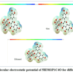 |
Figure 4: Molecular electrostatic potential of 5H3M1P1C4O for different solvents Click here to View Figure |
Table 6: Experimental and time-dependent DFT Study of the electronic excited states of 5H3M1P1C4O
| Exp. Wave length (nm) | Band Gap (eV) | Solve nts | Energy (cm⁻¹) | Theo. Wave length (nm) | Energy Gap (ΔE) (eV) | Oscillator Strength | Electronic State Symmetry | Major contribution |
| 330 | 3.76 | gas | 27983.40591 | 357.3546419 | 3.47 | 0.1664 | Singlet-A |
HOMO->L UMO (92%) |
| 31466.10794 | 317.8022531 | 3.9 | 0.2375 | Singlet-A | H-1->LUMO (92%) | |||
| 32422.68149 | 308.4260629 | 4.02 | 0.0006 | Singlet-A | H-6->LUMO (80%) | |||
| 350 | 3.54 | Me- OH | 27900.33081 | 358.4186893 | 3.46 | 0.3775 | Singlet-A | HO MO->L
UMO (93%) |
| 30500.66229 | 327.8617332 | 3.78 | 0.116 | Singlet-A | H-1->LUMO (93%) | |||
| 33283.27507 | 300.451202 | 4.13 | 0.0618 | Singlet-A |
H-5->LUMO (21%), H- 3->LUMO (15%), H- 2->LUMO (56%) |
|||
| 360 | 3.44 | Et- OH | 27900.33081 | 358.4186893 | 3.46 | 0.3775 | Singlet-A | HO MO->L
UMO (93%) |
| 30500.66229 | 327.8617332 | 3.78 | 0.116 | Singlet-A | H-1->LUMO (93%) | |||
| 33283.27507 | 300.451202 | 4.13 | 0.0618 | Singlet-A |
H-5->LUMO (21%), H- 3->LUMO (15%), H- 2->LUMO (56%) |
Investigation of UV–Visible absorption characteristics
The UV–Visible absorption behaviour of 5H3M1P1C4O was investigated experimentally and theoretically to elucidate its electronic excitation characteristics in different media (Fig. 5 and Table 6). Experimentally, the compound exhibits absorption maxima at 330 nm in the gas phase, 350 nm in methanol, and 360 nm in ethanol, corresponding to optical band gap energies of 3.76 eV, 3.54 eV and 3.44 eV, respectively, indicating a clear bathochromic shift with increasing solvent polarity. Time-dependent DFT calculations reproduce this trend well and predict dominant absorption bands at 357.35 nm (27983 cm⁻¹) in the gas phase, 358.42 nm (27900 cm⁻¹) in methanol, and 358.42 nm (27900 cm⁻¹) in ethanol, which are primarily associated with HOMO → LUMO transitions contributing more than 92–93%, confirming the π → π* nature of the excitation. Additional higher-energy transitions were observed around 317–328 nm and 300–308 nm, arising mainly from HOMO → LUMO and orbital contributions such as H-5, H-3, H-2, and H-6 to LUMO, with varying percentages depending on the medium. The oscillator strength values indicate enhanced transition probability in polar solvents, with the highest intensity observed in methanol (f = 0.3775), reflecting improved stabilization of the excited state. All computed transitions correspond to allowed singlet–singlet (singlet-A) excitations. The gradual reduction in band gap from gas to alcoholic solvents confirms solvent-induced electronic delocalization and stabilization of the excited states in 5H3M1P1C4O, leading to good agreement between experimental observations and theoretical predictions.
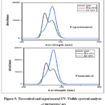 |
Figure 5; Theoretical and experimental UV–Visible spectral analysis of 5H3M1P1C4O Click here to View Figure |
Topological analysis (ELF and LOL)
Electron localization within the molecular framework of 5H3M1P1C4O was examined using the electron localization function (ELF) and the localized orbital locator (LOL), both of which are effective descriptors for understanding bonding characteristics and electron pairing behaviour. The ELF primarily highlights regions associated with covalent bonding and lone-pair electrons, while the LOL emphasizes areas of maximum orbital localization derived from kinetic energy density. As a result, ELF and LOL analyses are complementary and provide consistent insight into the electronic structure of the molecule. The two-dimensional ELF and LOL colour-filled maps along with their corresponding contour plots are illustrated in Figures 6 (a,b) and 6 (c,d). In both representations, regions of high electron localization are indicated by red colour, whereas blue regions correspond to low electron localization. Intermediate localization zones are shown by green-to-yellow shades.
From the ELF maps, strong electron localization is clearly observed around the hydrogen atoms and in the vicinity of σ-bond regions, indicating pronounced Pauli repulsion between electrons of identical spin. These regions appear as intense red patches, confirming the presence of localized bonding interactions. In contrast, comparatively lower ELF values (blue regions) are distributed around selected carbon and oxygen atoms within the heterocyclic and aromatic framework, reflecting reduced electron localization in these regions.
The LOL plots further support these observations. High LOL values, represented by red contours, are predominantly associated with non-bonding and localized valence regions, particularly around hydrogen atoms and specific bonding domains. Regions close to the inner valence shells, where electron density is relatively depleted, display lower LOL values and appear in blue. This distribution indicates that while the molecule contains localized bonding domains, extended delocalization is limited within the core framework.
Overall, the combined ELF and LOL analyses confirm that 5H3M1P1C4O exhibits distinct localized electron density around hydrogen atoms and selected bonding regions, along with comparatively weaker localization near certain carbon and oxygen centres. This electron distribution pattern reflects the balance between localized covalent interactions and regions of lower electron concentration within the molecular structure.
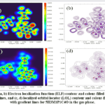 |
Figure 6: (a, b) Electron localization function (ELF) contour and colour-filled maps with gradient lines, and (c, d) localized orbital locator (LOL) contour and colour-filled maps with gradient lines for 5H3M1P1C4O in the gas phase. |
Table 7: Computational study of local softness and dual reactivity indices in 5H3M1P1C4O
| Mulliken atomic charges Fukui functions Fukui functions | ||||||||||
| Atom | 0, 1 (N) | N+1 (-1, 2) | N-1 (1,2) | fr+ | fr– | fr0 | Δfr | sr+ ƒr+ | sr–ƒr– | sr0 ƒr0 |
| 1O | -0.01068 | -0.00116 | – | 0.010 | -0.005 | 0.002 | 0.014 | 0.002 | -0.001 | 0.001 |
| 2O | 0.018039 | -0.00501 | 0.057984 | -0.023 | -0.040 | -0.031 | 0.017 | -0.005 | -0.009 | -0.007 |
| 3O | – | -0.227663 | – | -0.017 | -0.023 | -0.020 | 0.007 | -0.004 | -0.005 | -0.004 |
| 4O | – | -0.167446 | – | -0.006 | -0.008 | -0.007 | 0.002 | -0.001 | -0.002 | -0.002 |
| 5O | – | -0.243562 | – | -0.020 | -0.026 | -0.023 | 0.006 | -0.004 | -0.006 | -0.005 |
| 6O | – | -0.248266 | – | -0.015 | -0.022 | -0.018 | 0.007 | -0.003 | -0.005 | -0.004 |
| 7O | 0.049596 | 0.006853 | 0.091724 | -0.043 | -0.042 | -0.042 | -0.001 | -0.009 | -0.009 | -0.009 |
| 80 | – | -0.422319 | – | -0.113 | -0.053 | -0.083 | -0.060 | -0.025 | -0.012 | -0.018 |
| 9O | – | -0.282931 | – | -0.045 | -0.075 | -0.060 | 0.030 | -0.010 | -0.016 | -0.013 |
| 10O | – | -0.240479 | – | -0.037 | -0.045 | -0.041 | 0.008 | -0.008 | -0.010 | -0.009 |
| 11O | – | -0.250443 | – | -0.026 | -0.046 | -0.036 | 0.020 | -0.006 | -0.010 | -0.008 |
| 12O | – | -0.328074 | – | -0.037 | -0.052 | -0.045 | 0.015 | -0.008 | -0.011 | -0.010 |
| 13C | -0.29772 | -0.308481 | – | -0.011 | -0.002 | -0.007 | -0.008 | -0.002 | -0.001 | -0.001 |
| 14C | – | -0.356585 | – | 0.002 | -0.003 | -0.001 | 0.006 | 0.000 | -0.001 | 0.000 |
| 15C | – | -0.574056 | – | 0.026 | 0.011 | 0.019 | 0.015 | 0.006 | 0.002 | 0.004 |
| 16C | -0.01619 | 0.025243 | – | 0.041 | 0.042 | 0.042 | -0.001 | 0.009 | 0.009 | 0.009 |
| 17C | – | -0.29122 | – | 0.029 | 0.056 | 0.043 | -0.028 | 0.006 | 0.012 | 0.009 |
| 18C | – | -0.438446 | – | -0.032 | -0.028 | -0.030 | -0.004 | -0.007 | -0.006 | -0.007 |
| 19C | 0.394348 | 0.340841 | 0.473679 | -0.054 | -0.079 | -0.066 | 0.026 | -0.012 | -0.017 | -0.014 |
| 20C | -0.53715 | -0.577915 | – | -0.041 | -0.014 | -0.028 | -0.026 | -0.009 | -0.003 | -0.006 |
| 21C | 0.289052 | 0.197712 | 0.309788 | -0.091 | -0.021 | -0.056 | -0.071 | -0.020 | -0.005 | -0.012 |
| 22C | 0.708338 | 0.668081 | 0.768894 | -0.040 | -0.061 | -0.050 | 0.020 | -0.009 | -0.013 | -0.011 |
| 23C | – | -0.402689 | – | 0.010 | 0.011 | 0.010 | -0.001 | 0.002 | 0.002 | 0.002 |
| 24C | 0.388096 | 0.395721 | 0.414527 | 0.008 | -0.026 | -0.009 | 0.034 | 0.002 | -0.006 | -0.002 |
| 25C | – | -0.036581 | 0.013459 | -0.031 | -0.019 | -0.025 | -0.013 | -0.007 | -0.004 | -0.005 |
| 26C | -0.43701 | -0.455734 | – | -0.019 | -0.049 | -0.034 | 0.030 | -0.004 | -0.011 | -0.007 |
| 27C | 0.112514 | 0.087299 | 0.121774 | -0.025 | -0.009 | -0.017 | -0.016 | -0.005 | -0.002 | -0.004 |
| 28C | – | -0.348737 | – | -0.068 | -0.032 | -0.050 | -0.035 | -0.015 | -0.007 | -0.011 |
| 29C | – | -0.008255 | – | 0.014 | -0.011 | 0.001 | 0.026 | 0.003 | -0.002 | 0.000 |
| 30C | – | -0.534106 | – | 0.017 | 0.030 | 0.023 | -0.013 | 0.004 | 0.007 | 0.005 |
| 31C | – | -0.379118 | – | 0.025 | 0.022 | 0.024 | 0.003 | 0.006 | 0.005 | 0.005 |
| 32C | 0.132265 | 0.086159 | 0.164996 | -0.046 | -0.033 | -0.039 | -0.013 | -0.010 | -0.007 | -0.009 |
| 33C | – | -0.284319 | – | -0.048 | -0.028 | -0.038 | -0.020 | -0.010 | -0.006 | -0.008 |
| 34H | 0.220642 | 0.21275 | 0.23189 | -0.008 | -0.011 | -0.010 | 0.003 | -0.002 | -0.002 | -0.002 |
| 35H | 0.233727 | 0.208046 | 0.26025 | -0.026 | -0.027 | -0.026 | 0.001 | -0.006 | -0.006 | -0.006 |
| 36H | 0.310178 | 0.297186 | 0.323083 | -0.013 | -0.013 | -0.013 | 0.000 | -0.003 | -0.003 | -0.003 |
| 37H | 0.191947 | 0.183313 | 0.203497 | -0.009 | -0.012 | -0.010 | 0.003 | -0.002 | -0.003 | -0.002 |
| 38H | 0.266009 | 0.282077 | 0.272174 | 0.016 | -0.006 | 0.005 | 0.022 | 0.004 | -0.001 | 0.001 |
| 39H | 0.19258 | 0.181861 | 0.204588 | -0.011 | -0.012 | -0.011 | 0.001 | -0.002 | -0.003 | -0.002 |
| 40H | 0.191618 | 0.172675 | 0.211114 | -0.019 | -0.019 | -0.019 | 0.001 | -0.004 | -0.004 | -0.004 |
| 41H | 0.267403 | 0.267071 | 0.268521 | 0.000 | -0.001 | -0.001 | 0.001 | 0.000 | 0.000 | 0.000 |
| 42H | 0.260044 | 0.254509 | 0.265874 | -0.006 | -0.006 | -0.006 | 0.000 | -0.001 | -0.001 | -0.001 |
| 43H | 0.335272 | 0.349765 | 0.324732 | 0.014 | 0.011 | 0.013 | 0.004 | 0.003 | 0.002 | 0.003 |
| 44H | 0.264015 | 0.27421 | 0.254978 | 0.010 | 0.009 | 0.010 | 0.001 | 0.002 | 0.002 | 0.002 |
| 45H | 0.144256 | 0.110374 | 0.189544 | -0.034 | -0.045 | -0.040 | 0.011 | -0.007 | -0.010 | -0.009 |
| 46H | 0.245639 | 0.217867 | 0.262173 | -0.028 | -0.017 | -0.022 | -0.011 | -0.006 | -0.004 | -0.005 |
| 47H | 0.219224 | 0.191087 | 0.244437 | -0.028 | -0.025 | -0.027 | -0.003 | -0.006 | -0.005 | -0.006 |
| 48H | 0.218477 | 0.173681 | 0.262683 | -0.045 | -0.044 | -0.045 | -0.001 | -0.010 | -0.010 | -0.010 |
| 49H | 0.165247 | 0.118982 | 0.205164 | -0.046 | -0.040 | -0.043 | -0.006 | -0.010 | -0.009 | -0.009 |
| 50H | 0.331162 | 0.323895 | 0.341306 | -0.007 | -0.010 | -0.009 | 0.003 | -0.002 | -0.002 | -0.002 |
| 51H | 0.272274 | 0.256592 | 0.286162 | -0.016 | -0.014 | -0.015 | -0.002 | -0.003 | -0.003 | -0.003 |
| 52H | 0.284629 | 0.266622 | 0.299652 | -0.018 | -0.015 | -0.017 | -0.003 | -0.004 | -0.003 | -0.004 |
| 53H | 0.284986 | 0.263116 | 0.305226 | -0.022 | -0.020 | -0.021 | -0.002 | -0.005 | -0.004 | -0.005 |
Table 8: Computational Investigation of NLO Characteristics of 5H3M1P1C4O
| Parameter | B3LYP/6-311++G(d,p) | Parameter | B3LYP/6-311++G(d,p) |
| µx | 1.1598463 | β | -728.7821815 |
| µy | 3.3593608 | βxxy | 431.5888174 |
| µz | 0.2408685 | βxyy | 374.1637194 |
| µ(D) | 3.562101354 | βyyy | -2247.860405 |
| αxx | 213.028 | βzxx | -90.2845594 |
| αxy | -7.9288341 | βxyz | 39.8321483 |
| αyy | 399.9339353 | βzyy | 187.0148623 |
| αxz | 27.3846318 | βxzz | -22.5386529 |
| αyz | -6.5363008 | βyzz | -43.7891065 |
| αzz | 190.5032663 | βzzz | 24.1094624 |
| α (a.u) | 267.8217339 | βtot (a.u) | 1901.755905 |
| α (e.s.u) | 3.9691E-23 | βtot (e.s.u) | 1.6430E-29 |
| ∆α (a.u) | 419.2779329 | mz | 0.2408685 |
| ∆α (e.s.u) | 6.2137E-23 | m(D) | 3.562101354 |
Fukui function analysis and dual reactivity descriptors
The Fukui function, which is derived from Mulliken population analysis and used within the density functional theory framework, provides a good description of the electrophilic and nucleophilic properties of specific atomic sites. By characterizing a molecule’s electron acceptivity or electron donation, the Fukui indices quantify its chemical reactivity and selectivity.
Hence, fr− corresponds to the electrophilic attack index, and fr+ represents the nucleophilic attack index in the Fukui function. Positive values suggest nucleophilic character, and negative values signal electrophilic nature, which is further made easier to identify reactive sites by dual descriptor Δfr. Table 7 displays the molecule’s computed dual descriptor values and decreased Fukui functions. Amino acid 21C has the highest negative value (-0.071) in the data, suggesting that it is the electrophilic site in the molecule. Alternatively, the most strongly nucleophilic behavior is shown by the greatest positive value (0.034) for atom 24C. O7 and O8 are just a couple of the oxygen atoms; C13, C17, C20, C21, C25, C27, C28, C30, C32 and C33 are a bunch of carbon atoms; and numerous hydrogen atoms are electrophilic centers because they have negative values. However, some hydrogen atoms and atoms such as O1, O2, O3, O4, O5, O6, O9, O10, O11, O12, C14, C15, C19, C22, C24, C26, C29 and C31 show positive Δfr values, suggesting that they have nucleophilic potential. The existence of electrophilic and nucleophilic areas gives credence to the molecule’s strong chemical reactivity and implies that it can effectively interact with biological systems.
Hyperpolarizability investigations
Nonlinear optics (NLO) deals with phenomena that arise from the nonlinear interaction between electromagnetic radiation and matter, particularly when intense light propagates through an optical medium. In recent years, the development of molecular systems exhibiting enhanced NLO responses has attracted considerable attention because of their wide-ranging applications in optical switching, signal modulation, data storage and telecommunication technologies. Materials possessing strong nonlinear responses enable efficient manipulation of light, thereby extending the functional limits of modern optical science.27
From a theoretical perspective, molecular polarizability and hyperpolarizability parameters obtained through quantum chemical calculations are often found to correlate well with experimental observations. Consequently, comparison of the calculated NLO parameters with those of a standard reference material such as urea provides a reliable measure for assessing the NLO efficiency of newly designed molecules.28
Table 8 summarizes the calculated nonlinear optical properties of 5H3M1P1C4O in the gas phase, which include the dipole moment (μ), average polarizability (α), polarizability anisotropy (Δα), and first-order hyperpolarizability (β). Confirming the presence of charge asymmetry within the molecule and demonstrating favorable dipolar properties necessary for nonlinear optical activity, the optimized structure displays a non-zero dipole moment (μ = 3.56 D).29 The significant anisotropy of polarizability (Δα = 6.214 × 10⁻²³ esu) indicates that the electronic reaction is direction-dependent, while the computed mean polarizability value (α = 3.969 × 10⁻²³ esu) shows how easily the electron cloud may be warped by an external electric field. It is worth noting that the title molecule has a substantially higher total first-order hyperpolarizability (β = 1.643 × 10⁻²⁹ esu) than urea, a regularly used benchmark NLO material. The molecular framework’s efficient intramolecular charge transfer, robust donor-acceptor contacts, and prolonged π-conjugation are the reasons for this improvement.30
In addition to global NLO parameters, local reactivity descriptors derived from Mulliken atomic charges and Fukui functions (Table 7) provide deeper insight into the charge-transfer behavior governing the nonlinear response. The oxygen atoms exhibit comparatively larger negative charges and notable values of local softness, indicating their dominant role as electron-rich centers. Conversely, several carbon and hydrogen atoms display positive Fukui function values for electrophilic attack, highlighting potential charge migration pathways within the molecule. The dual descriptor analysis further confirms the coexistence of electrophilic and nucleophilic regions, reinforcing the push–pull electronic nature of the compound.31
Overall, the combined analysis of hyperpolarizability parameters and local softness descriptors clearly demonstrates that 5H3M1P1C4O possesses strong nonlinear optical characteristics. These findings establish the title compound as a promising candidate for future NLO applications and suggest its potential relevance in optoelectronic and medicinal chemistry domains where charge-transfer interactions play a crucial role.32
Integrated RDG analysis and structural stability
Using Reduced Density Gradient (RDG) analysis, which allows the visualization of non-covalent interactions (NCIs) based on electron density and its derivatives, the structural stability and conformational landscape of 5H3M1P1C4O were assessed.33,34 Figure 7 shows the RDG scatter plot, which divides the interaction regimes into three regions: the blue region represents strong attractive interactions, the green region represents Van der Waals forces, and the red region represents steric repulsion. The presence of strong intramolecular hydrogen bonding that supports the structural integrity of the molecule is indicated by the prominent blue spikes, which show that attractive stabilizing forces are dominant.35 These results are in good agreement with the experimental Raman spectra, which show that the localized strength of these H-bond networks is reflected by the high-frequency hydroxyl stretching modes (3200-3850 cm– 1) with large potential energy distribution (PED) values. Steric repulsion (λ2)ρ > 0) inside the aromatic rings and sugar moiety is crucial in defining the final 3D conformation, and the presence of dispersive Van der Waals interactions (λ2)ρ = 0) enhances binding efficiency in addition to these stronger forces.36,37
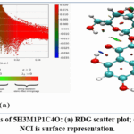 |
Figure 7: NCI analysis of 5H3M1P1C4O: (a) RDG scatter plot; (b) Corresponding 3D NCI is surface representation. Click here to View Figure |
Table 9: Evaluation of the Physicochemical Properties of 5H3M1P1C4O
| Physicochemical Properties | Drug-Likeness | ||
| Lipophilicity Index (WLOGP) | -0.54 | Bio availability score | 0.17 |
| Molecular weight | 464.38 g/mol | Pharmacokinetics | |
| Log S | -3.04 | GI absorption | LOW |
| Number of aromatic heavy | 16 | BBB permeant | NO |
| Fraction Csp3 | 0.29 | P-gp substrate | NO |
| Number of rotatable bonds | 4 | Log Kp (skin permeation) | -8.88 cm/ s |
| Number of H-bond acceptors | 12 | ||
| Number of H-bond donors | 8 | ||
| Molar refractivity | 110.16 | ||
| TPSA | 210.51 A2 | ||
TPSA: Topological Polar Surface Area; BBB: Blood-brain barrier penetration; log kp: skin permeability.
Pharmaceutical analysis of 5H3M1P1C4O
The oral bioavailability of the studied compound 5H3M1P1C4O was evaluated using Swiss ADME-predicted physicochemical, pharmacokinetic, and drug-likeness parameters, as summarized in Table 9. The compound exhibits a molecular weight of 464.38 g/mol and contains four rotatable bonds, both of which fall within the acceptable limits for orally administered drug candidates. Although the compound shows a relatively high number of hydrogen bond donors (8) and acceptors (12), it demonstrates favorable aqueous solubility across multiple predictive models (Log S_ESOL = −3.04; Log S_SILICOS-IT = −1.51), ensuring effective dissolution in the gastrointestinal environment. The consensus lipophilicity value (log P = −0.38) indicates a hydrophilic character that supports solubility while minimizing nonspecific tissue accumulation.38,39
The elevated topological polar surface area (TPSA = 210.51 Ų) accounts for the predicted low gastrointestinal absorption and the absence of blood–brain barrier permeation. Importantly, the compound is not predicted to be a P-glycoprotein substrate and does not inhibit major cytochrome P450 isoforms, suggesting a low risk of efflux-mediated transport and metabolic drug–drug interactions. The physicochemical and pharmacokinetic properties listed in Table 9 comply with the acceptable criteria for orally active drug candidates, with a predicted bioavailability score of 0.17. These results suggest that 5H3M1P1C4O is a potential candidate for further development as an oral therapeutic agent.40,41
Table 10: Molecular docking analysis of 5H3M1P1C4O with target proteins
| Protein (PDB ID) | Source Organism | Binding Energy (kcal/mol) | Inhibition Constant (µM) | Inter molecular energy (kcal/mol) | Electrostatic energy (kcal/mol) | Total internal energy (kcal/mol) | Referen ce RMSD (Å) |
| 3ERT | Homosapien | -4.29 | 712.31 | -7.87 | -0.28 | -8.48 | 37.26 |
| 6J90 | Homosapien | -4.25 | 766.68 | -7.83 | -0.62 | -6.96 | 58.83 |
| 7RPU | Homosapien | -5.23 | 147.73 | -8.81 | -0.48 | -6.18 | 94.78 |
| 8TSK | Homosapien | -3.67 | 2.03 | -7.25 | -0.14 | -6.48 | 40.69 |
Table 11: Molecular docking–derived binding energies and major non-covalent interactions of ligand 5H3M1P1C4O with selected antineoplastic target proteins.
| Protein | B.E energy (kcal/mol) | Interacting residues | Type of interaction | Distance (Å) |
| 3ERT | −4.29 | VAL12, LEU13 | Hydrophobic | 3.5–4.5 |
| ILE10, TYR5 | π–Alkyl / π–π | 3.8–4.2 | ||
| LYS11, LYS21 | Hydrogen bond | 2.0–2.8 | ||
| SER9 | Hydrogen bond | 2.1–2.6 | ||
| 6J90 | −4.25 | GLU31 | Hydrogen bond | 2.0–2.7 |
| TYR52, TRP38 | π–π stacking | 3.6–4.1 | ||
| LEU35, ALA32 | Hydrophobic | 3.7–4.6 | ||
| 7RPU | −5.23 | HIS41 | Hydrogen bond | 2.0–2.5 |
| LEU27, VAL25 | Hydrophobic | 3.6–4.4 | ||
| PHE48 | π–σ / π–Alkyl | 3.8–4.2 | ||
| 8TSK | −3.67 | SER88 | Hydrogen bond | 2.1–2.7 |
| TYR90, PHE92 | π–π / π–Alkyl | 3.7–4.3 | ||
| LEU85 | Hydrophobic | 3.8–4.6 |
Molecular docking analysis of 5H3M1P1C4O with four selected protein targets
The molecular docking performance of the ligand 5H3M1P1C4O against the four target proteins was comparatively evaluated using binding energy and interaction distance as the primary criteria. The quantitative docking parameters are summarized in Table 10 and Table 11, while the corresponding three-dimensional and two-dimensional binding conformations are illustrated in Fig. 8–11.
Table 10 summarizes the docking scores and matching binding parameters that were computed. Results showing negative binding energies for all four proteins show that the ligand interacts favourably inside their active regions. The binding energy of the ligand was −5.23 kcal/mol for 7RPU, −4.29 kcal/mol for 3ERT, −4.25 kcal/mol for 6J90 and −3.67 kcal/mol for 8TSK, out of all the targets that were studied. Distinct structural contexts and amino acid compositions of binding pockets among proteins account for the detected fluctuations in binding energy.
The protein-ligand interactions are examined in depth in Table 11, which lists the important amino acid residues that bind and the kind of interactions between them. Depending on the protein target, the ligand might establish electrostatic contacts, hydrophobic alkyl interactions, π-alkyl, carbon-hydrogen, and traditional hydrogen bonds as stabilizing interactions. These linked residues are essential for docked complex stability and for attaching the ligand to the active site.
Hydrogen bonding interactions were found to be particularly important in stabilizing the ligand within the binding cavity, while hydrophobic interactions further enhanced binding strength by promoting favourable van der Waals contacts. The presence of multiple interaction types suggests that the ligand is well accommodated within the active-site regions of the studied proteins. Overall, the docking results summarized in Tables 10 and 11 demonstrate that 5H3M1P1C4O exhibits a stable and energetically favourable binding profile, with 7RPU showing the most promising interaction characteristics among the evaluated targets.
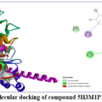 |
Figure 8: 3D and 2D Molecular docking of compound 5H3M1P1C4O with 3ERT protein Click here to View Figure |
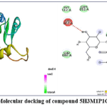 |
Figure 9: 3D and 2D Molecular docking of compound 5H3M1P1C4O with 6J90 protein Click here to View Figure |
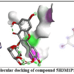 |
Fig.10 3D and 2D Molecular docking of compound 5H3M1P1C4O with 7RPU protein Click here to View Figure |
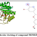 |
Figure 11: 3D and 2D Molecular docking of compound 5H3M1P1C4O with 8TSK protein Click here to View Figure |
Conclusion
The integrated molecular docking and density functional theory analyses provide mechanistic insight into the interaction profile of 5H3M1P1C4O with selected cancer-related protein targets. The investigated proteins represent key molecular systems involved in cancer- associated pathways, rather than being linked to a single cancer type. This approach enables evaluation of the general anticancer potential of the compound across diverse biological targets.42,43
Comparative docking analysis revealed noticeable differences in binding affinity and interaction patterns among the four protein structures. One target protein exhibited the most favorable binding energy, indicating stronger ligand–protein complex stability compared with the remaining proteins. This enhanced affinity can be attributed to the efficient accommodation of 5H3M1P1C4O within the active site, supported by a combination of π–π stacking, π–alkyl, hydrophobic and van der Waals interactions involving key amino acid residues lining the binding pocket.43-45
Proteins displaying less favourable docking scores showed comparatively weaker interaction networks, characterized by reduced hydrophobic complementarity and fewer stabilizing non- covalent contacts. These findings suggest that the anticancer activity of 5H3M1P1C4O is strongly influenced by binding site geometry, residue composition, and electronic compatibility, rather than disease specificity.45,46
From a structure-based design perspective, the aromatic and heterocyclic architecture of 5H3M1P1C4O plays a critical role in mediating hydrophobic and π-electron interactions, while limited hydrogen bonding contributes to positional stabilization. These features collectively highlight the importance of molecular planarity and electronic distribution in achieving favorable protein binding.46,47
Complementary DFT calculations further support the docking results by revealing delocalized electron density across the chromen-4-one framework, facilitating π-mediated interactions with aromatic residues of the proteins. The calculated HOMO–LUMO energy gap indicates adequate chemical stability combined with sufficient reactivity to support non-covalent binding. In addition, the frontier molecular orbital distribution identifies potential sites for rational functional modification aimed at enhancing interaction strength and target selectivity.48
Acknowledgement
The authors would like to state that there are no acknowledgements related to this work.
Funding Sources
The author(s) received no financial support for the research, authorship, and/or publication of this article.
Conflict of Interest
The author(s) do not have any conflict of interest.
Data Availability Statement
This statement does not apply to this article.
Ethics Statement
This research did not involve human participants, animal subjects, or any material that requires ethical approval.
Author Contributions
- Thangavelan Niruban Balu: Investigation, formal analysis, and preparation of the original draft.
- P. Venkatesh: Supervision and guidance of the research work.
- Rajakumari Subramaniyan: Concept development, manuscript review and editing, data curation, and visualization.
References
- World Health Organization. WHO Report, 2024.
- International Agency for Research on Cancer. IARC, 2024.
- Kilmister, E. J. Review Article, 2023.
- Lim, Z. F.; Ma, P. C. Journal of Hematology & Oncology, 2019, 12, , 134-150. https://doi.org/10.1186/s13045-019-0813-0
- Khan, S. U. Cancer Drug Resistance, 2022, 5, , 123-145. https://doi.org/10.20517/cdr.2022.12
CrossRef - Jain, A. Cancers, 2021, 13, 425. https://doi.org/10.3390/cancers13030425
CrossRef - Al-Karmalawy, A. A. Molecules, 2021, 26, 518. https://doi.org/10.3390/molecules26020518
- Meng, X. Y.; Zhang, H. X.; Mezei, M.; Cui, M. Current Computer-Aided Drug Design, 2011, 7, 46-157. https://doi.org/10.2174/157340911795677602
CrossRef - Agu, P. C. Scientific Reports, 2023, 13,13398. https://doi.org/10.1038/s41598-023-39921-3
- Magaji, B. Journal of Molecular Modeling, 2024, 30, 145. https://doi.org/10.1007/s00894-024-05521-0
- Paneru, T. R. Journal of Molecular Structure, 2024, 1290, 136-145. https://doi.org/10.1016/j.molstruc.2024.136145
- El-Shamy, N. T. Molecules, 2022, 27, 620. https://doi.org/10.3390/molecules27030620
CrossRef - Mellaoui, M. D. Journal of Molecular Structure, 2024, 1295, 137210. https://doi.org/10.1016/j.molstruc.2024. 137210
- Morris, G. M.; Huey, R.; Lindstrom, W.; Olson, A. J. Journal of Computational Chemistry, 2009, 30, 2785-2791. https://doi.org/10.1002/jcc.21256
CrossRef - RCSB Protein Data Bank. Protein Data Bank, 2024, 3ERT. https://doi.org/10.2210/pdb3ert/pdb
CrossRef - NCBI Structure Database. MMDB, 2024,3ERT.
- Morris, G. M.; Huey, R.; Lindstrom, W.; Olson, A. J. Journal of Computational Chemistry, 2009, 30, 2785-2791. https://doi.org/10.1002/jcc.21256
CrossRef - Scripps Research Institute. User Guide, 2023.
- Jebapriya, J. C.; Raj, R.; Kumar, S. Materials Today: Proceedings, 2022, 62, 2100- 2107. https://doi.org/10.1016/ j.matpr. 2022. 02.210
CrossRef - Potla, K. M. Vibrational Spectroscopy, 2023, 124, 103456. https://doi.org/10.1016/j.vibspec.2023.103456
- Govindammal, M.; Saravanan, R.; Kumar, V. Heliyon, 2020, 6, e03912. https://doi.org/10.1016/j. heliyon.2020. e03912
CrossRef - Saral, A.; Kumar, S.; Devi, R. Heliyon, 2024, 10, e24011. https://doi.org/10.1016/j.heliyon.2024.e24011
CrossRef - Edwin, B.; Joseph, P.; Thomas, K. Vibrational Spectroscopy, 2012, 60, 150-156. https://doi.org/10.1016/j.vibspec. 2012.02.005
CrossRef - Herzberg, G. Van Nostrand, 1950.
- Jamróz, M. H. Vibrational Spectroscopy, 2013, 114, 1-15. https://doi.org/10.1016/j.vibspec.2013.04.002
CrossRef - Çırak, Ç.; Karabacak, M.; Kurt, M. Vibrational Spectroscopy, 2011, 57, 123-130.https://doi.org/10.1016/j. vibspec. 2011.05.003
CrossRef - Çırak, Ç.; Karabacak, M.; Kurt, M. Journal of Molecular Structure, 2012, 1012, 1-8. https://doi.org/10.1016/j.molstruc.2011.12.021
CrossRef - Suvitha, A.; Kumar, P.; Devi, L. Spectrochimica Acta Part A, 2014, 120, 1-7. https://doi.org/10.1016/j.saa.2013.09.012
CrossRef - Dai, F.; Liu, Y.; Zhang, H. Fuel, 2023, 332, 125133. https://doi.org/10.1016/j.fuel.2022.125133
CrossRef - OpenStax. LibreTexts, 2024.
- Tsioptsias, C.; Pavlidou, E.; Panayiotou, C. Journal of Molecular Structure, 2022, 1267, 133614. https://doi.org/10.1016/j.molstruc.2022.133614
CrossRef - Malaganvi, S. S.; Patil, P.; Kumar, R. Journal of Molecular Structure, 2019, 1195, 1-9. https://doi.org/10.1016/j.molstruc.2019.05.021
CrossRef - Boyd, R. W. Academic Press, 2020.
- Prasad, P. N.; Williams, D. J. Wiley, 1991.
- Zyss, J.; Ledoux, I. Chemical Reviews, 1994, 94, 77-105. https://doi.org/10.1021/cr00025a003
CrossRef - Janani, J.; Kumar, S.; Devi, R. Journal of Molecular Modeling, 2023, 29, 210. https://doi.org/10.1007/s00894-023-05521-3
- Kumar, S.; Patel, R.; Singh, V. Optical Materials, 2023, 140, 113921. https://doi.org/10.1016/j.optmat.2023.113921
CrossRef - Geerlings, P.; De Proft, F.; Langenaeker, W. Chemical Reviews, 2003, 103, 1793-https://doi.org/10.1021/cr990 029p
CrossRef - Chakraborty, D.; Chattaraj, P. K. Chemical Science, 2021, 12, 6264-6279. https://doi.org/10.1039/D1SC01234A
CrossRef - Johnson, E. R.; Keinan, S.; Mori-Sánchez, P.; Contreras-García, J.; Cohen, A. J.; Yang, Journal of the American Chemical Society, 2010, 132, 6498-6506. https://doi.org/10.1021/ja100936w
CrossRef - Johnson, E. R. Manual, 2023.
- ChemTools Development Team. Documentation, 2023.
- LibreTexts, 2024.
- Tsioptsias, C. Journal of Molecular Structure, 2022, 1267, 133614.
- Daina, A.; Michielin, O.; Zoete, V. Scientific Reports, 2017, 7, 42717. https://doi.org/10.1038/srep42717
CrossRef - SwissADME Web Tool, 2024.
- Veber, D. F.; Johnson, S. R.; Cheng, H. Y.; Smith, B. R.; Ward, K. W.; Kopple, K. D. Journal of Medicinal Chemistry, 2002, 45, 2615-2623. https://doi.org/10.1021/jm020017n
CrossRef - Stegemann, S.; Leveiller, F.; Franchi, D. Drug Discovery Today, 2023, 28, 103456. https://doi.org/10.1016/j.drudis.2023.103456
CrossRef
Accepted on: 25 Apr 2026


ISSN Online: 2231-5039