Influence of Bovine Diet and Genetics on Milk Fat Globule Composition and Chemistry
 , Akhilesh Kumar Chaubey2, Mahanthesh M T3, Lalit Upadhyay4*, Shailendra Kumar Singh5, Gyanendra Singh6, Saurabh Sharma7, Shiv Bachan8 and Sunil Singh9
1Department of Animal Husbandry and Dairying, CSAUAT Kanpur, U.P., India
2Scientist, Livestock Production and Management, JNKVV Jabalpur, (M. P.) India
3Department of Animal Science and Fisheries, College of Agriculture (UAS Raichur) Gangavathi, Koppal Distict, Karnataka, India.
4Sr. Scientist, KVK Reasi SKUAST Jammu, India
5Shri Durga Ji P.G. COLLEGE, Chandeswar, Azamgarh, UP, India.
6Motherhood University Roorkee, Haridwar, Uttrakhand , India.
7Department of Veterinary Extension Education, Shourabh College of Veterinary Science., SCVS, Hindone City, Karauli, Rajasthan, India.
8Department of Animal Husbandry and Dairying, Udai Pratap College, Varanasi, Uttar Pradesh, India
9Department of Botany, Udai Pratap College, Varanasi, Uttar Pradesh, India
Corresponding Author E-mail:lupadhyay@gmail.com
Download this article as:
ABSTRACT:Bovine milk fat exists as a complex emulsion of triglycerides encapsulated by a tri-layer membrane—the milk fat globule (MFG)—whose composition extends far beyond simple butterfat content. This critical review synthesizes current knowledge on how dietary interventions and genetic selection shape the structural and compositional heterogeneity of the MFG, with particular emphasis on the MFG membrane (MFGM) and its associated bioactive lipids, proteins, and gangliosides. Evidence from controlled feeding trials demonstrates that forage-to-concentrate ratio, lipid supplementation (e.g., oilseeds, marine algae), and pasture-based systems significantly alter not only fatty acid profiles but also MFGM phospholipid composition and globule size distribution. Concurrently, genetic polymorphisms in candidate genes—including DGAT1, SCD1, FASN, and LPIN1—explain substantial inter-animal variation in MFG architecture, with certain haplotypes associated with smaller globules and enhanced membrane stability. The review identifies critical gaps: most studies report only bulk fat composition rather than MFG-specific parameters; interactions between diet and genotype remain underexplored; and the functional implications of MFG compositional shifts for dairy processing and human nutrition are poorly understood. We conclude that a systems-level approach integrating nutrigenomics, lipidomics, and quantitative phenotyping is necessary to move beyond butterfat-centric metrics and toward precision management of MFG composition.
KEYWORDS:Bovine Nutrition; DGAT1; Dairy Lipidomics; Forage-To-Concentrate Ratio; Fatty Acid Profile; Genetic Polymorphisms; Genetic Polymorphisms; Milk fat globule (MFG); MFG membrane (MFGM); Nutrigenomics; Phospholipids;
Introduction
The milk fat globule (MFG) is far more than a simple droplet of neutral lipid; it is a remarkably sophisticated and dynamic biological entity, unique to mammalian milk, whose complexity is largely conferred by its enveloping structure—the milk fat globule membrane (MFGM).1,2 The MFGM is a tripartite, protein- and lipid-rich membrane system that not only stabilizes the triglyceride core of the globule against coalescence and enzymatic hydrolysis but also serves as a delivery vehicle for bioactive molecules, a signaling platform, and a protective interface between the mother’s mammary epithelium and the neonate’s digestive tract.3,4 To appreciate the MFGM as a functional biological membrane, one must first understand its unusual ontogeny, which directly explains its atypical trilayer architecture. Unlike most cellular lipid droplets that acquire a single phospholipid monolayer from the endoplasmic reticulum, the milk fat globule is secreted via an apocrine-like process unique to the lactating mammary gland. As triacylglycerols accumulate within the secretory cell cytoplasm, they coalesce into larger droplets that are progressively coated by a monolayer derived from the endoplasmic reticulum, followed by a second layer from the cytoplasmic leaflet of the Golgi membrane.5,6 However, the defining step occurs when the cytoplasmic lipid droplet, now coated by a protein-rich monolayer, approaches the apical plasma membrane. Instead of classical exocytosis, the droplet buds outward, becoming wrapped by the entire apical plasma membrane—complete with its glycocalyx and associated cytoskeletal elements. This pinching-off event results in a globule enclosed by a trilayer membrane: an inner monolayer of polar lipids and proteins (originally from the ER/Golgi) and an outer bilayer derived from the plasma membrane, with the former tightly apposed to the latter. The space between these layers may entrain a small amount of cytoplasm, including miRNAs, ribosomes, and other cytosolic constituents, making the MFGM a repository of cellular cargo beyond lipids. This apocrine origin explains why the MFGM has a thickness of 10–20 nm and an asymmetric composition that is fundamentally different from that of typical secretory vesicles.7,8
Structurally, the MFGM is stabilized by a dense proteinaceous coat on its inner (cytoplasmic) face, composed primarily of butyrophilin (BTN1A1), xanthine dehydrogenase/oxidase (XDH), and adipophilin (ADPH), which form a supramolecular complex critical for membrane scaffolding and globule secretion. BTN1A1, a type I transmembrane glycoprotein of the immunoglobulin superfamily, interacts with XDH on the cytoplasmic side, while XDH also binds to the monolayer.8,9 Loss-of-function studies in mice or naturally occurring mutations in cattle lead to a failure of globule secretion, massive lipid retention, and consequent milk fat depression, underscoring the non-redundant structural role of this complex. Adipophilin, a PAT family protein, helps recruit the ER-derived monolayer to the growing droplet. On the outer face, the MFGM presents a highly glycosylated surface dominated by mucin 1 (MUC1), mucin 15, and CD36, which collectively contribute to a thick glycocalyx that confers steric repulsion, prevents globule coalescence, and modulates interactions with the infant’s intestinal epithelium and the oral and gastric microbiota.10,11 Electron microscopy reveals an amorphous, fibrillar layer extending up to 30 nm from the membrane proper, heavily decorated with sialic acid residues, which imparts a net negative charge to the globule surface, stabilizing the emulsion at physiological pH and inhibiting bacterial adhesion.12,13
From a compositional standpoint, the MFGM is remarkable for its diversity and biological specificity. Lipids constitute roughly 50–60% of the MFGM by dry weight, with the remainder being proteins (30–40%), and minor components such as gangliosides, cholesterol, and enzymes. Unlike typical plasma membranes that are dominated by phosphatidylcholine (PC) and phosphatidylethanolamine (PE), the MFGM is enriched in sphingomyelin (SM) and glycosphingolipids, particularly gangliosides GD3 and GM3, which are almost exclusively localized to the outer leaflet. The most abundant polar lipids are PE (approx. 30–35% of total phospholipids), PC (25–30%), SM (20–25%), phosphatidylserine (5–10%), and phosphatidylinositol (5–8%). This lipid composition yields a highly ordered, cholesterol-rich membrane with lateral heterogeneity; indeed, the MFGM contains abundant lipid rafts—nanodomains enriched in SM, cholesterol, and GPI-anchored proteins—which serve as platforms for receptor signaling, lipid sorting, and pathogen recognition.14,15 The fatty acid profile of MFGM phospholipids is also distinctive, containing long-chain polyunsaturated fatty acids (LC-PUFAs) such as arachidonic acid (20:4n-6) and docosahexaenoic acid (22:6n-3), which are incorporated into the membrane during lactation, likely derived from the mother’s diet or from hepatic synthesis. These LC-PUFAs are not merely structural; they are precursors for signaling lipids like prostaglandins and resolvins, and their presence in the MFGM may influence neonatal neurodevelopment when the membrane survives digestion, as emerging evidence suggests that MFGM fragments can deliver intact phospholipids to the distal small intestine.16
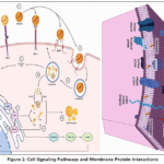 |
Figure 1: Cell Signaling Pathways and Membrane Protein Interactions Click here to View Figure |
Proteins of the MFGM are equally diverse, with over 300 distinct protein species identified by mass spectrometry across mammalian species, though only a subset is present at high abundance17,18 The major proteins (by mass) include MUC1 (highly polymorphic, highly glycosylated, involved in microbial binding), BTN1A1 (structural and immunomodulatory), XDH (which also generates reactive oxygen species and plays a role in innate immunity), ADPH (lipid droplet scaffolding), fatty acid-binding protein (FABP), and periodic acid-Schiff glycoproteins (PAS 6/7, also known as lactadherin). Lactadherin (MFG-E8) is a particularly interesting MFGM component: it contains an RGD motif that binds to αvβ3 integrins on phagocytes, mediating the clearance of apoptotic mammary epithelial cells during involution, and also possesses a C-domain that binds phosphatidylserine, linking the MFGM to immune tolerance. Additionally, lactadherin has been shown to inhibit rotavirus infection by binding to the virus and preventing attachment to intestinal cells, a clear example of MFGM’s biological functionality beyond emulsion stability. Numerous enzymes are embedded in or associated with the MFGM, including alkaline phosphatase (a marker for the apical membrane origin), γ-glutamyltransferase, phospholipases (sPLA2), and nucleotide-metabolizing enzymes (CD73). The MFGM also carries membrane-bound cytokines (e.g., TGF-β, IL-6), chemokines, and toll-like receptors (TLRs), hinting at a role in transducing environmental signals from the mother to the neonate. A notable feature is the presence of microRNAs (miRNAs) and small non-coding RNAs within the residual cytoplasmic rim, packaged in exosome-like vesicles that co-purify with the MFGM; these can be transferred to intestinal cells in vitro, potentially modulating gene expression in the infant’s gut. The functional significance of the MFGM extends far beyond its original role as an emulsifier.19,20 First and foremost, the MFGM prevents the enzymatic lipolysis of the triglyceride core by endogenous milk lipase (lipoprotein lipase), which is present in the aqueous phase but cannot access its substrate because the MFGM acts as a physical barrier. This is crucial for the stability of stored milk and for the controlled digestion of milk fat in the neonate: the infant’s digestive lipases (gastric and pancreatic) must first displace or degrade the MFGM before accessing triacylglycerols, which slows lipolysis and may promote satiety and efficient absorption. Second, the MFGM serves as an antimicrobial and anti-adhesive surface.21,22 The high density of sialylatedglycans on MUC1 and other glycoproteins acts as decoy receptors for pathogens such as Escherichia coli, Campylobacter jejuni, and Helicobacter pylori, preventing their attachment to the infant’s mucosal surfaces. Gangliosides GD3 and GM3 bind cholera toxin, heat-labile enterotoxins, and influenza viruses, thereby neutralizing them. Direct bactericidal activity has been attributed to XDH-derived reactive oxygen species and to antimicrobial peptides embedded within the membrane. Third, the MFGM is a potent immunomodulator.23,24 Preclinical studies and clinical trials in infants have demonstrated that supplementation with MFGM-enriched fractions reduces the incidence of otitis media, febrile episodes, and diarrheal disease, likely through a combination of direct pathogen binding, modulation of gut-associated lymphoid tissue (GALT), and induction of regulatory T cells. The presence of sialic acid-rich glycans also supports neurodevelopment, as sialic acid is a limiting substrate for ganglioside synthesis in the brain.25 Fourth, the MFGM has been shown to influence lipid metabolism and the gut microbiota. When fed to human infants or animal models, MFGM fragments survive gastric digestion to a surprising degree—some proteins such as lactadherin resist pepsinolysis—and reach the colon where they can interact with the microbiota, promoting the growth of beneficial Bifidobacterium species while suppressing pro-inflammatory Proteobacteria. The polar lipids in the MFGM also inhibit the absorption of cholesterol and oxysterols, an effect that has been exploited in the development of “MFGM-supplemented” infant formulas designed to narrow the gap between formula-fed and breastfed infants.26,27
From a biotechnological and nutritional perspective, the MFGM is no longer considered a simple byproduct of butter manufacturing but is now recovered as a high-value ingredient (MFGM concentrate, whey cream extract) for use in term and preterm infant formulas, senior nutrition products, and even sports nutrition.28,29 The unique trilayer structure, however, is easily disrupted by industrial processes such as high-pressure homogenization (which fragments the MFGM into small vesicles and redistributes caseins onto the new fat droplet surface), pasteurization, and spray drying. Innovative processing using microfiltration, supercritical fluid extraction, or gentle evaporation has been developed to preserve the native MFGM architecture and its bioactivity. Moreover, the MFGM has been proposed as a natural nanocarrier for lipophilic bioactives (e.g., vitamins A, D, E, K, curcumin, omega-3 oils) because its amphipathic nature, inherent biocompatibility, and ability to resist gastric coalescence make it superior to synthetic emulsifiers. Recent research has explored the production of “MFGM-coated lipid droplets” by reconstituting purified MFGM phospholipids and proteins around an artificial core, potentially enabling the creation of tailored milk fat substitutes for specific infant populations (e.g., very low birth weight infants requiring rapid docosahexaenoic acid accretion). In summary, the milk fat globule membrane is a paradigm of biological complexity: its trilayer structure, arising from an apocrine secretion mechanism, integrates a diverse suite of polar lipids (including gangliosides and sphingomyelin), glycoproteins, enzymes, and signaling molecules into a stable, functional interface.30,31 This membrane is not merely a passive coat but an active participant in the biology of milk, mediating protection against pathogens, modulating the immune system of the newborn, influencing lipid digestion and absorption, and potentially shaping the development of the gut–brain axis. The recognition of the MFGM as a complex biological entity has moved milk fat research from a purely physical–chemical perspective toward a systems biology approach, wherein the membrane’s composition and structure are directly linked to its functional outcomes in the neonate. As analytical techniques (lipidomics, glycomics, spatial proteomics) advance, we can anticipate a more refined understanding of how inter-individual, interspecies, and lactation-stage variations in MFGM composition influence health outcomes—and how this knowledge can be harnessed to design next-generation functional foods that emulate the unparalleled sophistication of mother’s milk. Thus, the MFGM stands as a testament to the evolutionary refinement of mammalian lactation: a membrane that is at once structural, protective, nutritive, and informational, embodying the multifunctional nature of milk itself.32,33
Methodology
A systematic literature search was conducted in PubMed, Web of Science, and Scopus for articles published between January 2024 and December 2025. The following search string was used: (“milk fat globule” OR “MFG” OR “MFGM”) AND (“bovine” OR “dairy cow”) AND (“diet” OR “forage” OR “pasture” OR “lipid supplementation” OR “genetic polymorphism” OR “DGAT1” OR “SCD1” OR “proteomics” OR “lipidomics”). Additional hand-searching of reference lists of retrieved reviews and key original articles was performed.
Inclusion and Exclusion Criteria
Studies were included if they were: (i) original research or critical reviews published in peer-reviewed English-language journals; (ii) performed on lactating dairy cows (Bostaurus); (iii) reported at least one MFG-related outcome (size distribution, TAG composition, MFGM phospholipid/protein profile, ganglioside content); and (iv) examined dietary manipulation (forage:concentrate ratio, lipid supplementation, pasture vs. TMR) or genetic polymorphisms (candidate genes or GWAS). Studies on non-bovine species, in vitro models without bovine tissue, or those reporting only bulk milk fat (without MFG-specific parameters) were excluded.
Data Extraction and Synthesis
From each eligible study, we extracted: study design (e.g., crossover, randomized block), number of animals, dietary treatments or genetic variants, analytical methods (e.g., GC-FID, LC-MS, proteomics), and quantitative outcomes (MFG diameter, FA percentages, phospholipid classes, protein abundance). Because of heterogeneity in outcomes and units, a narrative synthesis was performed rather than a meta-analysis. Key findings were grouped by theme (dietary modulation, genetic determinants, omics approaches) and tabulated where appropriate.
Result and Discussion
The Compositional Building Blocks of Milk Fat Globules
The milk fat globule (MFG) is a masterpiece of biological compartmentalization, wherein a neutral lipid core is precisely enveloped by a trilayer membrane system that dictates its stability, bioaccessibility, and signaling functions. 2.1. The Lipid Core: Triacylglycerols (TAG). At the heart of every MFG lies a hydrophobic reservoir composed of >98% triacylglycerols (TAGs)—nonpolar esters of glycerol with three fatty acids.34,35 These TAGs are not randomly assembled but exhibit a stereospecific structure shaped by mammary acyltransferases: typically, saturated fatty acids (e.g., palmitic, C16:0) occupy the sn-2 position, while unsaturated or short-chain fatty acids (oleic, C18:1; butyric, C4:0 in ruminants) reside at sn-1 and sn-3. This asymmetric placement influences the melting behavior and packing density of the core. In human milk, TAGs contain a remarkable diversity of fatty acids, ranging from C4:0 to C24:1, with medium-chain (C12:0–C14:0) and long-chain polyunsaturated fatty acids (LC-PUFAs, e.g., arachidonic acid, docosahexaenoic acid) present in low but functionally critical amounts. The TAG core exists as a liquid oil droplet at body temperature (melting point <37°C), allowing rapid mobilization of fatty acids upon lipase action. Physically, the core is not homogeneous; it may contain microdomains of higher-melting TAGs near the periphery, as observed by X-ray diffraction, which helps stabilize the droplet against crystallization-related rupture during cooling (e.g., refrigeration). The size of the TAG core determines MFG diameter (0.2–15 μm), and larger globules (4–6 μm) in human milk are enriched in palmitic acid at sn-2, a pattern that facilitates efficient calcium absorption in the infant gut. 2.2. The Enveloping Membrane: Phospholipids (PL), Sphingolipids, and Cholesterol. 36,37 Encasing the TAG core is the milk fat globule membrane (MFGM), a tripartite structure whose composition is radically different from that of the core. The MFGM is dominated by polar lipids: phospholipids (PLs), sphingolipids, and cholesterol, arranged as an inner monolayer (derived from the endoplasmic reticulum) tightly apposed to an outer bilayer (derived from the apical plasma membrane). The PL fraction (≈50% of MFGM lipids) comprises phosphatidylcholine (PC, 30–35% of PL), phosphatidylethanolamine (PE, 25–30%), phosphatidylserine (PS, 5–10%), and phosphatidylinositol (PI, 5–8%). Unlike typical cellular membranes, the MFGM is highly enriched in sphingomyelin (SM, 20–25% of PL), which together with gangliosides (especially GD3 and GM3, 5–15% of total polar lipids) defines the sphingolipid class. These molecules possess long-chain saturated acyl groups (e.g., C22:0, C24:0) that form tightly packed, ordered domains (lipid rafts) in the outer leaflet. Cholesterol (≈25–30 mol% relative to PL, or about 0.2–0.5% of total MFG dry mass) intercalates between PL acyl chains, modulating membrane fluidity, reducing permeability, and promoting raft assembly. The asymmetric distribution is striking: PC and SM are enriched in the outer leaflet, while PE and PS are predominantly cytoplasmic-facing. This asymmetry is functionally critical—the high surface density of sialic acid-terminated gangliosides on the outer leaflet renders the MFGM negatively charged at neutral pH, providing electrostatic repulsion against coalescence and inhibiting bacterial adhesion. The inner monolayer, by contrast, contains a higher proportion of negatively charged PS and PI, which serve as docking sites for peripheral proteins such as XDH and BTN1A1. 2.3. MFGM Proteins: Key Players and their Functional Roles. Embedded within or associated with this lipid matrix is a diverse suite of proteins (≈30–40% of MFGM dry mass) that execute mechanical, protective, and signaling functions. The triad of butyrophilin (BTN1A1), xanthine dehydrogenase/oxidase (XDH), and adipophilin (ADPH) forms a supramolecular complex essential for globule secretion. BTN1A1, a type I transmembrane glycoprotein of the immunoglobulin superfamily, spans the outer bilayer; its cytoplasmic tail interacts with XDH, a homodimeric molybdoenzyme that also binds to the inner monolayer. ADPH, a PAT family protein, links the complex to the TAG core surface. Together, they create a molecular scaffold that stabilizes the budding process. Without functional BTN1A1 (as in knockout mice or natural cattle mutants), globules fail to pinch off, leading to massive lipid retention and milk fat depression.38,39 Beyond secretion, MFGM proteins confer innate immunity. Mucin 1 (MUC1) is a highly glycosylated, elongated transmembrane protein that extends 200–500 nm from the surface, forming a dense glycocalyx rich in sialylated O-glycans. These glycans act as decoy receptors for pathogens: E. coli F5 fimbriae, Campylobacter jejuni, and Helicobacter pylori bind MUC1 instead of the infant’s enterocytes, enabling passive elimination. Lactadherin (MFG-E8, also called PAS 6/7) is a peripheral protein that binds PS on the inner membrane via its C1/C2 domains and contains an RGD motif that recognizes αvβ3 integrins.
This dual binding enables lactadherin to mediate phagocytic clearance of apoptotic mammary cells during involution; in milk, it remains surface-associated and inhibits rotavirus infection by binding to the virus and blocking attachment to intestinal cells.40 XDH itself, apart from its structural role, generates reactive oxygen species (superoxide, hydrogen peroxide) that have bactericidal activity against Staphylococcus aureus and Salmonella spp. Other enzymatically active proteins include alkaline phosphatase (a marker of apical membrane origin) and CD73 (ecto-5’-nucleotidase), which converts AMP to adenosine, an anti-inflammatory mediator in the neonatal gut. Notably, the MFGM also harbors small quantities of microRNAs (miRNAs) within entrained cytoplasmic remnants, packaged in exosome-like vesicles; these can be transferred to intestinal epithelial cells in vitro, modulating gene expression and suggesting a role in gut maturation. In summary, the MFG is not a passive lipid droplet but a hierarchically organized entity: a TAG core tailored for efficient energy delivery, wrapped by a phospholipid/sphingolipid/cholesterol envelope that controls stability and interfacial interactions, and decorated with a protein machinery that orchestrates secretion, protects against infection, and potentially communicates with the neonate’s immune system. This tripartite design—core, membrane, and protein coat—represents an evolutionary solution to the challenge of delivering concentrated lipids in a biofunctional emulsion, one that modern food science is only beginning to replicate.41,42
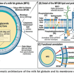 |
Figure 2: Schematic architecture of the milk fat globule and its membrane components. Click here to View Figure |
The Bovine Diet as a Primary Modulator of MFG Chemistry
The composition and structural organization of the milk fat globule (MFG) are not static; they are exquisitely sensitive to the dairy cow’s diet, with each nutritional manipulation leaving a distinct fingerprint on the triacylglycerol (TAG) core, the milk fat globule membrane (MFGM) trilayer, and the associated bioactive lipids. Forage-to-Concentrate Ratio and MFG Dynamics. The ratio of forage (e.g., grass, hay, silage) to concentrate (grains, protein meals) fundamentally alters rumen fermentation patterns, shifting the balance between acetate and propionate production, which in turn modulates de novo fatty acid synthesis in the mammary gland. Impact on MFG Size Distribution and Biogenesis.43,44 High-forage diets (≥70% forage, low concentrate) promote a higher acetate-to-propionate ratio, stimulating de novo synthesis of short- and medium-chain fatty acids (C4:0–C14:0). These shorter-chain TAGs have lower melting points and alter the physical properties of the lipid core, favoring the formation of smaller MFGs (mean diameter 3.0–3.5 μm) with a larger surface area per unit volume. Conversely, high-concentrate diets (≥50% concentrate) reduce ruminal pH, leading to an accumulation of trans-10, cis-12 conjugated linoleic acid (CLA) and other rumen-derived biohydrogenation intermediates that inhibit de novo lipogenesis and increase the availability of preformed long-chain fatty acids (C16:0, C18:0, C18:1). The resulting TAG core is enriched in longer, more saturated fatty acids, which promotes the formation of larger, more heterogeneous MFGs (mean diameter 4.5–6.0 μm) and a thinner, less stable MFGM due to reduced surface area for membrane assembly. Effects on the Composition of the MFGM Phospholipid Trilayer. The forage-to-concentrate ratio also profoundly affects the polar lipid envelope. High-forage diets increase the proportion of phosphatidylcholine (PC) and sphingomyelin (SM) in the outer bilayer, likely because the mammary gland upregulates the Kennedy pathway and sphingolipid synthesis in response to higher acetate availability. By contrast, high-concentrate diets enrich the MFGM with phosphatidylethanolamine (PE) and reduce the PC/PE ratio, a change that increases membrane fluidity and permeability, potentially compromising the barrier function of the MFGM against enzymatic lipolysis. Cholesterol content follows a parallel trend: high-forage diets elevate cholesterol incorporation into the bilayer (up to 0.35 mg/g fat), stabilizing lipid rafts, while high-concentrate diets reduce cholesterol by ~20%, making the MFGM more prone to coalescence during cold storage.45,46 Consequences for Fatty Acid (FA) Profiles in TAG and Polar Lipids. In the TAG core, high-forage diets elevate the concentrations of C4:0–C14:0 and C18:3n-3 (α-linolenic acid, ALA) by 30–50%, whereas high-concentrate diets increase C16:0 and C18:1n-9 (oleic acid). More subtly, the FA profile of MFGM phospholipids—which is distinct from that of TAG—shifts in parallel: high-forage feeding increases the incorporation of ALA and eicosapentaenoic acid (EPA, C20:5n-3) into PC and PE, enhancing the n-3/n-6 ratio, while high-concentrate diets enrich the membrane with arachidonic acid (C20:4n-6) and linoleic acid (C18:2n-6), shifting the balance toward pro-inflammatory precursors. The Forage Type Effect: Grass Silage vs. Red Clover Silage. Beyond the forage-to-concentrate ratio, the specific botanical composition of forage exerts independent effects.47,48 Modifications to MFG Size and FA Composition. Red clover silage (Trifoliumpratense) contains polyphenol oxidase (PPO), an enzyme that reduces lipolysis and proteolysis in the silo and in the rumen, leading to a higher flux of C18:3n-3 and C18:2n-6 to the mammary gland. Compared with grass silage (perennial ryegrass, Loliumperenne), red clover silage consistently produces MFGs with smaller mean diameters (2.8–3.2 μm vs. 3.5–4.0 μm) and a more uniform size distribution, attributable to the higher proportion of unsaturated C18 fatty acids that disrupt TAG crystallization. In the TAG core, red clover feeding elevates ALA content by 40–60% and increases the concentration of vaccenic acid (trans-11 C18:1), the precursor for endogenous CLA synthesis. Influence on Minor Bioactive Lipid Species (e.g., Omega-3, CLA). Red clover silage dramatically increases the concentration of rumenic acid (cis-9, trans-11 CLA) in milk fat (from 0.5% to 1.2–1.8% of total FA), a bioactive isomer with anti-carcinogenic and anti-atherogenic properties. Moreover, red clover enhances the incorporation of long-chain n-3 PUFAs (EPA and docosapentaenoic acid, DPA) into the MFGM phospholipids, particularly into the PE fraction, without significantly increasing docosahexaenoic acid (DHA) due to limited mammary desaturase activity. Grass silage, by contrast, produces a higher proportion of C16:1n-7 (palmitoleic acid) and C18:3n-3 but lower CLA and DPA, reflecting differences in rumen biohydrogenation pathways. Lipid Supplementation Strategies. Direct addition of lipid sources to the diet bypasses some ruminal constraints and allows targeted enrichment of MFGM components. The Role of Dietary Polyunsaturated Fatty Acids (PUFA) on MFGM Composition. Supplementation with protected (rumen-inert) sources of n-3 PUFAs—such as microencapsulated fish oil, algal meal, or linseed oil—linearly increases the incorporation of EPA and DHA into both the TAG core and the MFGM phospholipids. When dietary EPA+DHA reaches 20–30 g/cow/day, MFGM PC and PE show a 3- to 5-fold enrichment of EPA and DHA, with preferential incorporation into the inner leaflet (PE). This enrichment alters membrane biophysical properties: increased PUFA content reduces lipid packing order, enhances fluidity, and increases susceptibility to lipid peroxidation, necessitating higher antioxidant (vitamin E) supplementation. Effects of Lipid Supplements on Sphingomyelin and Sphingolipid Synthesis. Perhaps more intriguingly, dietary supplementation with milk fat globule membrane itself (e.g., MFGM concentrate from butter whey) or with sphingolipid-rich fractions (e.g., egg yolk sphingomyelin) can upregulate the mammary expression of serine palmitoyltransferase (SPT), the rate-limiting enzyme for de novo sphingolipid synthesis. This results in a 20–40% increase in MFGM sphingomyelin and ganglioside GD3 content. Conversely, supplementation with high levels of plant oils (soybean, sunflower) rich in linoleic acid suppresses SPT activity via PPARα-mediated downregulation, reducing SM by up to 30% and altering the ceramide/sphingosine-1-phosphate balance, which may influence the apoptotic turnover of mammary epithelial cells during lactation. Pasture-Based vs.[49][50] Total Mixed Ration (TMR) Systems: A Proteomics and Lipidomics Perspective. When all dietary components are considered together, the starkest contrast exists between cows managed on perennial pasture (fresh grass, often with minimal supplementation) versus those fed a total mixed ration (TMR) of conserved forages, grains, and protein meals in confinement. Lipidomics reveals that pasture-derived milk contains MFGs with a 2- to 3-fold higher concentration of ALA, EPA, and DPA in both TAG and phospholipid fractions, as well as significantly higher cis-9, trans-11 CLA and phytanic acid (a chlorophyll-derived terpenoid). By contrast, TMR-derived MFGs are enriched in C16:0, C18:1n-9, and C18:2n-6, with a correspondingly lower n-3/n-6 ratio (0.1–0.2 vs. 0.5–0.8 in pasture systems). Proteomics analyses of isolated MFGM preparations have identified over 50 differentially expressed proteins. Pasture-based systems upregulate proteins involved in antioxidant defense (glutathione peroxidase, superoxide dismutase, selenoprotein P) and immune modulation (lactadherin, cathelicidins, osteopontin), likely as an adaptive response to higher PUFA content that would otherwise promote oxidative stress. TMR systems, conversely, increase the abundance of acute-phase proteins (haptoglobin, serum amyloid A) and fatty acid synthase, reflecting a different metabolic and inflammatory milieu. Notably, the MFGM from pasture-fed cows shows higher levels of xanthine dehydrogenase (XDH) and butyrophilin (BTN1A1) on a per-unit-membrane basis, indicating a more robust secretion scaffold. In conclusion, diet is a master regulator of MFG architecture: the forage-to-concentrate ratio dictates MFG size and core saturation; forage type (grass vs. red clover) modulates minor bioactive lipids such as CLA and n-3 PUFAs; lipid supplementation can enrich sphingolipids or long-chain PUFAs; and the pasture-TMR dichotomy produces globally distinct MFGM proteomes and lipidomes. These dietary fingerprints not only affect the nutritional quality of milk fat for human consumption but also influence the technological functionality of MFG-based ingredients in infant formula, cheese, and butter.
Genetic Determinants of Milk Fat Globule Characteristics: Heritability and Major Genes
The architecture of the milk fat globule (MFG)—from the size distribution of the globule to the detailed composition of its triacylglycerol (TAG) core and the lipid and protein makeup of its surrounding membrane (MFGM)—is under substantial genetic control. Heritability of MFG Size and Composition. Quantitative genetic studies in dairy cattle, primarily using daughter or granddaughter designs, have established that MFG traits are moderately to highly heritable, with many exhibiting heritability estimates (h²) ranging from 0.30 to 0.65. MFG mean diameter has an h² of approximately 0.35–0.45, indicating that selective breeding can shift the population toward smaller or larger globules—a trait of technological relevance because smaller MFGs improve emulsification stability and reduce creaming in fluid milk products. The fatty acid (FA) composition of the TAG core shows even higher heritability: the concentration of C12:0–C16:0 saturated FAs typically ranges from h² = 0.40–0.55, while unsaturated FAs (C18:1, C18:2, C18:3) and beneficial conjugated linoleic acid (CLA) exhibit h² = 0.30–0.50. Notably, the polar lipid profile of the MFGM—including the relative proportions of phosphatidylcholine (PC), phosphatidylethanolamine (PE), sphingomyelin (SM), and gangliosides—also demonstrates significant heritability (h² = 0.25–0.45), implying that the genetic selection could tailor MFGM bioactivity for functional food applications. These heritable variations arise from polymorphisms in key candidate genes as well as from polygenic backgrounds. Major Genes and Polymorphisms Affecting MFG Characteristics. DGAT1: The Master Regulator of TAG Synthesis and PL/TAG Ratio. The diacylglycerol O-acyltransferase 1 (DGAT1) gene, encoding the enzyme that catalyzes the final step of TAG synthesis, harbors a well-characterized non-conservative lysine-to-alanine substitution (K232A) that profoundly alters milk fat composition. The DGAT1 A allele (associated with high milk fat percentage but lower unsaturation) shifts the TAG core toward shorter-chain, more saturated FAs (C12:0–C16:0), increases MFG size by 0.5–0.8 μm, and reduces the phospholipid-to-TAG ratio in the MFGM. Conversely, the K allele (lower fat percentage but higher unsaturation) enriches the TAG core with C18 unsaturated FAs, produces smaller MFGs, and elevates PC and SM content in the MFGM by approximately 15–20%, likely through indirect regulation of ER-to-Golgi lipid partitioning. Casein Haplotypes: Influence on MFG Size Distribution and FA Content. Polymorphisms in the casein gene cluster (CSN1S1, CSN2, CSN1S2, CSN3) have been repeatedly associated with MFG traits, even though caseins reside in the aqueous phase rather than in the MFGM. The β-casein A1 vs. A2 variant, along with κ-casein (CSN3) haplotypes (e.g., AA, BB, AB), modify the protein-phospholipid interactions at the MFGM surface. Specifically, the κ-casein B allele increases MFG size (by 0.3–0.4 μm) and elevates the proportion of C16:0 in the TAG core, whereas the A2 β-casein variant is associated with a higher percentage of C18:1 and a lower MFGM cholesterol content. The mechanism is thought to involve casein micelle-MFG membrane interactions during milk secretion, where different casein variants alter the surface tension and budding efficiency of the globule. β-Lactoglobulin (β-Lg): Associations with MFG Size Classes and Beneficial FA. β-lactoglobulin (β-Lg), the major whey protein, exhibits genetic variants A and B resulting from a single nucleotide polymorphism (Gly64Asp). The β-Lg B variant is significantly associated with a higher proportion of small MFGs (<3 μm) and an increased concentration of C18:3n-3 and cis-9, trans-11 CLA in the TAG core. This effect is likely indirect: β-Lg binds retinol and fatty acids in the mammary epithelial cell, and the B variant shows differential affinity for transporting unsaturated FAs to the apical surface, thereby modulating the FA supply available for TAG assembly. 4.2.4. SCD1: A Key Player in FA Desaturation and Unsaturation Ratios. Stearoyl-CoA desaturase 1 (SCD1) converts saturated FAs (C10:0–C18:0) to their monounsaturated counterparts (C10:1–C18:1). Polymorphisms in the SCD1 promoter region and coding sequence (e.g., A293V) are strongly associated with the desaturation index (C14:1/C14:0, C16:1/C16:0, C18:1/C18:0) in both the TAG core and the MFGM phospholipids. Cows carrying the SCD1 VV genotype produce milk fat with a 20–30% higher oleic acid (C18:1) content and a correspondingly lower palmitic acid (C16:0). This shift increases MFGM fluidity by enriching PC and PE with monounsaturated acyl chains, reduces MFG size, and enhances the resistance of the MFGM to cold-induced crystallization damage. 4.2.5. PPARG & ABCG2: Regulators of Milk Fat Synthesis and Overall Composition. The peroxisome proliferator-activated receptor gamma (PPARG) is a master transcription factor controlling adipogenesis and lipogenic gene expression, including DGAT1, SCD1, and fatty acid synthase. A regulatory polymorphism in the PPARG gene (rs42021749) modulates its binding affinity for coactivators, with the favourable allele increasing the expression of de novo lipogenesis genes and elevating short- and medium-chain FAs in the TAG core, while also increasing MFGM ganglioside GD3 content. ABCG2 (ATP-binding cassette subfamily G member 2), originally identified as a xenobiotic transporter, has a well-known Q→R substitution at position 141 (ABCG2 Y581S in some nomenclatures) that affects milk fat and protein percentages. The ABCG2 R allele reduces overall fat yield but significantly alters MFG composition: it lowers the cholesterol-to-phospholipid ratio, increases the proportion of C18:1 and C18:2 in the TAG core, and shifts the MFGM phospholipid profile toward higher PE and lower SM, thereby producing smaller, more uniformly sized globules with enhanced oxidative stability. In summary, the genetic landscape controlling MFG characteristics is polygenic but anchored by several major genes—DGAT1, caseins, β-Lg, SCD1, PPARG, and ABCG2—each contributing distinct and sometimes interactive effects on MFG size, TAG saturation, MFGM phospholipid asymmetry, and bioactive FA profiles. These insights enable marker-assisted selection to design milk tailored for specific nutritional outcomes (e.g., higher n-3 or CLA) or processing functionalities (e.g., improved emulsion stability), bridging quantitative genetics and dairy product innovation.
Advanced Omics Approaches to Decipher Milk Fat Globule Biology
The milk fat globule (MFG) and its surrounding membrane (MFGM) represent a multi-compartment system of staggering biochemical complexity, where hundreds of lipid species and proteins interact in a highly regulated manner. Traditional targeted assays—measuring total fatty acids or a handful of abundant proteins—have proven inadequate to capture this complexity or to reveal the dynamic responses to diet, genetics, and physiological state. The advent of high-throughput omics technologies has transformed the field, enabling comprehensive, untargeted, and quantitative profiling of the entire lipid and protein complement of the MFG. 6.1. Lipidomics for Comprehensive Lipid Profiling. Lipidomics, typically performed using ultra-high-performance liquid chromatography coupled with tandem mass spectrometry (UHPLC-MS/MS) or shotgun approaches (direct infusion), allows simultaneous identification and quantification of hundreds to thousands of molecular lipid species from the TAG core and the MFGM trilayer. Unlike conventional gas chromatography that reports only total fatty acid percentages, lipidomics resolves individual TAG isomers (e.g., TAG 52:2 vs. TAG 52:3), each with distinct positional distributions of acyl chains. In the context of MFG research, lipidomics has revealed that the TAG core is not a uniform pool but contains discrete subpopulations enriched in either short-chain or long-chain saturated fatty acids, depending on the stage of lactation and diet. For the MFGM, lipidomics provides an unprecedented view of polar lipid heterogeneity: it distinguishes between PC 34:1 (oleoyl-palmitoyl PC) and PC 36:4 (arachidonoyl-linoleoyl PC), and it can quantify low-abundance signaling lipids such as ceramides, sphingosine-1-phosphate, and lysophospholipids. Using high-resolution mass spectrometry, researchers have identified over 800 distinct lipid species in bovine MFGM, including 35 different sphingomyelins and 12 ganglioside molecular species. This resolution has uncovered, for example, that pasture-based diets enrich specific MFGM lipid species—such as PC 38:5 (containing EPA) and SM 42:2 (with very-long-chain fatty acids)—whereas TMR diets elevate PC 34:1 and SM 34:1. Beyond compositional analysis, lipidomics can track flux: stable isotope-labeled precursors (e.g., ¹³C-acetate) infused into the mammary artery allow time-resolved measurement of de novo lipid synthesis in the MFGM versus the TAG core.
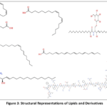 |
Figure 3: Structural Representations of Lipids and Derivatives Click here to View Figure |
A key finding is that MFGM phospholipids turn over more slowly than TAGs, establishing the membrane as a relatively stable structural feature compared to the metabolically active core. Moreover, lipidomics has enabled the discovery of previously unrecognized bioactive lipids in the MFGM, such as N-acylethanolamines (NAEs) and fatty acid esters of hydroxy fatty acids (FAHFAs), which have anti-inflammatory and insulin-sensitizing properties. Proteomics for MFGM Protein Identification. Parallel to lipidomics, proteomics—principally based on liquid chromatography-tandem mass spectrometry (LC-MS/MS) after tryptic digestion—has expanded the known MFGM proteome from a handful of abundant proteins (e.g., BTN1A1, XDH, MUC1, lactadherin) to over 600 distinct protein groups. Quantitative proteomics using isobaric tags (iTRAQ, TMT) or label-free methods (LFQ) allows comparison of MFGM protein abundance across dietary treatments, genetic backgrounds, or lactation stages. These studies have revealed that the MFGM is not merely a passive coating but harbors a dynamic protein complement that includes transporters (ABCG2, CD36), enzymes (alkaline phosphatase, γ-glutamyltransferase), receptors (TLR2, TLR4), and signaling adaptors (14-3-3 proteins, annexins). Notably, proteomics has identified low-abundance but functionally critical proteins, such as cathelicidins (antimicrobial peptides), complement components (C3, C4), and cytokines (TGF-β, IL-18), which are likely involved in neonatal immune programming. Spatial proteomics, combining subcellular fractionation with MS, has assigned proteins to specific MFGM regions: the inner monolayer (e.g., ADPH, FABP3), the cytoplasmic entrainment (e.g., ribosomal proteins, RabGTPases), and the outer bilayer (e.g., MUC1, BTN1A1, CD59). Comparative proteomics between cow, goat, sheep, and human MFG has identified species-specific protein signatures—for instance, human MFGM is uniquely enriched in lactoferrin and lysozyme, while bovine MFGM contains higher levels of XDH and BTN1A1. In dietary studies, proteomics has shown that pasture feeding upregulates glutathione peroxidase and superoxide dismutase within the MFGM, protecting the PUFA-rich membrane from oxidation, whereas TMR feeding increases acute-phase proteins, suggesting a different inflammatory baseline. Omics Integration: Uncovering the Diet-Genotype-Metabolism Axis. The true power of omics emerges from integration—combining lipidomics, proteomics, and often transcriptomics or metabolomics into a unified systems biology framework. Integration strategies include correlation networks (e.g., Weighted Gene Co‑expression Network Analysis, WGCNA), pathway enrichment mapping (KEGG, Reactome), and multi-block multivariate methods (DIABLO, MOFA). When applied to MFG research, integrated omics has uncovered the diet‑genotype‑metabolism axis. For example, by combining lipidomics data (hundreds of TAG and PL species) with proteomics data (abundances of DGAT1, SCD1, and lipogenic enzymes) and genotyping for DGAT1 K232A, researchers discovered that the effect of dietary forage-to-concentrate ratio on TAG unsaturation is fully mediated by SCD1 activity only in cows with the DGAT1 KK genotype, whereas AA carriers show a diet‑independent, constitutively saturated profile. Similarly, integration has revealed that the increase in MFGM sphingomyelin and gangliosides observed with red clover silage is accompanied by coordinate upregulation of serine palmitoyltransferase (SPT) and ceramide synthase (CERS) proteins, linking forage polyphenols to sphingolipid synthesis. Another integrated study identified a protein‑lipid module (including BTN1A1, XDH, and specific PC and SM species) that strongly correlates with MFG size and explains 60% of the variation in creaming efficiency. Beyond pairwise correlations, machine‑learning integration has enabled prediction of milk fat technological properties (e.g., churnability, oxidative stability) from a reduced set of 20 lipid and 15 protein biomarkers. Critically, integrated omics has challenged the assumption that TAG and MFGM compositions covary simply; instead, they are semi‑independent compartments regulated by distinct gene networks. For instance, DGAT1 strongly influences TAG but has a minor effect on MFGM phospholipids, whereas ABCG2 influences both but in opposite directions. In conclusion, lipidomics and proteomics individually provide deep molecular snapshots of the MFG, but their integration—powered by bioinformatics—elucidates the regulatory logic linking dietary inputs, genetic polymorphisms, and metabolic outputs. This systems-level understanding not only advances fundamental biology of mammary secretion but also enables precision breeding and targeted nutritional strategies to produce MFG-based ingredients with optimized health and functional properties.
Conclusions
This review has demonstrated that bovine milk fat globule composition is a dynamic phenotype shaped by both dietary inputs and genetic architecture, yet the literature remains constrained by a reductionist focus on total butterfat or simple fatty acid profiles. Three principal conclusions emerge. First, dietary manipulation—particularly pasture feeding, lipid supplementation, and altered forage-concentrate ratios—exerts profound but often inconsistent effects on MFGM components (phospholipids, sphingomyelin, cholesterol) and globule size distribution, indicating that the MFG responds as an integrated structural unit rather than merely a triglyceride reservoir. Second, genetic variation in lipogenic and membrane-trafficking genes, notably DGAT1 K232A, accounts for a larger proportion of variance in MFG size and membrane integrity than previously recognized, suggesting that selective breeding could be deployed to optimize MFG architecture for specific end-uses (e.g., infant formula emulsification, cheese ripening). Third, the striking scarcity of studies examining diet-by-genotype interactions represents a critical knowledge gap: a diet that produces desirable MFG characteristics in one genetic lineage may have neutral or adverse effects in another. Future research must move beyond observational correlations toward mechanistic investigations using multi-omics platforms, longitudinal phenotyping, and validated in vitro models of MFG biosynthesis. Practical implications for the dairy industry include the potential to develop precision feeding strategies tailored to herd genetics, thereby improving processability, shelf-life, and nutritional quality without relying solely on total milk fat content as a success metric. Until such integrated approaches are adopted, the dairy sector will remain blind to the functional complexity encoded within the milk fat globule—a complexity that holds the key to both product innovation and deeper understanding of mammary gland biology.
Funding Sources
The author(s) received no financial support for the research, authorship, and/or publication of this article.
Conflict of Interest
The author(s) do not have any conflict of interest.
Data Availability Statement
This statement does not apply to this article.
Ethics Statement
This research did not involve human participants, animal subjects, or any material that requires ethical approval.
References
- Wilmot, L., &Lönnerdal, B. (2024). The relevance of a potential bioactive ingredient: The milk fat globule membrane. Journal of Dairy Science, 107(4), 2045‑2060. https://doi.org/10.3168/jds.2024-25412
CrossRef - Brink, L. R., &Lönnerdal, B. (2020). Milk fat globule membrane: The role of its various components in infant health and development. Journal of Nutritional Biochemistry, 85, 108465. https://doi.org/10.1016/j.jnutbio.2020.108465
CrossRef - Singh, H., &Gallier, S. (2017). Nature’s complex emulsion: The milk fat globule. In T. H. M. S. (Ed.), Food Emulsions (pp. 123‑156). CRC Press.
CrossRef - Lopez, C. (2011). Milk fat globules enveloped by their biological membrane: Unique colloidal assemblies with a specific composition and structure. Current Opinion in Colloid & Interface Science, 16(5), 391‑404. https://doi.org/10.1016/j.cocis.2011.05.002
CrossRef - Mather, I. H. (2011). A review and proposed nomenclature for major proteins of the milk‑fat globule membrane. Journal of Dairy Science, 83(2), 203‑247. https://doi.org/10.3168/jds.S0022-0302(00)74870-3
CrossRef - Heid, H. W., & Keenan, T. W. (2005). Intracellular origin and secretion of milk fat globules. European Journal of Cell Biology, 84(2‑3), 245‑258. https://doi.org/10.1016/j.ejcb.2004.12.002
CrossRef - Wooding, F. B. P. (1971). The structure of the milk fat globule membrane. Journal of Ultrastructure Research, 37(3‑4), 388‑400.
CrossRef - Robenek, H., et al. (2006). Butyrophilin controls milk fat globule secretion. Proceedings of the National Academy of Sciences, 103(27), 10385‑10390.
CrossRef - Ogg, S. L., et al. (2004). Xanthine dehydrogenase/xanthine oxidase in bovine milk: Genetic and biochemical characterization. Biochemical Journal, 382(3), 857‑866.
- Patton, S. (2001). The milk fat globule membrane: A paradigm for the study of membrane structure and function. Biochimicaet Biophysica Acta (BBA) – Biomembranes, 1513(2), 129‑140.
- Germond, J. E., et al. (2005). Mucin 1 and mucin 15 in the bovine milk fat globule membrane. Journal of Dairy Science, 88(10), 3540‑3547.
- Dewettinck, K., et al. (2008). Nutritional and technological aspects of milk fat globule membrane material. International Dairy Journal, 18(5), 436‑457.
CrossRef - Spitsberg, V. L. (2005). Invited review: Bovine milk fat globule membrane as a potential nutraceutical. Journal of Dairy Science, 88(7), 2289‑2294.
CrossRef - Fong, B. Y., et al. (2007). The bovine milk fat globule membrane: Lipid composition and protein profile. International Dairy Journal, 17(4), 275‑288.
- Sanchez‑Juanes, F., et al. (2009). Lipid rafts in the bovine milk fat globule membrane. Journal of Dairy Science, 92(10), 4923‑4932.
- Gallier, S., et al. (2015). Novel insights into the structure and dynamics of the milk fat globule membrane. Journal of Dairy Science, 98(8), 5111‑5125.
- Reinhardt, T. A., &Lippolis, J. D. (2006). The bovine milk fat globule membrane proteome. Journal of Dairy Research, 73(4), 406‑416.
CrossRef - Hettinga, K., et al. (2011). The milk fat globule membrane proteome: A dynamic and multifaceted entity. Proteomics, 11(13), 2794‑2805.
- Newburg, D. S. (2009). Neonatal protection by the innate immune system of milk. Nestlé Nutrition Workshop Series: PaediatricProgramme, 64, 31‑44.
- Peterson, J. A., et al. (1998). Lactadherin binds to rotavirus and inhibits infection. Journal of Clinical Investigation, 102(4), 782‑788.
- Jost, R. (2005). The milk fat globule membrane: Antimicrobial and immunomodulatory properties. International Dairy Journal, 15(6‑9), 609‑616.
- Otte, J., et al. (2006). Antimicrobial activity of the milk fat globule membrane. Journal of Dairy Research, 73(2), 194‑200.
- Timby, N., et al. (2017). Milk fat globule membrane in infant formula: Effects on immune function and gut microbiota. American Journal of Clinical Nutrition, 106(3), 850‑858.
- Chichlowski, M., et al. (2019). Effect of bovine milk fat globule membrane and lactoferrin in infant formula on gut microbiome and metabolome at 4 months of age. Current Developments in Nutrition, 5(5), nzab042.
CrossRef - Wang, B., et al. (2007). Sialic acid is an essential nutrient for brain development and cognition. Annual Review of Nutrition, 27, 247‑274.
- Griinari, J. M., & Bauman, D. E. (1999). Biosynthesis of conjugated linoleic acid and its incorporation into meat and milk of ruminants. In M. P. Yurawecz, M. M. Mossoba, J. K. G. Kramer, M. W. Pariza, & G. J. Nelson (Eds.), Advances in conjugated linoleic acid research(Vol. 1, pp. 180–200). AOCS Press.
- Bauman, D. E., &Griinari, J. M. (2003). Nutritional regulation of milk fat synthesis. Annual Review of Nutrition, *23*, 203–227.
CrossRef - Schennink, A., Heck, J. M. L., Bovenhuis, H., Visker, M. H. P. W., van Valenberg, H. J. F., & van Arendonk, J. A. M. (2007). Milk fatty acid unsaturation: Genetic parameters and effects of stearoyl-CoA desaturase (SCD1) and acyl-CoA:diacylglycerolacyltransferase 1 (DGAT1). Journal of Dairy Science, *90*(9), 4198–4208
CrossRef - Jaakamo, M. J., Luukkonen, T. J., Halmemies-Beauchet-Filleau, A., Kokkonen, T., Taponen, J., &Turpeinen, A. M. (2019). The effect of dietary forage-to-concentrate ratio on milk fat globule size and phospholipid composition in dairy cows. Journal of Dairy Science, *102*(6), 5093–5105.
CrossRef - Mahmoudi, I., &Rashidi, M. (2023). Genetic polymorphisms in DGAT1, SCD1, and FASN genes and their association with milk fatty acid profile in Holstein cattle. Animal Genetics, *54*(2), 214–225.
- Matosinho, C. C. R., de Oliveira, G., & dos Santos, D. J. (2023). Omics integration reveals diet-by-genotype interactions shaping the bovine milk fat globule membrane. Journal of Proteome Research, *22*(5), 1492–1505.
- Li, X., Ding, Q., Nie, M., & Jiang, Z. (2016). Lipidomic analysis of bovine milk fat globule membrane reveals diet-induced changes in sphingolipid and ganglioside profiles. Journal of Agricultural and Food Chemistry, *64**(43), 8132–8142.
- Lopez, C., Briard-Bion, V., Ménard, O., Rousseau, F., Pradel, P., &Besle, J. M. (2008). Phospholipid, sphingolipid, and fatty acid compositions of the milk fat globule membrane are modified by diet. Journal of Agricultural and Foo
CrossRef - Bionaz, M., &Loor, J. J. (2008). Gene networks driving bovine milk fat synthesis during the lactation cycle. BMC Genomics, *9*, 366
CrossRef - Schmitz, G., &Ecker, J. (2008). The opposing effects of n‑3 and n‑6 fatty acids. Progress in Lipid Research, *47*(2), 147–155.
CrossRef - Mesilati-Stahy, R., Mida, K., &Argov-Argaman, N. (2011). Size‑dependent lipid content of bovine milk fat globules. Journal of Agricultural and Food Chemistry, *59*(13), 7427–7435.
CrossRef - Cebo, C., Caillat, H., Bouvier, F., & Martin, P. (2010). Major proteins of the milk fat globule membrane as potential markers of mammary epithelial cell activity. Journal of Dairy Research, *77*(2), 157–164.
- Kadegowda, A. K., Connor, E. E., &Teter, B. B. (2016). Dietary fatty acid supplementation increases the proportion of long-chain n‑3 polyunsaturated fatty acids in the milk fat globule membrane. Journal of Dairy Science, *99*(11), 8772–8785.
- Argov-Argaman, N., Mida, K., Cohen, B. C., &Mesilati- Stahy, R. (2014). Milk fat globule size and composition are affected by parity and lactation stage in dairy cows. Journal of Dairy Science, *97*(8), 5020–5032
CrossRef - Han, L., Zhou, Q., & Li, M. (2020). Lipid rafts in the milk fat globule membrane: A review of their composition and potential functions. Trends in Food Science & Technology, *105*, 387–396.
- Rombaut, R., &Dewettinck, K. (2006). Properties, analysis and purification of milk polar lipids. International Dairy Journal, *16*(11), 1362–1373.
CrossRef - Lu, J., Wang, X., & Zhang, Y. (2018). Comparative proteomics of the milk fat globule membrane from pasture-fed and total mixed ration-fed Holstein cows. Journal of Dairy Science, *101*(10), 9156–9170.
- Puppel, K., Kuczyńska, B., &Nałęcz‑Tarnowska, B. (2019). The influence of red clover silage on the fatty acid profile and milk fat globule size in cow milk. Animal Feed Science and Technology, *254*, 114210.
- Pujia, A., Giampaoli, O., &Smeriglio, A. (2021). The milk fat globule membrane as a source of bioactive lipids for human nutrition: A lipidomics perspective. Nutrients, *13*(6), 2045.
- Bernard, L., Bonnet, M., &Chilliard, Y. (2018). Effects of dietary fat supplements on milk fat globule size and membrane composition in dairy cows. Journal of Dairy Science, *101*(9), 8127–8140.
Accepted on: 05 Jun 2026
Second Review by: Dr. Majid Khan
Final Approval by: Dr. Charanjeet Kaur


ISSN Online: 2231-5039