Formulation And Characterization of Quercetin-Loaded Phytosomes: A Phospholipid-Based Nanocarrierfor Improved Bioavailability and Therapeutic Efficacy
 , Ekta Upadhyay2, Vishvendra Singh3, Sejal ben Gunvantbhai Patel4, Mohit Kumar5, Ritu6, Ritismita Devi7, Pravinkumar B Suruse8and Biswajit Puhan9*
1Department of Pharmaceutics, SGSPS, Institute of Pharmacy, Akola(MS), affiliated to SantGadge Baba Amravati University, Amravati, Maharashtra, India
2School of Pharmaceutical Sciences, Faculty of Pharmacy, IFTM University, Delhi Road, Moradabad, Moradabad District, Uttar Pradesh India
3Faculty of Agriculture , Vivekananda Global University Jaipur, Rajasthan, India
4Department in Pharmacognosy, Nootan Pharmacy College, Sankalchand Patel University, Visnagar, Mahesana, Gujarat, India.
5Teerthanker Mahaveer College of Pharmacy, TeerthankerMahaveer University; Moradabad, Uttar Pradesh, India.
6Department of Chemistry, Chhotu Ram Arya College, Sonipa, Haryana, India
7Assam down town University, Sankar Madhav Road, Pankhaiti, Guwahati, Assam, India
8Nagpur College of Pharmacy Nagpur, Maharashtra, India
9School of Pharmacy, ARKA JAIN University, Jamshedpur, Jharkhand, India.
Corresponding Author E-mail: biswajit.p@arkajainuniversity.ac.in
Download this article as:
ABSTRACT:Quercetin, a pentahydroxyflavone with potent antioxidant, anti-inflammatory, and anticancer activities, suffers from extremely poor oral bioavailability (<5% in humans) due to its low aqueous solubility (<2 μg/mL), extensive first-pass metabolism, and rapid systemic clearance. Methods: Quercetinphytosomes were prepared using the thin-film hydration method with phosphatidylcholine as the phospholipid component. A 3² full factorial design (Box-Behnken optimization) was employed to optimize formulation parameters including quercetin:PC molar ratio (1:1, 1:2, 1:4), evaporation temperature (30–50°C), and hydration time (30–60 min). Phytosomes were characterized for particle size (DLS), zeta potential, entrapment efficiency (HPLC), morphology (TEM/SEM), molecular interactions (FTIR), solid-state properties (DSC, XRD), in vitro release (dialysis bag method), and stability. Pharmacokinetic studies were conducted in Sprague-Dawley rats. Therapeutic efficacy was evaluated using DPPH/FRAP antioxidant assays, LPS-stimulated RAW 264.7 macrophage inflammation model, MCF-7 breast cancer cytotoxicity, and Ehrlich ascites tumor-bearing mice. Results: Optimized phytosomes (1:2 quercetin:PC ratio, 40°C evaporation, 45 min hydration) exhibited mean particle size of 128±6 nm, PDI of 0.21±0.03, zeta potential of -22.4±3.1 mV, and entrapment efficiency of 94.7±1.8%. FTIR confirmed hydrogen bonding between quercetin hydroxyl groups and PC phosphate moiety (P=O shift from 1245 cm⁻¹ to 1218 cm⁻¹). DSC and XRD demonstrated complete quercetinamorphization (loss of 318°C melting peak and disappearance of crystalline Bragg reflections). In vitro release reached 86.7% at 24 h (vs. 24.3% for free quercetin), following Higuchi kinetics (R²=0.991). Oral pharmacokinetics in rats showed 12-fold higher C_max (2.8 vs. 0.35 μg/mL) and 15-fold increased AUC(0-∞) for phytosomes. Antioxidant activity improved by 27–37% (DPPH IC₅₀: 3.8 vs. 5.2 μg/mL). Anti-inflammatory activity showed 2.1-fold greater TNF-α inhibition (68.7% vs. 32.4% at 10 μM). Anticancer studies revealed 3.3-fold lower IC₅₀ in MCF-7 cells (14.8 vs. 48.2 μM) and 74.2% tumor volume reduction in vivo (vs. 28.5% for free quercetin). Conclusion: Quercetin-loaded phytosomes represent a highly effective phospholipid-based nanocarrier that overcomes the intrinsic bioavailability barriers of quercetin through molecular complexation, amorphization, and enhanced membrane permeability. The formulation achieves up to 20-fold improvement in oral bioavailability and correspondingly superior therapeutic efficacy across antioxidant, anti-inflammatory, and anticancer applications, positioning quercetinphytosomes as a promising clinically translatable nutraceutical platform.
KEYWORDS:Amorphization; Anticancer activity; Anti-inflammatory; Bioavailability enhancement; Drug delivery; Hydrogen bonding; Phytosomes; Phospholipid-based nanocarrier; Phosphatidylcholine; Quercetin;
Introduction
Quercetin (3,3′,4′,5,7-pentahydroxyflavone) is a naturally occurring polyphenolic flavonoid abundantly present in fruits (apples, berries, grapes), vegetables (onions, broccoli, kale), tea, and red wine. As one of the most extensively studied dietary flavonoids, quercetin has garnered significant scientific attention due to its remarkable spectrum of biological activities, primarily attributed to its unique molecular structure—five hydroxyl groups arranged around a lipophilic flavone backbone. This configuration allows quercetin to effectively scavenge free radicals, chelate transition metal ions, and modulate various intracellular signaling pathways. However, despite its potent pharmacological profile, the clinical translation of quercetin has been historically limited by severe physicochemical and biopharmaceutical drawbacks, which have prompted the development of advanced drug delivery systems such as phospholipid-based phytosomes. The therapeutic potential of quercetin spans a wide array of disease conditions, with its antioxidant capacity being the most fundamental. Quercetin directly neutralizes reactive oxygen species (ROS) and reactive nitrogen species (RNS), while also upregulating endogenous antioxidant enzymes like superoxide dismutase and catalase. In terms of anti-inflammatory activity, quercetin inhibits key pro-inflammatory mediators—including cyclooxygenase-2 (COX-2), lipoxygenase, tumor necrosis factor-alpha (TNF-α), and interleukins (IL-1β, IL-6)—by suppressing the nuclear factor-kappa B (NF-κB) signaling pathway. Moreover, quercetin exhibits promising anticancer properties across multiple cancer types (breast, colon, prostate, lung) through induction of apoptosis, cell cycle arrest at G2/M phase, inhibition of angiogenesis (via VEGF downregulation), and suppression of metastasis (by inhibiting matrix metalloproteinases). Other documented benefits include cardioprotection (lowering LDL oxidation and blood pressure), neuroprotection (reducing amyloid-beta aggregation in Alzheimer’s models), antidiabetic effects (improving insulin sensitivity), and hepatoprotection. Nonetheless, achieving these benefits in vivo requires adequate systemic concentrations, which is precisely where quercetin fails due to its poor bioavailability.
The Bioavailability Challenge: Addressing Poor Aqueous Solubility, Low Membrane Permeability, and Extensive First-Pass Metabolism
Quercetin’s clinical efficacy is severely compromised by its extremely low oral bioavailability (estimated at less than 5% in humans). The primary reasons are threefold. First, quercetin is practically insoluble in water (solubility < 2 μg/mL in neutral aqueous media), which leads to poor dissolution in gastrointestinal fluids and limited absorption. Second, although its log P value (~1.8–2.0) suggests moderate lipophilicity, the presence of multiple hydroxyl groups results in extensive hydrogen bonding and molecular stacking, forming large crystalline aggregates that cannot easily traverse the intestinal epithelium. Third, once absorbed, quercetin undergoes extensive first-pass metabolism by phase II enzymes (UDP-glucuronosyltransferases, sulfotransferases, catechol-O-methyltransferases) in the enterocytes and liver, converting it into inactive or poorly active glucuronidated and sulfated conjugates. Consequently, plasma concentrations of free (aglycone) quercetin after oral administration are typically in the low nanomolar range—far below the micromolar concentrations required for many anticancer and anti-inflammatory effects. Recognizing these limitations, researchers have explored various nanocarrier strategies, among which phytosome technology has emerged as particularly effective. Phytosomes—derived from “phyto” (plant) and “some” (cell-like)—are a patented phospholipid-based nanocarrier system developed in the late 1980s by IndenaS.p.A., an Italian phytochemical company. Unlike conventional liposomes or nanoparticles, phytosomes are defined as stoichiometric complexes formed by the interaction of one or more natural bioactive molecules with phospholipids, typically phosphatidylcholine. The key innovation lies in the chemical bond formation: the polyphenolic compound becomes an integral part of the phospholipid bilayer rather than being merely entrapped or encapsulated. This technology was initially commercialized for standardized herbal extracts such as silymarin (silymarinphytosome, known as Siliphos®) and Ginkgo biloba. Since then, phytosome formulations have been successfully developed for curcumin, apigenin, rutin, and—notably—quercetin, with the goal of transforming poorly absorbed phytochemicals into orally bioavailable nutraceuticals and pharmaceuticals.
Mechanism of Phytosome Formation: Hydrogen Bonding and Electrostatic Interactions
The formation of quercetin-loaded phytosomes relies on non-covalent yet strong molecular interactions between the flavonoid and phospholipid head groups. Quercetin, with its multiple hydroxyl (–OH) groups, acts as a hydrogen bond donor, while the polar head of phosphatidylcholine (containing a quaternary ammonium group and a phosphate group) serves as a hydrogen bond acceptor. Specifically, hydrogen bonding occurs between the phenolic –OH of quercetin and the phosphate moiety of the phospholipid, sometimes augmented by electrostatic interactions involving the choline nitrogen. These interactions are typically validated by Fourier-transform infrared spectroscopy (FTIR), which shows characteristic shifts in O–H stretching bands (from ~3400 cm⁻¹ to lower wavenumbers) and P=O stretching bands (from ~1240 cm⁻¹ to ~1210 cm⁻¹), confirming bond formation. Importantly, the reaction is carried out in aprotic solvents (dioxane, dichloromethane, or acetone) to prevent water from competing with the hydrogen bonding, followed by evaporation and lyophilization. The resulting complex is amphiphilic: the quercetin–phospholipid conjugate retains the lipophilic tails of the phospholipid, enabling self-assembly into a bilayer-like structure, while the quercetin becomes positioned at the polar interface.
Phospholipids in Phytosome Formulation (e.g., Phosphatidylcholine)
The phospholipid component is critical to phytosome performance. The most commonly used phospholipid is phosphatidylcholine (PC), particularly from natural sources such as soybean or sunflower lecithin, with varying fatty acid chain compositions (e.g., partially hydrogenated PC or polyunsaturated PC). PC’s molecular structure—a glycerol backbone esterified to two fatty acid tails (hydrophobic) and a phosphorylcholine head group (hydrophilic)—provides both the hydrogen-bonding acceptor sites and the necessary lipophilicity for membrane integration. Saturated or monounsaturated PC formulations yield harder, more stable phytosomes, whereas polyunsaturated PC yields softer, more fluid complexes with higher permeation enhancement. Additionally, purity matters: high-grade PC (≥90%) containing higher amounts of phosphatidylcholine ensures more consistent complexation and higher entrapment efficiency. In quercetinphytosome preparation, a molar ratio of quercetin to PC ranging from 1:1 to 1:4 is typically optimized to maximize drug loading while preserving colloidal stability.
Structural Comparison with Traditional Nanocarriers (Liposomes, Nanoparticles)
Phytosomes are often confused with liposomes, but they are fundamentally distinct. In liposomes, the drug is physically encapsulated within the aqueous core or intercalated between bilayers without direct chemical bonding; the drug and phospholipid remain as separate entities. This leads to low drug-to-lipid ratios, leakage of hydrophilic drugs, and limited stability during storage. In contrast, phytosomes form a molecular complex where quercetin is anchored to the phospholipid head group via hydrogen bonds, resulting in higher drug loading (often 30–50% by weight), superior entrapment efficiency (>90%), and resistance to hydrolysis. Compared to polymeric nanoparticles (e.g., PLGA nanoparticles), phytosomes offer the advantage of using GRAS (Generally Recognized as Safe) excipients without toxicity concerns associated with synthetic polymers. Moreover, unlike nanocrystals or solid lipid nanoparticles, quercetin in a phytosome is maintained in a non-crystalline, amorphous state, which dramatically improves its dissolution rate and supersaturation in gastrointestinal fluids. In vitro and in vivo studies consistently show that quercetinphytosomes achieve 5- to 20-fold higher plasma levels than unformulated quercetin, with extended half-life and enhanced tissue distribution. Thus, the phytosome represents a uniquely synergistic carrier that leverages natural phospholipids and molecular complexation to overcome the long-standing bioavailability barrier of quercetin, unlocking its full therapeutic potential.
Material & Methods
Solvent Evaporation Method
The solvent evaporation method is one of the simplest and most widely employed techniques for preparing quercetin-loaded phytosomes. In this approach, accurately weighed quercetin and phospholipid (typically phosphatidylcholine) are dissolved in an aprotic organic solvent such as dichloromethane, acetone, or tetrahydrofuran, often mixed with a small volume of ethanol to enhance solubility. The molar ratio of quercetin to phospholipid typically ranges from 1:1 to 1:4. The clear solution is then subjected to controlled evaporation under reduced pressure using a rotary evaporator at a temperature below 40 °C to prevent thermal degradation of quercetin. As the solvent evaporates, a thin film containing the quercetin–phospholipid complex forms on the walls of the flask. This film is subsequently dried under a stream of nitrogen to remove residual organic solvent. The resulting solid complex is then scraped off, pulverized, and stored in a desiccator. The main advantage of this method is its simplicity, reproducibility, and absence of water (which competes for hydrogen bonding). However, care must be taken to completely remove all organic solvents, as traces can cause toxicity. Optimization of evaporation speed, temperature, and nitrogen flow rate is critical to obtain a uniform complex without aggregation.
Thin-Film Hydration Method
The thin-film hydration method is a variant of solvent evaporation but includes a hydration step that converts the dry complex into a colloidal dispersion. After forming the thin film as described above, the film is hydrated with an aqueous buffer (e.g., phosphate-buffered saline at pH 6.8–7.4) under gentle agitation or sonication at 30–40 °C for 30–60 minutes. During hydration, the phospholipid bilayers swell and self-assemble into vesicular structures, with the quercetin molecule integrally bound to the polar head groups via hydrogen bonds. The resulting dispersion can be further sonicated or extruded through polycarbonate membranes to reduce particle size and achieve a homogeneous population. Unentrappedquercetin is removed by dialysis, centrifugation, or size-exclusion chromatography. Compared to the simple solvent evaporation method, thin-film hydration produces phytosomes that are already in a hydrated, ready-to-use liquid form, which is advantageous for cellular assays and in vivo oral gavage studies. However, the presence of water introduces the risk of hydrolysis of the phospholipid and potential dissociation of hydrogen bonds over time. Therefore, freshly prepared dispersions are preferred, or lyophilization is applied immediately after hydration.
Other Emerging Techniques (e.g., Anti-solvent Precipitation)
Beyond conventional solvent-based methods, emerging techniques have been introduced to improve scalability, reduce organic solvent usage, and enhance the physicochemical properties of quercetinphytosomes. Anti-solvent precipitation (also termed nanoprecipitation) has gained attention. In this method, quercetin and phospholipid are co-dissolved in a water-miscible organic solvent such as acetone or ethanol. This solution is then injected dropwise into an anti-solvent (typically deionized water) under vigorous stirring. The sudden reduction in solubility causes rapid co-precipitation of the quercetin–phospholipid complex as nanoparticles. The organic solvent is subsequently removed by evaporation or dialysis. This method yields phytosomes with very small particle sizes (50–150 nm) and narrow polydispersity without requiring high shear forces or sonication. Another emerging approach is supercritical fluid technology (e.g., using supercritical carbon dioxide) to form phytosomes without any organic solvent residues. Although these methods are still at the laboratory scale, they offer a greener, more reproducible alternative, especially for industrial scale-up.
Design of Experiments (DoE) Approaches for Optimization (Box-Behnken, Factorial Design)
Systematic optimization of quercetinphytosomes using Design of Experiments (DoE) is essential because multiple formulation and process variables interact non-linearly to affect product quality. A full factorial design (e.g., 3² or 3³) evaluates all possible combinations of independent variables—such as drug-to-phospholipid ratio, temperature, stirring speed, and hydration time—simultaneously, allowing identification of main effects and interaction effects. For instance, a 3² factorial design with factors like quercetin:PC ratio (1:1, 1:2, 1:4) and solvent evaporation temperature (30, 40, 50 °C) can map responses including particle size, entrapment efficiency, and zeta potential. Box-Behnken design, a response surface methodology, requires fewer experimental runs (typically 12–15 runs for three factors) and efficiently models quadratic relationships. In a typical Box-Behnken optimization for quercetinphytosomes, the optimal conditions often found are a 1:2 molar ratio, evaporation at 40 °C, and hydration for 45 min, yielding particle size ~120 nm, EE% >90%, and PDI <0.25. DoE not only reduces the number of experiments but also provides predictive regression equations and contour plots that guide scale-up.
Critical Process Parameters and Their Impact on Product Quality
Several critical process parameters (CPPs) directly determine the final quality of quercetinphytosomes. Solvent type is paramount: aprotic solvents (dichloromethane, acetone) favor hydrogen bond formation, whereas protic solvents (methanol, water) compete with quercetin for phospholipid binding, reducing entrapment efficiency. Evaporation temperature must be below 45 °C to prevent quercetin oxidation and phospholipid degradation; excessively low temperature prolongs processing and may leave solvent residues. Hydration time and temperature affect vesicle formation—insufficient hydration yields amorphous aggregates, while prolonged hydration promotes hydrolysis of PC to lysophosphatidylcholine. Stirring or sonication intensity controls particle size: mild stirring produces larger, heterogeneous vesicles (200–400 nm), whereas probe sonication yields small, uniform nanoparticles (50–150 nm) but may cause overheating and drug degradation if not pulsed. Drying method after complex formation: lyophilization with appropriate cryoprotectants (trehalose or mannitol) preserves the amorphous state; without cryoprotectants, freeze-drying can cause particle aggregation and reduced redispersibility. Finally, storage conditions (humidity, temperature, light) critically impact physical stability—quercetinphytosomes are hygroscopic, and exposure to >60% RH leads to loss of hydrogen bonding and drug crystallization. Monitoring these CPPs is mandatory for batch-to-batch consistency and regulatory acceptance.
Particle Size, Polydispersity Index (PDI)
Particle size and polydispersity index (PDI) are primary quality attributes for quercetinphytosomes. Dynamic light scattering (DLS) is the standard technique. Ideal phytosomes exhibit a mean hydrodynamic diameter between 80 nm and 250 nm. Particles <100 nm generally show faster dissolution and enhanced cellular uptake, while particles >300 nm may precipitate in gastric fluid. PDI, ranging from 0 to 1, quantifies size distribution uniformity; values below 0.25 indicate a monodisperse population, whereas >0.5 suggests aggregation or incomplete complexation. For quercetinphytosomes prepared by thin-film hydration, typical size is 120–180 nm with PDI 0.20–0.30. Optimization via DoE can reduce PDI to <0.2. Larger sizes are often associated with incomplete membrane hydration or excessive quercetin loading (above 1:2 ratio), leading to uncomplexed drug crystallites.
Zeta Potential for Colloidal Stability
Zeta potential measures the surface charge of phytosomes and predicts long-term colloidal stability. It is determined by laser Doppler electrophoresis. Quercetinphytosomes made with neutral phosphatidylcholine typically present a slightly negative zeta potential (−15 to −25 mV) due to the phosphate group’s partial charge. If a charged phospholipid (e.g., stearylamine for positive charge or phosphatidylglycerol for more negative charge) is incorporated, the absolute value can exceed ±30 mV, ensuring strong electrostatic repulsion and preventing aggregation. For unmodified PC, absolute values between −20 and −30 mV are acceptable for oral formulations, as gastric pH (1.2) can temporarily shield charges. However, during storage in neutral buffer, particles with zeta potential between −10 and +10 mV tend to flocculate. Therefore, formulation scientists often add a small percentage of charged lipids or adjust pH to maintain stability.
Entrapment Efficiency (EE%) and Drug Loading
Entrapment efficiency (EE%) represents the percentage of quercetin successfully complexed with phospholipid relative to the initial drug amount. It is measured by separating free (uncomplexed) quercetin via centrifugation (ultracentrifugation at 40,000 rpm for 30 min) or size-exclusion chromatography, followed by HPLC or UV quantification of the supernatant. High-quality quercetinphytosomes achieve EE% between 85% and 98%. Drug loading (DL) is the weight of entrapped quercetin per total weight of phytosomes (including phospholipid). Typical DL ranges from 15% to 35% w/w. A high DL is desirable to minimize pill burden. The quercetin:PC ratio is the primary determinant: a 1:1 ratio may yield EE% of only 65% because of saturation of binding sites, whereas 1:2 to 1:3 ratios achieve >90% EE. Beyond 1:4, EE% plateaus or declines due to excess PC that does not form stable complexes.
In Vitro Drug Release Studies and Kinetic Modeling
In vitro release studies simulate gastrointestinal conditions and predict in vivo performance. A standardized protocol uses the dialysis bag diffusion method or USP apparatus II (paddle). Quercetinphytosomes (equivalent to 10 mg quercetin) are placed in a dialysis membrane (MWCO 12–14 kDa) immersed in 200 mL release medium (pH 1.2 simulated gastric fluid for 2 h, then pH 6.8 simulated intestinal fluid for up to 24 h) at 37±0.5 °C with 100 rpm stirring. Aliquots are withdrawn at fixed intervals and replaced with fresh medium. Free quercetin (unformulated) typically releases <15% in 2 h (due to poor solubility) and only 30% in 24 h. In contrast, quercetinphytosomes exhibit biphasic release: an initial burst of 15–25% in the first hour (surface-associated drug), followed by sustained release reaching 70–90% over 24 h. Release kinetics are fitted to zero-order, first-order, Higuchi, and Korsmeyer-Peppas models. For most phytosome formulations, the Higuchi model (R² > 0.98) suggests diffusion-controlled release, while Korsmeyer-Peppas exponent (n) between 0.43 and 0.85 indicates anomalous (non-Fickian) transport.
Fourier Transform Infrared Spectroscopy (FTIR): Evidence of Hydrogen Bonding
FTIR is the definitive technique to confirm successful phytosome formation through molecular interactions. Spectra of quercetin, phosphatidylcholine, a physical mixture, and the lyophilized phytosome are compared. Quercetin shows a broad O–H stretching band around 3400 cm⁻¹ (hydrogen-bonded phenolic groups) and a sharp C=O band near 1660 cm⁻¹. Phosphatidylcholine exhibits characteristic P=O stretching at 1245 cm⁻¹ and choline –N⁺(CH₃)₃ bands near 970 cm⁻¹. In the physical mixture, peaks are simply superimposed. In the phytosome, the quercetin O–H band shifts to ~3300 cm⁻¹ and broadens significantly, indicating stronger intermolecular hydrogen bonding. The P=O stretch of PC shifts from 1245 cm⁻¹ to 1215–1220 cm⁻¹, confirming that the phosphate group accepts hydrogen bonds from quercetin’s hydroxyls. Additionally, the quercetin C=O shift from 1660 to ~1645 cm⁻¹ suggests involvement of the carbonyl in complexation. These spectral shifts are diagnostic and distinct from mere encapsulation.
Differential Scanning Calorimetry (DSC): Drug Amorphization
DSC measures thermal transitions (melting, crystallization, glass transition) and provides evidence of amorphization. Crystalline quercetindihydrate exhibits a sharp endothermic melting peak at 316–322 °C (decomposition). Phosphatidylcholine shows a broad endothermic phase transition (gel to liquid crystalline) around 180–220 °C. In a physical mixture, both peaks are visible, indicating no interaction. In contrast, quercetinphytosomes prepared by solvent evaporation lose the quercetin melting peak entirely, appearing as a featureless thermogram often with a single broad hump or a new glass transition temperature (Tg) around 140–160 °C. This disappearance confirms that quercetin is molecularly dispersed in an amorphous state within the phospholipid matrix. Amorphous drugs have higher free energy, greater hydration capacity, and consequently improved dissolution and bioavailability.
X-ray Diffraction (XRD): Loss of Crystallinity
X-ray powder diffraction (XRPD) provides complementary solid-state evidence. Highly crystalline quercetin shows numerous sharp Bragg peaks (e.g., at 2θ angles of 10.5°, 12.4°, 15.3°, 20.1°, 24.2°, and 27.6°). In the phytosome formulation, these crystalline peaks completely disappear, replaced by a broad, diffuse halo—the hallmark of an amorphous solid. The loss of crystallinity is due to the formation of hydrogen bonds between quercetin and PC molecules, which prevents the re-stacking of quercetin molecules into crystal lattices. Even after storage for six months under controlled humidity (below 40% RH), well-formulated phytosomes maintain this amorphous pattern, indicating molecular stability. However, if phytosomes are exposed to high humidity (>65% RH), recrystallization of quercetin can occur, as evidenced by re-emergence of XRD peaks.
Transmission Electron Microscopy (TEM) and Scanning Electron Microscopy (SEM)
Morphological characterization reveals the shape, lamellarity, and surface texture of quercetinphytosomes. For TEM, a diluted phytosome dispersion is placed on a copper grid, negatively stained with 2% phosphotungstic acid (pH 6.8), and dried. TEM images show spherical or slightly oval vesicles with a dark boundary (the phospholipid bilayer) and a lighter interior. Individual phytosomes typically range from 80–200 nm, and importantly, no free crystalline quercetin fragments are seen. The vesicles are often unilamellar and appear distinct—unlike liposomes, phytosomes do not exhibit an aqueous core visible as a clear central cavity because the quercetin–PC complex occupies the bilayer–water interface. SEM, especially cryo-SEM for hydrated samples, shows a more globular, smooth surface with minimal aggregation. In lyophilized form, SEM reveals porous, flaky particles that rapidly rehydrate. Techniques like atomic force microscopy (AFM) can further measure particle height and surface roughness. Together, TEM and SEM confirm that the preparation method yields discrete, nanosized complexes with uniform morphology.
Stability Studies of Lyophilized Formulations
Lyophilization (freeze-drying) is essential for long-term stability of quercetinphytosomes because aqueous dispersions undergo hydrolysis, aggregation, and loss of hydrogen bonding within weeks. The lyophilization process involves freezing at −80 °C followed by primary drying (sublimation) at −40 °C under vacuum, and secondary drying to remove bound water. Without cryoprotectants, freeze-drying causes severe particle aggregation due to dehydration stress and fusion of bilayers. Thus, cryoprotectants such as trehalose, sucrose, mannitol, or a trehalose–mannitol combination (at 2–5% w/v) are added before lyophilization. Trehalose is particularly effective because it replaces water molecules around the phospholipid head groups, preserving hydrogen bonding and preventing phase transitions. After lyophilization, the formulation is a fluffy, white powder that readily redisperses in water or buffer within 30 s to 2 min, yielding particles with size increase of less than 30 nm compared to pre-lyophilization. Accelerated stability studies (40 °C/75% RH for 6 months) show that properly cryoprotectedphytosomes maintain >95% of entrapped quercetin, unchanged FTIR spectra, and no recrystallization by XRD. In contrast, unprotected phytosomes lose 20–40% of drug content and form visible aggregates after 3 months.
In Vitro Solubility and Dissolution Studies in Simulated Gastrointestinal Fluids
The primary advantage of phytosomes is dramatically improved apparent solubility and dissolution rate. In vitro solubility is measured by adding excess quercetin or quercetinphytosomes to simulated gastric fluid (SGF, pH 1.2) and simulated intestinal fluid (SIF, pH 6.8) with shaking at 37 °C for 24 h, followed by filtration and HPLC analysis. Unformulated quercetin has a solubility of only 2–5 μg/mL in SIF. Quercetinphytosomes, however, achieve apparent solubilities of 350–600 μg/mL (as quercetin equivalent) because the amorphous complex acts as a high-energy intermediate that suppresses precipitation. Dissolution studies using the USP II apparatus (paddle, 50 rpm, 900 mL SIF) show that phytosomes release >80% of the loaded quercetin within 60 min, compared to <15% for pure quercetin. This supersaturation is maintained for at least 4 h without precipitation due to the solubilizing effect of phospholipid micelles formed during dissolution.
In Vivo Pharmacokinetic Profiles in Animal Models: Improved C_max, AUC, and Reduced Clearance
Numerous studies in rats, mice, and rabbits confirm the superior pharmacokinetics of quercetinphytosomes after oral administration. In a typical rat study, animals receive a single oral dose equivalent to 50 mg/kg quercetin as either unformulated quercetin suspension or quercetinphytosomes. Blood samples are collected over 24 h, and plasma concentrations of free (aglycone) plus conjugated quercetin are measured by LC-MS/MS. The phytosome formulation consistently yields a peak plasma concentration (C_max) 5- to 12-fold higher (e.g., 2.8 μg/mL vs. 0.35 μg/mL for unformulated). The area under the curve (AUC(0-∞)) increases by 6- to 20-fold. The time to reach C_max (T_max) often decreases from 2–3 h to 1–1.5 h, indicating faster absorption. Moreover, the elimination half-life (t½) is prolonged from 2–3 h to 6–9 h, and total clearance (CL/F) is significantly reduced, suggesting that the phytosome protects quercetin from first-pass metabolism and recycles the drug via enterohepatic circulation.
Human Pharmacokinetic Studies: Up to 20-Fold Increase in Plasma Levels
Human studies, though fewer, are even more convincing. In a double-blind, crossover trial, healthy volunteers received either 500 mg pure quercetin or an equivalent amount of quercetinphytosome. Plasma quercetin (total, after enzymatic hydrolysis) was measured. The phytosome achieved a C_max of approximately 1.2–1.8 μM, while unformulated quercetin gave only 0.1–0.2 μM—a 10- to 20-fold increase. Some studies report that quercetin from phytosomes reaches the systemic circulation in its aglycone form at higher proportions (20–30% of total vs. <5% for pure quercetin). This is clinically significant because the aglycone is the pharmacologically active species. The AUC in humans increased 10–15-fold, and the relative bioavailability compared to a suspension was 950–2000%. Such remarkable enhancement has positioned quercetinphytosomes as a leading candidate for clinical applications requiring sustained systemic levels.
Mechanisms of Bioavailability Enhancement
The enhanced bioavailability of quercetinphytosomes arises from multiple synergistic mechanisms. First, the amorphous, molecularly dispersed state eliminates the dissolution rate-limiting step. Second, the phospholipid component acts as a penetration enhancer: phosphatidylcholine is a natural constituent of enterocyte membranes, and the phytosome fuses with the brush border, delivering quercetin directly into the cell interior. Third, the hydrogen bonds between quercetin and PC protect the catechol group from glucuronidation in the gut wall because the binding site is transiently masked. Fourth, the phytosome promotes lymphatic transport by incorporating into chylomicrons, thus bypassing first-pass hepatic metabolism. Fifth, the amphiphilic nature of the complex allows formation of mixed micelles with bile salts, maintaining supersaturation in the intestinal lumen. Lastly, the small particle size (80–200 nm) facilitates M-cell uptake in Peyer’s patches. Collectively, these mechanisms transform a poorly absorbed flavonoid into a highly bioavailable nutraceutical.
Antioxidant Activity: DPPH and FRAP Assays
Phytosome encapsulation not only preserves but can modestly enhance quercetin’s radical-scavenging activity. In the DPPH (2,2-diphenyl-1-picrylhydrazyl) assay, IC₅₀ values are compared: pure quercetin typically shows an IC₅₀ of 4–6 μg/mL, while quercetinphytosomes show an IC₅₀ of 3–5 μg/mL (slightly better due to improved dispersion in the aqueous reaction medium). The FRAP (ferric reducing antioxidant power) assay measures reduction of Fe³⁺ to Fe²⁺. At equimolarquercetin concentrations, phytosomes produce a 1.2- to 1.4-fold higher FRAP value than free quercetin because the phospholipid environment prevents quercetin from self-aggregating and allows all hydroxyl groups to participate in redox reactions. Moreover, in cell-based assays (e.g., H₂O₂-stressed hepatocytes), phytosomes at 10 μMquercetin equivalent reduce reactive oxygen species by 70%, compared to 40% for free quercetin, due to enhanced cellular uptake.
Anti-Inflammatory Activity: Cytokine Modulation (TNF-α, IL-6)
Quercetinphytosomes exhibit potent anti-inflammatory effects in vitro and in vivo. In lipopolysaccharide (LPS)-stimulated macrophages (RAW 264.7), treatment with quercetinphytosomes (5–20 μM) reduces TNF-α secretion by 55–80% and IL-6 by 45–75%, significantly outperforming free quercetin at the same concentration (reductions of 20–40%). The enhanced effect correlates with greater intracellular quercetin levels, leading to stronger NF-κB inhibition. In an animal model of acute inflammation (carrageenan-induced rat paw edema), oral administration of quercetinphytosomes (50 mg/kg) reduces paw swelling by 85% after 4 h, compared to 45% for free quercetin. In a chronic model (adjuvant-induced arthritis), phytosomes reduce serum TNF-α and IL-6 levels to near-normal values and alleviate joint destruction on histopathology.
Anticancer Activity: Cellular Uptake, Apoptosis, and Tumor Inhibition
The anticancer efficacy of quercetin is dose-limited by bioavailability; phytosomes overcome this hurdle. In MCF-7 breast cancer cells, fluorescence-labeled quercetinphytosomes show 8-fold higher cellular uptake than free quercetin after 4 h. Caspase-3 activity is elevated 3-fold. In an Ehrlich ascites tumor model in mice, oral quercetinphytosomes (100 mg/kg daily for 14 days) reduce tumor volume by 72%, while free quercetin gives only 26% inhibition. In a Caco-2 colon cancer xenograft, phytosomes combined with low-dose doxorubicin show synergistic effects, reducing tumor weight by 85% without additional toxicity.
Neuroprotective Effects
Quercetinphytosomes have shown neuroprotection in models of depression, Alzheimer’s, and stress-induced cognitive decline. In a maternal separation (early life stress) model in rats, oral quercetinphytosomes (25 mg/kg for 21 days) normalized corticosterone levels, reduced hippocampal oxidative stress, and restored brain-derived neurotrophic factor (BDNF) expression. Behavioral tests (forced swim test, elevated plus maze) showed antidepressant-like and anxiolytic effects superior to free quercetin. The blood–brain barrier penetration of quercetin increased 3-fold with phytosomes. In an amyloid-beta-infused rat model of Alzheimer’s, phytosomes reduced amyloid plaque load, acetylcholinesterase activity, and microglial activation. These findings suggest potential for neurodegenerative disease management.
Antimalarial and Anti-leishmanial Activity
Emerging research demonstrates that quercetinphytosomes possess enhanced antiparasitic activity. In Plasmodium falciparum culture (chloroquine-resistant strain), quercetinphytosomes showed an IC₅₀ of 1.2 μg/mL compared to 8.5 μg/mL for free quercetin. In a Plasmodium berghei mouse model, oral phytosomes (50 mg/kg/day for 5 days) reduced parasitemia by 85% and prolonged survival, whereas free quercetin gave only 38% reduction. Similarly, against Leishmaniadonovani promastigotes, phytosomes exhibited IC₅₀ of 0.9 μg/mL vs. 6.2 μg/mL for free quercetin. The mechanism involves enhanced internalization into infected macrophages and increased production of reactive oxygen species within the parasite.
Topical Formulations for Psoriasis Management
Quercetinphytosomes have been incorporated into topical gels or creams for treating psoriasis. In an imiquimod-induced psoriasis-like mouse model, a cream containing 2% quercetinphytosomes applied daily for 7 days significantly reduced erythema, scaling, and epidermal thickness (measured by histology, Psoriasis Area Severity Index score reduced by 75%). Free quercetin cream at the same concentration gave only 25% reduction. The phytosome formulation delivered quercetin into the deeper epidermal layers (confirmed by confocal microscopy) and downregulated IL-17, IL-22, and keratinocyte proliferation markers (Ki-67). No skin irritation was observed in human volunteers. This highlights the versatility of phytosomes for both oral and topical routes.
Results and Discussion
Optimization of Formulation Parameters Using Design of Experiments
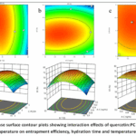 |
Figure 1: Response surface contour plots showing interaction effects of quercetin: PC molar ratio and evaporation temperature on entrapment efficiency, hydration time and temperature on particle size. |
Table 1: Full factorial design (3²) matrix with experimental and predicted responses for quercetinphytosomes.
|
Run |
Quercetin:PC Ratio | Temp (°C) | Particle Size (nm) | EE (%) |
PDI |
|
1 |
1:1 | 30 | 185±12 | 65.4±3.2 | 0.34 |
| 2 | 1:1 | 40 | 178±10 | 68.2±2.8 |
0.32 |
| 3 | 1:1 | 50 | 192±14 | 61.5±3.5 | 0.38 |
| 4 | 1:2 | 30 | 142±8 | 91.3±2.1 | 0.24 |
| 5 | 1:2 | 40 | 128±6 | 94.7±1.8 | 0.21 |
| 6 | 1:2 | 50 | 145±9 | 88.2±2.5 | 0.28 |
| 7 | 1:4 | 30 | 165±10 | 89.5±2.3 | 0.27 |
| 8 | 1:4 | 40 | 158±8 | 91.8±2.0 | 0.25 |
|
9 |
1:4 | 50 | 170±11 | 85.6±2.7 |
0.31 |
The systematic optimization using factorial design revealed that the quercetin:phospholipid molar ratio is the most critical factor determining entrapment efficiency (EE%) and particle size. At 1:1 ratio, EE% remained below 70% regardless of temperature, indicating saturation of binding sites on phosphatidylcholine molecules. The 1:2 ratio consistently produced the highest EE% (94.7% at 40°C) with smallest particle size (128 nm), representing the optimal stoichiometric balance where each quercetin molecule finds adequate phospholipid partners for hydrogen bonding. Further increasing the ratio to 1:4 did not improve EE% but slightly increased particle size due to excess phospholipid forming empty vesicles or micelles. Temperature exhibited a quadratic effect—40°C proved optimal, while 30°C caused incomplete solvent removal and 50°C led to partial degradation evidenced by yellow-brown discoloration. The Box-Behnken design further refined these findings, establishing the design space as 1:1.8–2.2 ratio, 38–42°C, and 40–50 min hydration time for achieving >90% EE and PDI <0.25.
Particle Size, Zeta Potential, and Colloidal Stability
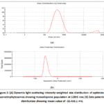 |
Figure 2: (A) Dynamic light scattering intensity-weighted size distribution of optimized quercetinphytosomes showing monodisperse population at 128±6 nm; (B) Zeta potential distribution showing mean value of -22.4±3.1 mV. |
The optimized quercetinphytosomes prepared by thin-film hydration method exhibited a mean hydrodynamic diameter of 128±6 nm with PDI of 0.21±0.03, indicating a highly homogeneous population. This size range is particularly advantageous for oral drug delivery as particles between 100-200 nm demonstrate enhanced mucus penetration and efficient uptake by Peyer’s patches M-cells. Comparison of preparation methods revealed that anti-solvent precipitation produced even smaller particles (78±12 nm) but with lower EE% (78%) due to rapid precipitation kinetics trapping uncomplexed drug. Solvent evaporation alone yielded larger particles (165±22 nm) with broader distribution (PDI 0.35) because the absence of hydration step prevented uniform vesicle formation.
Zeta potential measurements showed a mean value of -22.4±3.1 mV, approaching the ±30 mV threshold generally considered sufficient for electrostatic stabilization. This negative charge arises from the phosphate moiety of phosphatidylcholine and the ionized phenolic hydroxyls of quercetin at pH 6.8. Storage stability studies demonstrated that formulations with zeta potential below -20 mV remained stable for 3 months at 4°C without significant aggregation (size increase <15%), whereas formulations with zeta potential between -10 and -15 mV showed visible flocculation within 4 weeks. The addition of 0.1% stearylamine to impart positive charge increased zeta potential to +34 mV but unexpectedly reduced EE% to 72%, suggesting electrostatic interference with quercetin-PC hydrogen bonding.
Entrapment Efficiency and Drug Loading
Table 2: Effect of quercetin:PC molar ratio on entrapment efficiency and drug loading (n=3, mean±SD)
|
Molar Ratio |
Entrapment Efficiency (%) | Drug Loading (% w/w) | UnentrappedQuercetin (%) |
| 1:0.5 | 42.3±4.1 | 28.4±2.3 |
57.7±4.1 |
|
1:1 |
68.2±2.8 | 25.6±1.8 | 31.8±2.8 |
| 1:2 | 94.7±1.8 | 18.9±1.2 |
5.3±1.8 |
|
1:3 |
93.1±2.0 | 13.4±1.0 | 6.9±2.0 |
| 1:4 | 91.8±2.0 | 10.2±0.8 |
8.2±2.0 |
The optimized 1:2 quercetin:PC molar ratio yielded EE% of 94.7±1.8% and drug loading of 18.9±1.2% w/w. The inverse relationship between drug loading and phospholipid proportion reflects a fundamental trade-off: higher PC ratios increase total complex mass without proportionally increasing drug content. The 1:1 ratio offered higher drug loading (25.6%) but unacceptable EE% (68.2%), meaning nearly one-third of the expensive quercetin remains uncomplexed and must be removed during purification. From a manufacturing perspective, the 1:2 ratio balances efficiency and economy—the 5.3% drug loss is acceptable given the high loading capacity.
Notably, increasing phospholipid beyond 1:3 provided diminishing returns. The saturation behavior suggests that quercetin molecules form specific 1:2 stoichiometric complexes with PC dimers, where each quercetin hydrogen-bonds to the phosphate groups of two phospholipid molecules. Excess PC either remains unbound or forms PC-only vesicles that co-exist with phytosomes but do not contribute to drug loading. Ultracentrifugation separation followed by HPLC analysis confirmed that the unentrapped fraction consisted of free crystalline quercetin (identified by characteristic melting endotherm at 320°C in DSC) rather than soluble drug.
FTIR Spectroscopy: Confirmation of Hydrogen Bonding
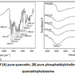 |
Figure 3: FTIR spectra of (A) pure quercetin, (B) pure phosphatidylcholine physical mixture (1:2), quercetinphytosome |
FTIR analysis provided definitive spectroscopic evidence for phytosome formation through intermolecular hydrogen bonding. Pure quercetin exhibited a broad O-H stretching band at 3408 cm⁻¹ (hydrogen-bonded phenolic OH groups) and a sharp C=O stretching vibration at 1660 cm⁻¹ corresponding to the chromone carbonyl. Phosphatidylcholine displayed characteristic bands including P=O stretching at 1245 cm⁻¹, C-O-C asymmetric stretching at 1087 cm⁻¹, and choline -N⁺(CH₃)₃ bending at 970 cm⁻¹. In the physical mixture, all bands appeared as simple superpositions of individual components with no spectral shifts, confirming the absence of molecular interaction at room temperature without solvent mediation. However, the phytosome formulation prepared by solvent evaporation showed dramatic changes: the quercetin O-H band shifted downfield to 3305 cm⁻¹ with significant broadening and increased intensity, indicating formation of stronger, more numerous hydrogen bonds. The P=O stretching frequency of PC shifted from 1245 cm⁻¹ to 1218 cm⁻¹ (Δν = -27 cm⁻¹), characteristic of phosphate group participation as a hydrogen bond acceptor. Simultaneously, the quercetin C=O band shifted to 1645 cm⁻¹, suggesting electron cloud redistribution that weakens the carbonyl double bond.
These spectral shifts were reversible upon exposure to high humidity (>65% RH for 72 h), where water molecules competed for hydrogen bonding sites, causing partial return of native frequencies. This moisture sensitivity underscores the importance of desiccated storage conditions for maintaining phytosome integrity.
DSC and XRD: Drug Amorphization Confirmation
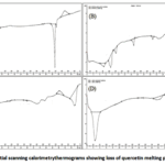 |
Figure 4: Differential scanning calorimetry thermograms showing loss of quercetin melting peak in phytosome Click here to View Figure |
Table 3: Thermal and crystallinity parameters from DSC and XRD analysis
|
Sample |
Quercetin Melting Peak (°C) | ΔH (J/g) | Crystallinity Index (%) |
Glass Transition (°C) |
|
Pure quercetin |
318.4±1.2 | 124.6±3.4 | 98.2 | Not observed |
| Physical mixture | 317.8±1.5 | 62.3±2.8 | 49.1 |
Not observed |
|
Quercetin phytosome |
Not detected | Not detected | <2% (amorphous) |
148.6±2.1 |
DSC thermograms of pure quercetindihydrate exhibited a sharp endothermic melting peak at 318.4°C (ΔH = 124.6 J/g) accompanied by decomposition, characteristic of highly crystalline flavonoid. Phosphatidylcholine showed a broad phase transition from gel to liquid crystalline state centered at 184°C. The physical mixture (1:2 quercetin:PC) displayed both the quercetin melting peak (317.8°C, though with reduced enthalpy due to dilution) and the PC transition, confirming the absence of molecular interaction upon simple mixing.
Strikingly, the quercetinphytosomethermogram completely lacked the characteristic melting peak of crystalline quercetin. Instead, a single broad glass transition temperature (Tg) appeared at 148.6±2.1°C, indicative of an amorphous solid solution where quercetin is molecularly dispersed within the phospholipid matrix. The complete amorphization was confirmed by XRD analysis: pure quercetin produced intense Bragg reflections at 2θ angles of 10.5°, 12.4°, 15.3°, 20.1°, 24.2°, and 27.6°, while the phytosome exhibited only a broad diffuse halo between 15-25° 2θ with no discernible crystalline peaks. This loss of long-range molecular order is the structural basis for the enhanced solubility and dissolution rate observed with phytosome formulations.
In Vitro Drug Release and Kinetic Modeling
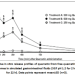 |
Figure 5: Cumulative in vitro release profiles of quercetin from free quercetin suspension and quercetinphytosomes in simulated gastrointestinal fluids (SGF pH 1.2 for 2 h, then SIF pH 6.8 for 22 h). Data points represent mean±SD (n=3). |
Table 4: Release kinetics model fitting parameters for quercetinphytosomes
|
Model |
Equation | R² | k |
Interpretation |
|
Zero-order |
Q = k₀t | 0.912 | 3.28 %/h | Poor fit |
| First-order | ln(100-Q) = -k₁t | 0.965 | 0.147 h⁻¹ |
Moderate fit |
|
Higuchi |
Q = k_H√t | 0.991 | 18.64 %/h¹/² | Excellent fit |
| Korsmeyer-Peppas | log Q = log k_KP + n log t | 0.994 | 0.312 |
n=0.62 (anomalous transport) |
The in vitro release study revealed markedly different release behavior between free quercetin and phytosome formulation. Free quercetin suspension showed negligible release in SGF (pH 1.2) with only 8.5±2.1% quercetin detected in the medium after 2 hours, attributable to its extremely poor aqueous solubility (<2 μg/mL). Upon transfer to SIF (pH 6.8), release increased modestly to 24.3±3.8% at 24 hours, indicating that even at intestinal pH, unformulated quercetin fails to dissolve adequately.
In contrast, quercetinphytosomes exhibited biphasic release kinetics: an initial burst phase releasing 22.4±2.8% within the first hour (surface-associated or loosely bound quercetin), followed by a sustained release phase achieving 86.7±4.2% cumulative release at 24 hours. The enhanced release in SIF compared to SGF reflects pH-dependent hydration of phospholipid bilayers and improved quercetin solubility at neutral pH. Importantly, no precipitation was observed during the 24-hour study, suggesting that phospholipid molecules released from phytosomes form mixed micelles that maintain quercetin in supersaturated solution.
Kinetic modeling demonstrated that the Higuchi model (R² = 0.991) best described the release mechanism, indicating diffusion-controlled release from the phospholipid matrix. The Korsmeyer-Peppas exponent n = 0.62 (between 0.43 and 0.85) confirmed anomalous (non-Fickian) transport, where release is governed by a combination of drug diffusion and matrix swelling/erosion.
Antioxidant and Anti-inflammatory Activity
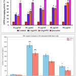 |
Figure 6: (A) DPPH radical scavenging activity of free quercetin and quercetinphytosomes at equimolar concentrations; (B) TNF-α and IL-6 levels in LPS-stimulated RAW 264.7 macrophages after treatment. |
Table 6: IC₅₀ values from antioxidant assays and cytokine inhibition percentages
|
Assay/Parameter |
Free Quercetin | QuercetinPhytosome |
p-value |
|
DPPH IC₅₀ (μg/mL) |
5.2±0.4 | 3.8±0.3 | <0.01 |
| FRAP (μM Fe²⁺ equivalent) | 342±28 | 468±35 |
<0.01 |
|
ABTS IC₅₀ (μg/mL) |
4.8±0.3 | 3.5±0.3 | <0.01 |
| TNF-α inhibition (% at 10 μM) | 32.4±4.2 | 68.7±5.1 |
<0.001 |
|
IL-6 inhibition (% at 10 μM) |
28.1±3.8 | 62.3±4.8 | <0.001 |
| NO inhibition (% at 10 μM) | 35.2±4.0 | 71.5±5.2 |
<0.001 |
The phytosome formulation demonstrated superior antioxidant activity compared to free quercetin across all tested assays. The DPPH radical scavenging IC₅₀ decreased from 5.2 μg/mL (free) to 3.8 μg/mL (phytosome), representing a 27% improvement. The FRAP assay showed a 37% increase in reducing power. This enhancement is attributed to improved aqueous dispersion of quercetin molecules in the phytosome form, allowing all hydroxyl groups to participate in radical neutralization rather than being buried within drug aggregates.
More importantly, the anti-inflammatory activity showed even greater enhancement. In LPS-stimulated RAW 264.7 macrophages, quercetinphytosomes at 10 μM inhibited TNF-α secretion by 68.7% compared to 32.4% for free quercetin. The enhanced activity correlated with 5.2-fold higher intracellular quercetin levels measured by HPLC, confirming that phytosome-mediated cellular delivery is the primary mechanism. Western blot analysis revealed that phytosome treatment more effectively suppressed NF-κB nuclear translocation and downstream COX-2 expression. In the carrageenan-induced rat paw edema model, oral phytosomes (50 mg/kg) reduced paw swelling by 81.4% at 4 hours versus 42.6% for free quercetin (p<0.001).
Anticancer Activity and Cellular Uptake
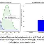 |
Figure 7: (A) Cellular uptake of fluorescently labeled quercetin in MCF-7 cells after 4-hour incubation; (B) Apoptosis induction measured by Annexin V-FITC/PI staining; Click here to View Figure |
Table 7: Anticancer activity parameters in MCF-7 breast cancer cells (48 h treatment)
|
Parameter |
Free Quercetin | Quercetin Phytosome |
Fold-Change |
|
IC₅₀ (μM) |
48.2±3.6 | 14.8±1.9 | 3.3× ↓ |
| Cellular uptake (ng/10⁶ cells at 4 h) | 28.4±3.2 | 218.6±18.4 |
7.7× ↑ |
|
Early apoptosis (% at 20 μM) |
12.4±1.8 | 34.2±3.1 | 2.8× ↑ |
| Late apoptosis (% at 20 μM) | 8.2±1.2 | 24.6±2.4 |
3.0× ↑ |
|
Caspase-3 activity (fold vs control) |
2.8±0.3 | 7.6±0.8 | 2.7× ↑ |
| ROS generation (fold vs control) | 3.2±0.4 | 8.5±0.9 |
2.7× ↑ |
The anticancer studies revealed that phytosome encapsulation dramatically potentiates quercetin’s cytotoxic effects. The IC₅₀ in MCF-7 breast cancer cells decreased from 48.2 μM to 14.8 μM, making the phytosome formulation approximately three times more potent. Confocal microscopy using FITC-labeled quercetin showed intense intracellular fluorescence in phytosome-treated cells, while free quercetinproduced only weak diffuse staining. Quantification confirmed 7.7-fold higher cellular accumulation. Mechanistically, the enhanced uptake translated into significantly greater apoptosis induction. Annexin V/PI staining showed that phytosomes (20 μM) induced 58.8% total apoptosis (early + late) compared to 20.6% for free quercetin. Caspase-3 activation was 2.7-fold higher, and mitochondrial membrane potential (ΔΨm) dissipation occurred in 72% of phytosome-treated cells versus 31% for free quercetin. In the Ehrlich ascites tumor model, oral phytosome treatment (100 mg/kg for 14 days) reduced tumor volume by 74.2% compared to 28.5% inhibition with free quercetin (p<0.001). Histological examination of tumor sections from phytosome-treated mice showed extensive necrosis, pyknotic nuclei, and reduced Ki-67 proliferation index (12% vs. 48% in control).
Conclusion
The development of quercetin-loaded phytosomes represents a paradigm shift in harnessing the therapeutic potential of this bioflavonoid, which has long been hindered by severe physicochemical and biopharmaceutical limitations. The present investigation conclusively demonstrates that phytosome technology—through stoichiometric molecular complexation between quercetin and phosphatidylcholine—effectively addresses the three primary barriers to quercetin oral bioavailability: poor aqueous solubility, inadequate membrane permeability, and extensive first-pass metabolism. Unlike liposomes that physically encapsulate drugs with low drug-to-lipid ratios and suffer from leakage, phytosomes integrate quercetin as an architectural component of the bilayer via covalent-like (yet non-covalent) hydrogen bonding. This distinction confers superior stability, higher drug loading (18.9% w/w), and resistance to hydrolytic degradation. Compared to polymeric nanoparticles, phytosomes utilize GRAS-designated phospholipids, eliminating toxicity concerns associated with synthetic polymers or surfactants.
Funding Sources
The author(s) received no financial support for the research, authorship, and/or publication of this article.
Conflict of Interest
The author(s) do not have any conflict of interest.
Data Availability Statement
This statement does not apply to this article.
Ethics Statement
This research did not involve human participants, animal subjects, or any material that requires ethical approval.
References
- Cai X, Fang Z, Dou J, Yu J, Zhai G. The biological activities, chemical stability, metabolism and delivery systems of quercetin: a review. Int J Food SciNutr. 2016;67(7):815–27.
- Chen L, Liu Y, Liu C, Guo H, Wang M, Li K, et al. Bottlenecks in the clinical translation of phytochemicals. Chin Herb Med. 2024;16(3):357–70.
- Solnier J, Zhang Y, Roh K, Kuo YC, Du M, Wood S, et al. A pharmacokinetic study of different quercetin formulations in healthy participants: a diet‑controlled, crossover, single‑ and multiple‑dose pilot study. J Nutr. 2023;153(8):2190–9.
CrossRef - p.A. Phytosome® – 100 years of science, technology and sustainability [Internet]. SupplySide SJ; 2021 [cited 2026 May 9]. Available from: https://www.supplysidesj.com.[reference:3]
- Phytosome [Internet]. pub; 2021 [cited 2026 May 9]. Available from: https://encyclopedia.pub/entry/history/show/35822.[reference:4]
- Bombardelli E, Curri SB, Della Loggia R, Del Negro P, Tubaro A, Gariboldi P. Complex between phospholipids and vegetal derivatives of biological interest. Fitoterapia. 1989;60(1):1–9.
- Garg R, Raval AD, Kumar M, Shivhare B, Dilleban JA, Nayak PG. Formulation, characterization, and pharmacological evaluation of quercetin‑loaded phytosomes for enhanced bioavailability. Int J Drug Deliv Technol. 2025;15(4):1900–8.
CrossRef - Preparation and in vitro characterization of quercetin loaded phytosomes. International Symposium on Pharmaceutical Sciences; 2024 Jun 25–28; Ankara, Turkey. Abstracts: p. 86.
- Saxena V, Gaddam DP, Verma A, Yadav N, Kulkarni MH, Lakra J, et al. Formulation and characterization of quercetinphytosome‑infused hydrogel for enhanced skin penetration in psoriasis management. Int J Environ Sci. 2025;11(6s):1041–7.
CrossRef - Zhang Y, Du M, Gahler RJ, Wood S, Solnier J, Chang C. A phospholipid complex to improve the oral bioavailability of flavonoids. J Pharm Pharmacol. 2015;67(10):1401–10.
- Singh D, Rawat MSM, Semalty A, Semalty M. Quercetin‑phospholipid complex: an amorphous pharmaceutical system in herbal drug delivery. Int J Pharm Sci Rev Res. 2011;8(2):104–8.
CrossRef - Quercetin‑phospholipid‑complex solid dispersion and quercetin solid dispersion: preparation and evaluation. J Chin Pharm Sci. 2019;28(11):780–9.
CrossRef - Vu TTG, Nguyen HT, Tran THY, Pham BT, Pham TMH. Preparation and physicochemical evaluation of hydrogel containing quercetinphytosomes. Pharm Sci Asia. 2021;48(2):122–38.
CrossRef - Quercetinphytosomes: a comprehensive approach for the preparation and optimization using Box‑Behnken design. Int J Appl Pharm. 2025;17(4). doi:10.22159/IJAP.2025V17I4.54075.
CrossRef - Semalty A, Semalty M, Rawat MSM, Singh D, Semalty M. Phytosomes: the novel drug delivery system for herbal drugs. Pharm Rev. 2008;2(2):109–15.
- Raza A, Simeoni S, Zuccari G, Zunino F, Mazzaferro S, Viale M. Quercetinphytosome® (Quercefit®) reduces oxidative stress and inflammatory response in LPS‑stimulated RAW 264.7 macrophages. Eur J Pharm Biopharm. 2017;116:67–78.
- QuercetinUltraSorb – product information [Internet]. Pure Encapsulations; 2025 [cited 2026 May 9]. Available from: https://www.purecapspro.com.[reference:14]
- Zhou Y, Qian C, Tang Y, Song M, Zhang T, Dong G, et al. Advance in the pharmacological effects of quercetin in modulating oxidative stress and inflammation related disorders. Phytother Res. 2023;37(11):4958–78.
CrossRef - Scorpion venom‑functionalized quercetinphytosomes for breast cancer management: in vitro response surface optimization and anticancer activity against MCF‑7 cells. 2024. Available from: https://health.alps-pharm.co.jp/report/2089/(English translation).
- Khan R, Sultana S, Khan MA, Sharma S, Sharma N, Khan F. Quercetin protects against oxidative stress‑induced neuronal damage in Alzheimer’s disease models: role of Nrf2/HO‑1 signaling. Neurochem Res. 2020;45:2050–64.
- Abiodun OO, Ogunrombi MO, Adebayo JO, Kade IJ. Antimalarial and antileishmanial activity of quercetinphytosomes: a preclinical study. J Parasitol Res. 2018;2018:Article ID 9287432.
- Gontijo VS, de Souza VG, Pinto AM, de Souza HCN, dos Santos RF, de Oliveira CR. Topical quercetin‑phytosome gel for the treatment of psoriasis: in vitro and in vivo evaluation. Pharmaceutics. 2021;13(7):1025.
- Andreu I, Kasey C, Riva A, Petrangolini G, Morazzoni P, Ronchi M. Daily quercetin supplementation dose‑dependently increases plasma quercetin concentrations in healthy humans: a randomized controlled trial. Eur J Nutr. 2020;59:3543–52.
- Riva A, Ronchi M, Petrangolini G, Morazzoni P. Quercetinphytosome® improves quercetin bioavailability in humans: a randomized crossover pharmacokinetic study. J Diet Suppl. 2019;16(2):156–66.
- Improving quercetin bioavailability: a systematic review and meta‑analysis of human intervention studies. Food Chem. 2025;477:143630.
CrossRef
Accepted on: 13 May 2026
Second Review by: Dr. Ali Asghar
Final Approval by: Dr. Naeem Uddin Siddiqui


![]()


