Chemistry of Celastrus paniculatus Seed Oil role in the Management of Parkinson’s Disease
 and Ravikant Kushwaha
Maharishi School of Pharmaceutical Sciences, Maharishi University of Information Technology (MUIT), IIM Road, Indrapuri Colony, Diguria, Aziz Nagar, Lucknow, Uttar Pradesh, India
Corresponding Authoe E-mail: vaibhav22mpharma@gmail.com
Download this article as:
ABSTRACT:Parkinson’s disease (PD) is a progressive neurodegenerative disorder characterized by tremors, rigidity, bradykinesia, and postural instability, primarily resulting from dopaminergic neuronal loss and oxidative stress in the substantia nigra. The present study investigates the neuroprotective and antioxidant potential of Celastrus paniculatus seed oil (CPS oil) in experimental models of PD. Gas chromatography–mass spectrometry (GC–MS) analysis revealed the presence of bioactive phytoconstituents such as sesquiterpenoids, alkaloids, phenolic triterpenoids, and essential fatty acids, which may underlie its pharmacological effects. CPS oil exhibited moderate DPPH free radical scavenging activity compared to ascorbic acid, indicating intrinsic antioxidant potential. Pharmacological evaluations, including assessments of body weight, grip strength (rotarod test), locomotor activity (actophotometer), and skilled motor coordination (staircase test), demonstrated significant improvement in CPS oil-treated groups relative to the disease control. Biochemical assays further revealed elevated levels of antioxidant enzymes—superoxide dismutase (SOD), catalase (CAT), and reduced glutathione (GSH)—alongside reduced lipid peroxidation (LPO), confirming oxidative stress attenuation. These findings suggest that CPS oil confers neuroprotection by enhancing endogenous antioxidant defense and motor function. The study scientifically validates the traditional Ayurvedic use of Celastrus paniculatus as a nervine tonic and highlights its potential as a natural therapeutic candidate for managing Parkinson’s disease.
KEYWORDS:Antioxidant; Celastrus paniculatus; Dopaminergic neurons; GC–MS; Neuroprotection; Oxidative stress; Parkinson’s disease
Introduction
Parkinson’s disease (PD) is a progressive neurodegenerative disorder characterized by tremor, rigidity, bradykinesia, and gait disturbances. These motor impairments primarily result from the gradual loss of dopaminergic neurons in the substantia nigra (SN). Current therapeutic strategies for PD largely focus on dopamine replacement therapy, which offers symptomatic relief and improves quality of life. However, long-term administration often leads to motor complications that are challenging to manage. In addition to motor dysfunction, patients experience several non-motor symptoms such as olfactory loss, sleep disturbances, autonomic dysfunction, and cognitive impairment, which are generally resistant to available pharmacological treatments and contribute significantly to disability. This highlights the urgent need for neuroprotective strategies capable of delaying disease progression and mitigating the onset of these complications.1
Neuropathological studies have revealed that dopaminergic neuronal loss is part of a broader neurodegenerative process that begins in the brainstem and gradually extends to cortical and subcortical regions. Thus, the course of PD spans several decades, with motor symptoms appearing only at intermediate stages. This slow progression provides a critical therapeutic window in which interventions targeting oxidative stress, mitochondrial dysfunction, and neuroinflammation could prevent or delay irreversible neuronal loss. Despite advances in symptomatic treatment, a major challenge remains—the lack of an effective neuroprotective therapy that can be implemented early in the disease course.2
Celastrus paniculatus Willd. (Celastraceae), commonly known as Jyotishmati, Malkangni, or Kangani, is classified in Ayurveda as a Medhya Rasayana (nervine tonic). Traditionally, its seeds and seed oil have been used for enhancing memory and cognitive function. Phytochemical investigations have identified numerous bioactive constituents in the seeds and oil, including sesquiterpenoid polyalcohols and esters (malkanguniol, malkangunin, polyalcohol A–D, celapnin), alkaloids (paniculatine, celastrine), phenolic triterpenoids (celastrol, paniculatadiol), fatty acids (oleic, linoleic, linolenic, palmitic, stearic, lignoceric acid), and agarofuran derivatives. Experimental studies have reported C. paniculatus to possess neuroprotective, antioxidant, anti-inflammatory, anxiolytic, and memory-enhancing properties, attributed largely to its free-radical-scavenging and mitochondrial-protective mechanisms.3
In the present study, CPS oil was evaluated for its neuroprotective and antioxidant potential in experimental models of PD. The study aimed to (i) characterize the phytochemical profile of CPS oil using gas chromatography–mass spectrometry (GC–MS), (ii) assess its antioxidant activity in vitro, and (iii) evaluate its neuroprotective effects through behavioral and biochemical assessments in PD-induced rats. This work provides preclinical evidence supporting the traditional Ayurvedic use of CPS oil and its potential role as a natural neuroprotective agent for the management of Parkinson’s disease.
Materials and Methods
Materials
Seeds of Celastrus paniculatus were procured from a local herbal market in Lucknow, India. The seeds were authenticated and their oil extracted by cold pressing. All other reagents and chemicals used were of analytical grade and procured from standard suppliers.
Experimental Animals
Male Wistar rats (220–250 g) were used for the study. Animals were obtained from a CPCSEA-registered breeder and acclimatized for one week under standard laboratory conditions (temperature: 22 ± 2 °C; relative humidity: 55–65%; 12 h light/dark cycle). Standard pellet diet and water were provided ad libitum. All experimental protocols were approved by the Institutional Animal Ethics Committee (IAEC) of Bilwal Medchem and Research Laboratory Pvt. Ltd., Jaipur (Approval Ref. No.: BMRL/IAEC/2024-37). All behavioral experiments were conducted during the light phase (09:00–17:00 h).4
Gas Chromatography–Mass Spectrometry (GC–MS) Analysis
The chemical composition of C. paniculatus seed oil was determined using GC–MS (Agilent 7890A GC system coupled with a 5975C mass selective detector). Separation was performed on a DB-5MS capillary column (30 m × 0.25 mm × 0.25 µm film thickness). The sample was diluted with n-hexane (1:10 v/v), and 1 µL was injected in split mode using helium as the carrier gas at a constant flow rate of 1.0 mL/min. The injector temperature was maintained at 250 °C. The oven temperature was programmed as follows: initial 60 °C (held for 2 min), increased to 200 °C at 3 °C/min, and then to 300 °C at 5 °C/min, with a final hold of 10 min. The mass spectrometer operated in electron impact (EI) mode at 70 eV with an ion source temperature of 230 °C and a mass scan range of m/z 40–650.
Chromatographic peaks were identified by comparing their mass spectra with those in the NIST and Wiley libraries. A total of 109 peaks were detected, representing diverse phytoconstituent classes, including sesquiterpenes, alkaloids, triterpenoids, and fatty acids. The relative abundance (%) of each component was calculated from peak area normalization. Representative chromatograms and corresponding mass spectra are presented in Figures 1–2, and major compounds are listed in Table 1.5-8
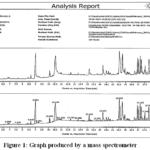 |
Figure 1: Graph produced by a mass spectrometer Click here to View Figure |
Table 1: Antioxidant activity
|
Conc. (µg/ml) |
% Of inhibition AS |
% Of inhibition CPS OIL |
|
100 |
90.69 | 46.04 |
| 200 | 91.93 |
52.34 |
|
300 |
93.02 | 60.44 |
| 400 | 95.19 |
70.85 |
|
500 |
96.74 | 80.93 |
| IC50 | 5.73 |
26.44 |
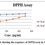 |
Figure 2: Graph showing the responses of DPPH assayin AS and CPS Oil Click here to View Figure |
Methods and Results
Pharmacological Studies
Body weight was recorded for each animal at the beginning and at the end of the experimental period to assess general health status and the effect of CPS oil treatment. The percentage change in body weight was calculated using the formula:
Percentage change in body weight=(Final weight−Initial weight)/ Initial weight×100
As shown in Table 2 and Figure 3, the disease control group exhibited a significant reduction in body weight compared to the normal control group, indicating systemic stress and dopaminergic neuronal damage associated with Parkinsonian pathology. In contrast, treatment with CPS oil produced a dose-dependent improvement in body weight, reflecting enhanced metabolic and neuromuscular function. The improvement in weight gain among CPS oil-treated groups suggests overall physiological recovery and neuroprotection, comparable to the standard treatment group.9
Table 2: Assessment of body weight
|
Weight parameter Initial |
|||||||||
|
|
I | I | I | I | I | I | MEAN | SD |
SE |
|
NC |
175 | 178 | 182 | 179 | 184 | 170 | 178.00 | 5.02 | 2.05 |
| DC | 171 | 173 | 178 | 185 | 170 | 172 | 174.83 | 5.71 |
2.33 |
|
SD |
180 | 185 | 187 | 190 | 195 | 180 | 186.17 | 5.85 | 2.39 |
| TLD | 190 | 175 | 179 | 188 | 180 | 176 | 181.33 | 6.25 |
2.55 |
| THD | 180 | 173 | 181 | 187 | 170 | 171 | 177.00 | 6.72 |
2.74 |
|
Weight parameter Final |
|||||||||
| F | F | F | F | F | F | MEAN | SD |
SE |
|
|
NC |
186 | 187 | 179 | 171 | 185 | 180 | 181.33 | 6.02 | 2.46 |
| DC | 145 | 148 | 140 | 135 | 150 | 155 | 145.50 | 7.18 |
2.93 |
|
SD |
175 | 160 | 175 | 180 | 175 | 176 | 173.50 | 6.89 | 2.81 |
| TLD | 169 | 165 | 170 | 179 | 160 | 175 | 169.67 | 6.80 |
2.78 |
|
THD |
174 | 176 | 172 | 178 | 168 | 165 | 172.17 | 4.92 |
2.01 |
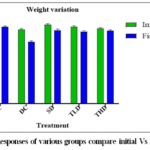 |
Figure 3: Responses of various groups compare initial Vs final weight Click here to View Figure |
Grip strength
Grip strength was assessed to evaluate muscle tone, balance, and motor coordinationkey indicators of neuromuscular integrity affected in Parkinson’s disease. The test was performed using a rotarod apparatus, following the method described by Dunham and Miya.10 Each rat was placed on a rotating rod (20 rpm), and the time (in seconds) that the animal remained on the rod without falling was recorded. Prior to testing, animals were trained for three consecutive days to minimize variability. The mean latency to fall was calculated for each group.
Data presented in Table 3 and Figure 4 demonstrate a marked reduction in grip strength and fall latency in the disease control group, indicating severe motor impairment. Conversely, animals treated with CPS oil exhibited a significant, dose-dependent improvement in grip strength and retention time on the rotarod compared to the disease control. This improvement reflects enhanced neuromuscular coordination, balance, and resistance to fatigue. The higher dose of CPS oil (e.g., 400 mg/kg) produced effects comparable to those observed with the standard drug treatment, supporting its neuroprotective efficacy.
Table 3: Activity of muscle coordination using the Rota Rod apparatus
|
ROTA ROD |
MEAN | SD |
SE |
||||||
|
NC |
164 | 161 | 151 | 157 | 162 | 167 | 160.33 | 5.65 | 2.30 |
| DC | 64 | 52 | 56 | 61 | 72 | 60 | 60.83 | 6.88 |
2.81 |
|
SD |
165 | 154 | 150 | 156 | 160 | 168 | 158.83 | 6.82 | 2.79 |
| TLD | 122 | 129 | 138 | 126 | 133 | 135 | 130.50 | 5.96 |
2.43 |
|
THD |
156 | 148 | 141 | 152 | 147 | 160 | 150.67 | 6.80 |
2.78 |
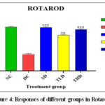 |
Figure 4: Responses of different groups in Rotarod Click here to View Figure |
Locomotor Activity (Actophotometer Test)
Spontaneous locomotor activity was evaluated using a digital actophotometer, which measures the movement of animals based on the interruption of infrared light beams positioned above the chamber floor.11 Each rat was placed individually in the activity cage, and the total number of beam interruptions was automatically recorded for a period of 5 minutes. The apparatus was cleaned with 70% ethanol between trials to eliminate odor cues. Decreased locomotor activity reflects bradykinesia and reduced exploratory behavior typical of Parkinsonian symptoms.
As shown in Table 4 and Figure 5, the disease control group demonstrated a significant reduction (p < 0.01) in locomotor activity compared to the normal control, confirming motor impairment due to dopaminergic neurodegeneration. Treatment with CPS oil significantly enhanced locomotor activity in a dose-dependent manner, indicating improvement in spontaneous movement and overall motor function. The higher dose of CPS oil (400 mg/kg) restored activity levels close to those of the standard-treated group, suggesting effective neuroprotection and partial restoration of dopaminergic tone.
Table 4: Locomotor activity
|
ACTOPHOTOMETER |
MEAN | SD | SE | ||||||
| NC | 129 | 119 | 122 | 129 | 125 | 136 | 126.67 | 6.02 |
2.46 |
|
DC |
40 | 34 | 38 | 42 | 35 | 48 | 39.50 | 5.13 | 2.09 |
| SD | 118 | 122 | 128 | 125 | 120 | 124 | 122.83 | 3.60 |
1.47 |
|
TLD |
100 | 97 | 89 | 99 | 85 | 98 | 94.67 | 6.15 | 2.51 |
|
THD |
116 | 121 | 122 | 120 | 112 | 118 | 118.17 | 3.71 |
1.51 |
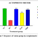 |
Figure 5: Responses of various groups in Actophotometer Click here to View Figure |
Skilled Motor Coordination (Staircase Test)
The staircase test was employed to assess fine motor coordination, skilled forelimb use, and paw dexterity in experimental animals.12-15 Each rat was placed in the central compartment of the staircase apparatus, which consisted of two descending staircases on either side, each containing recessed wells holding food pellets. The setup allows for independent evaluation of the left and right forepaws, as pellets can only be retrieved with the corresponding paw.
Animals were subjected to mild food restriction (maintaining 90% of their normal body weight) to enhance motivation. Prior to testing, each rat underwent three training sessions to familiarize them with the apparatus. During the test session, the number of pellets retrieved, displaced, and left untouched within a 15-minute period was recorded. This provides a quantitative measure of reaching ability, coordination, and fine motor performance. As shown in Tables 5.1 and 5.2 and Figure 6, the disease control group exhibited a significant reduction in the number of pellets retrieved and a higher number of pellets displaced or untouched compared to the normal control group (p < 0.01). This indicates pronounced impairment in skilled forelimb function, typical of Parkinsonian motor deficits.
Treatment with CPS oil markedly improved performance in a dose-dependent manner, evidenced by an increased number of pellets successfully retrieved and a decrease in pellets displaced. The higher dose of CPS oil (400 mg/kg) showed results comparable to the standard-treated group, demonstrating substantial restoration of motor coordination and paw dexterity. These findings confirm the therapeutic potential of CPS oil in improving fine motor control and mitigating PD-related neuromotor dysfunctions.
Table 5.1: Test of muscle coordination using a staircase
|
Stair case (15 min) |
|||||||||
|
|
I | I | I | I | I | I | MEAN | SD |
SE |
| NC | 36 | 39 | 40 | 45 | 47 | 48 | 42.50 | 4.85 |
1.98 |
|
DC |
110 | 108 | 106 | 112 | 110 | 104 | 108.33 | 2.94 | 1.20 |
| SD | 45 | 44 | 50 | 48 | 53 | 46 | 47.67 | 3.39 |
1.38 |
|
TLD |
58 | 54 | 59 | 56 | 50 | 58 | 55.83 | 3.37 | 1.38 |
| THD | 50 | 49 | 53 | 50 | 58 | 44 | 50.67 | 4.63 |
1.89 |
Table 5.2: Responses of different groups in Initiation test
|
Stair case (15 min) |
|||||||||
| F | F | F | F | F | F | MEAN | SD |
SE |
|
|
NC |
39 | 49 | 49 | 48 | 49 | 50 | 47.33 | 4.13 | 1.69 |
| DC | 113 | 111 | 115 | 118 | 111 | 108 | 112.67 | 3.50 |
1.43 |
|
SD |
60 | 70 | 64 | 61 | 62 | 70 | 64.50 | 4.46 | 1.82 |
| TLD | 70 | 68 | 70 | 72 | 76 | 73 | 71.50 | 2.81 |
1.15 |
|
THD |
62 | 65 | 69 | 70 | 71 | 71 | 68.00 | 3.69 |
1.51 |
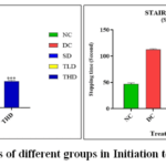 |
Figure 6: Responses of different groups in Initiation test and Stepping Click here to View Figure |
Biochemical Estimations
Biochemical estimations were performed to evaluate the antioxidant potential of the test formulations by quantifying endogenous antioxidant enzyme levels and oxidative stress markers in brain tissue homogenates. The assays included SOD, CAT, and GSH, which represent key components of the enzymatic antioxidant defense system. In addition, LPO levels were measured to assess the extent of oxidative damage to membrane lipids.
These biochemical parameters were determined using standard spectrophotometric methods following established protocols.16-19 The results were expressed as mean ± SEM and are presented in Table 6 and Figure 7, which demonstrate the comparative antioxidant status among different treatment groups. Collectively, these estimations provided a comprehensive insight into the ability of the test formulation to mitigate oxidative stress and restore redox homeostasis in experimental animals.
Table 6: Assay for antioxidant enzymes in anti-parkinson’s disease Various antioxidant measurements in the context of antiparkinsonian effect SOD, CAT, GSH, LPO.
| SOD | MEAN | SD | SE | ||||||
|
NC |
60 | 63 | 65 | 67 | 62 | 58 | 62.50 | 3.27 | 1.34 |
|
DC |
21 | 23 | 20 | 19 | 18 | 25 | 21.00 | 2.61 | 1.06 |
| SD | 50 | 55 | 49 | 48 | 53 | 51 | 51.00 | 2.61 |
1.06 |
|
TLD |
45 | 38 | 39 | 40 | 42 | 43 | 41.17 | 2.64 |
1.08 |
| THD | 46 | 52 | 51 | 48 | 47 | 42 | 47.67 | 3.61 |
1.48 |
|
CAT |
MEAN | SD | SE | ||||||
| NC | 58 | 63 | 62 | 58 | 57 | 63 | 60.17 | 2.79 |
1.14 |
|
DC |
30 | 28 | 26 | 29 | 25 | 29 | 27.83 | 1.94 | 0.79 |
| SD | 51 | 49 | 51 | 53 | 50 | 48 | 50.33 | 1.75 |
0.71 |
|
TLD |
35 | 37 | 35 | 39 | 42 | 39 | 37.83 | 2.71 | 1.11 |
| THD | 48 | 45 | 47 | 46 | 45 | 42 | 45.50 | 2.07 |
0.85 |
|
GSH |
MEAN | SD | SE | ||||||
| NC | 55 | 58 | 54 | 52 | 57 | 58 | 55.67 | 2.42 |
0.99 |
| DC | 20 | 21 | 25 | 23 | 20 | 26 | 22.50 | 2.59 | 1.06 |
| SD | 47 | 49 | 46 | 45 | 40 | 48 | 45.83 | 3.19 | 1.30 |
| TLD | 39 | 37 | 30 | 32 | 35 | 38 | 35.17 | 3.54 | 1.45 |
| THD | 42 | 46 | 41 | 43 | 44 | 48 | 44.00 | 2.61 | 1.06 |
| LPO | MEAN | SD | SE | ||||||
| NC | 102 | 108 | 101 | 109 | 103 | 107 | 105.00 | 3.41 | 1.39 |
| DC | 240 | 238 | 242 | 245 | 237 | 234 | 239.33 | 3.88 | 1.58 |
| SD | 175 | 180 | 182 | 170 | 181 | 176 | 177.33 | 4.55 | 1.86 |
| TLD | 200 | 206 | 204 | 198 | 197 | 204 | 201.50 | 3.67 | 1.50 |
| THD | 197 | 196 | 186 | 194 | 195 | 185 | 192.17 | 5.27 | 2.15 |
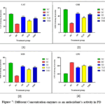 |
Figure 7: Different Concentration enzymes as an antioxidant’s activity in PD Click here to View Figure |
Discussion
The present study demonstrated that CPS oil possesses significant neuroprotective and antioxidant potential in experimental models of PD. GC–MS analysis revealed the presence of bioactive constituents such as sesquiterpenoids, alkaloids, phenolic triterpenoids, and essential fatty acids, all of which are known for their neuroprotective and antioxidative activities. The DPPH assay confirmed the free radical scavenging capacity of CPS oil, which, although lower than the standard ascorbic acid, still indicated a moderate yet pharmacologically relevant antioxidant activity.
Pharmacological assessments including body weight, grip strength (rotarod test), locomotor activity, and skilled paw use (staircase test) showed marked improvements in motor coordination, muscle tone, and dexterity in CPS oil-treated groups compared to the disease control. These findings suggest that CPS oil effectively ameliorates dopaminergic dysfunction and alleviates Parkinsonian motor deficits.
Biochemical estimations further substantiated these observations by showing a significant restoration of endogenous antioxidant defense systems. Elevated levels of SOD, CAT, and GSH, along with reduced lipid peroxidation (LPO), indicate that CPS oil attenuates oxidative stress, a central pathogenic mechanism in Parkinson’s disease. The observed neuroprotection may be attributed to the synergistic action of phytoconstituents, which not only scavenge reactive oxygen species but also enhance mitochondrial function and promote neuronal survival.
Collectively, these findings corroborate the traditional Ayurvedic use of CPS oil as a Medhya Rasayana (memory and intellect enhancer) and provide a scientific rationale for its neuroprotective potential in PD models.20-24
Conclusion
The study concludes that CPS oil exhibits promising neuroprotective and antioxidant properties, effectively improving motor coordination, muscle strength, and locomotor activity while reducing oxidative stress in experimental models of Parkinson’s disease. Its rich phytoconstituent profile contributes to the enhancement of endogenous antioxidant defenses, thereby mitigating neurodegeneration. These findings highlight the therapeutic potential of CPS oil as a natural adjunct in the management of PD. However, further investigations focusing on its molecular mechanisms of action, long-term safety, and clinical validation are essential to translate these preclinical findings into viable therapeutic applications.
Acknowledgement
The authors would like to thank Maharishi School of Pharmaceutical Sciences, Maharishi University of Information Technology (MUIT), IIM Road, Indrapuri Colony, Diguria, Aziz Nagar, Lucknow, Uttar Pradesh 226013, India
Funding Sources
The author(s) received no financial support for the research, authorship, and/or publication of this article.
Conflict of Interest
The author(s) do not have any conflict of interest.
Data Availability Statement
This statement does not apply to this article.
Ethics Statement
This research is requires ethical Approval Ref. No.: BMRL/IAEC/2024-37
References
- Sankaramourthy D, Sankaranarayanan L, Subramanian K, Sadras SR. Neuroprotective potential of Celastrus paniculatus seeds against common neurological ailments: a narrative review. Journal of Complementary and Integrative Medicine. 2023 Aug 14;20(3):530-6.
CrossRef - Pujari GR, Subramanian V, Rao SR. Neuroprotective role of Celastrus paniculatus Willd and Sida cordifolia Linn on kainic acid-induced neuronal damage in neurodegenerative diseases. Pharmaceutical Sciences Asia. 2022 May 1;49(3).
CrossRef - Kandikattu HK, Amruta N, Khanum F, Narayana VV, Srinivasulu D. Phytochemical composition, pharmacological properties, and therapeutic applications of celastrus paniculatus. Current Traditional Medicine. 2021 Feb 1;7(1):107-24.
CrossRef - van Harten AC, Smits LL, Teunissen CE, Visser PJ, Koene T, Blankenstein MA, Scheltens P, van der Flier WM. Preclinical AD predicts decline in memory and executive functions in subjective complaints. Neurology. 2013 Oct 15;81(16):1409-16.
CrossRef - Elawad MA, Ayaz M, Mosa OF, Usman A, Hamdoon AA, Almawash S, Salim LH, Ahmed A, Elkhalifa ME. Polyphenols and Their Biogenic Nano‐Formulations Targeting BACE1 as Anti‐Amyloid Therapies; Meeting the Challenges of Bioavailability, Safety, and Specificity for the Treatment of Alzheimer’s Disease. Molecular Nutrition & Food Research. 2024:2400525.
CrossRef - Zhang H, Liang J, Chen N. The potential role of miRNA-regulated autophagy in Alzheimer’s disease. International Journal of Molecular Sciences. 2022 Jul 14;23(14):7789.
CrossRef - Saleh O, Albakri K, Altiti A, Abutair I, Shalan S, Mohd OB, Negida A, Mushtaq G, Kamal MA. The Role of Non-coding RNAs in Alzheimer’s Disease: Pathogenesis, Novel Biomarkers, and Potential Therapeutic Targets. CNS & Neurological Disorders-Drug Targets (Formerly Current Drug Targets-CNS & Neurological Disorders). 2024 Jun 1;23(6):731-45.
CrossRef - Nahálková J. Finding new ways how to control BACE1. The Journal of Membrane Biology. 2022 Jun;255(2):293-318.
CrossRef - Keable R, Hu S, Pfundstein G, Kozlova I, Su F, Du X, Yang H, Gunnersen J, Schachner M, Leshchyns’ ka I, Sytnyk V. The BACE1-generated C-terminal fragment of the neural cell adhesion molecule 2 (NCAM2) promotes BACE1 targeting to Rab11-positive endosomes. Cellular and Molecular Life Sciences. 2022 Nov;79(11):555.
CrossRef - Sabaie H, Amirinejad N, Asadi MR, Jalaiei A, Daneshmandpour Y, Rezaei O, Taheri M, Rezazadeh M. Molecular insight into the therapeutic potential of long non-coding RNA-associated competing endogenous RNA axes in alzheimer’s disease: a systematic scoping review. Frontiers in Aging Neuroscience. 2021 Nov 25;13:742242.
CrossRef - Lee KH, Kim UJ, Cha M, Lee BH. Chronic treatment of ascorbic acid leads to age-dependent neuroprotection against oxidative injury in hippocampal slice cultures. International Journal of Molecular Sciences. 2021 Feb 5;22(4):1608.
CrossRef - Singh D, Hembrom S, Raj A. Neuroprotective effect of flavonoids: A systematic review. J Pharmacogn Phytochem. 2019;8(1):699-707.
- Aleem M. Phytochemistry and pharmacology of Celastrus paniculatus Wild.: a nootropic drug. Journal of Complementary and Integrative Medicine. 2023 Mar 14;20(1):24-46.
CrossRef - Phattanakiatsakul T, Chaemsawang W, Athipornchai A, Thongon N, Chamniansawat S. Celastrus paniculatus seed extract exhibits neuroprotective effects against MPP+‑induced apoptotic cell death via GSK‑3β in a Parkinson’s disease model. Biomedical reports. 2024 Jan 23;20(3):46.
CrossRef - Pujari GR, Subramanian V, Rao SR. Neuroprotective role of Celastrus paniculatus Willd and Sida cordifolia Linn on kainic acid-induced neuronal damage in neurodegenerative diseases. Pharmaceutical Sciences Asia. 2022 May 1;49(3).
CrossRef - Kandikattu HK, Amruta N, Khanum F, Narayana VV, Srinivasulu D. Phytochemical composition, pharmacological properties, and therapeutic applications of celastrus paniculatus. Current Traditional Medicine. 2021 Feb 1;7(1):107-24.
CrossRef - Faldu KG, Patel SS, Shah JS. Celastrus paniculatus oil ameliorates NF-KB mediated neuroinflammation and synaptic plasticity in the scopolamine-induced cognitive impairment rat model. Metabolic Brain Disease. 2023 Apr;38(4):1405-19.
CrossRef - Manoharan N, Jayamurali D, Sridhar A, Govindarajulu SN. Neuroprotective capacity of Celastrus paniculatus on rotenone-induced parkinsonism in zebrafish model. Molecular Biology Reports. 2025 Dec;52(1):1-4.
CrossRef - Nagpal K, Garg M, Arora D, Dubey A, Grewal AS. An extensive review on phytochemistry and pharmacological activities of Indian medicinal plant Celastrus paniculatus Willd. Phytotherapy Research. 2022 May;36(5):1930-51.
CrossRef - Patel R, Jain NS. Stimulation of central histaminergic transmission attenuates diazepam-induced motor disturbance on rota-rod and beam walking tests in mice. Behavioural Pharmacology. 2024 Sep 1;35(6):351-65.
CrossRef - Keane SP, Chadman KK, Gomez AR, Hu W. Pros and cons of narrow-versus wide-compartment rotarod apparatus: An experimental study in mice. Behavioural Brain Research. 2024 Apr 12;463:114901.
CrossRef - Ogita H, Taura A, Nishimura K, Tona Y, Tateya T, Ohnishi H, Omori K, Ito J. Development of the New Rota-Rod Treadmill System of Guinea Pigs. AINO JOURNAL. 2025 Jun 30;22(1):49-52.
- Halliwell B. Understanding mechanisms of antioxidant action in health and disease. Nature Reviews Molecular Cell Biology. 2024 Jan;25(1):13-33.
CrossRef - Hu B, Ouyang Y, Zhao T, Wang Z, Yan Q, Qian Q, Wang W, Wang S. Antioxidant hydrogels: antioxidant mechanisms, design strategies, and applications in the treatment of oxidative stress‐related diseases. Advanced healthcare materials. 2024 Apr;13(11):2303817.
CrossRef
Accepted on: 08 Jan 2026
Second Review by: Dr. Andrew J.
Final Approval by: Dr. Tanay Pramanik


![]()


