Green Synthesis and Characterization of Silver Nanoparticles from Aerva lanata Leaf Extract: A Medicinal Plant for Bone Fracture Healing in Tribal Communities of Chhattisgarh
 and Pratush Jaiswal*
Department of Chemistry, Dr. C. V. Raman University, Kota, Bilaspur, Chhattisgarh, India.
Corresponding Author E-mail:pratyush.jaiswal08shanu@gmail.com
DOI : http://dx.doi.org/10.13005/ojc/420240
Download this article as:
![]()
Medicinal plants are the most common biosource of medications in traditional medical systems. The current study used aqueous leaf extracts of the Amaranthaceae plant Aerva lanata to synthesize silver nanoparticles (AgNPs) via green synthesis. The crystalline nature, size, shape, and elemental composition of the biosynthesized AgNPs are analyzed using FTIR, EDAX, Zeta Potentials, FESEM, XRD, and UV-Visible spectroscopy. FESEM and EDAX examination confirmed the green-synthesized AgNPs, which werespherical in shape, having a size range of 66.70 ±0.15 to 98.40 ±0.05 nm.A prominent absorbance peak confirmed the presence of AgNPs at 344 nm in UV-Visible spectra.XRD spectra evaluate the AgNPs were of FCC crystal having 111, 200, 220, and 311 planes. AgNPs' moderate stability is confirmed by a -22.0 ±0.01 mV zeta potential value.
KEYWORDS:Aerva lanata; FESEM; FTIR; Nanoparticles; UV-Visible spectroscopy
Introduction
With its potential applications in material engineering, environmental research, and health, nanotechnology has become a ground-breaking area of contemporary science.1 One kind of nanomaterial that has created a lot of interest is AgNPs, which have broad-spectrum antibacterial, anti-inflammatory, and wound-healing properties.2 Traditional techniques for creating silver nanoparticles frequently use high-energy procedures and hazardous chemicals, endangering both human health and the environment.3 On the other hand, environmentally benign, economical, and sustainable alternatives are provided by green synthesis methods that use plant extracts.4
Aerva lanata (Linn.), a medicinal herb commonly found in India, particularly in the tribal regions of Chhattisgarh, is traditionally used for treating various ailments.5 Phytochemicals, including flavonoids, alkaloids, and phenolic compounds, which are abundant in plants, are essential for decreasing and stabilizing metal ions during NPs synthesis.6
This plant has been widely researched forits medicinal properties through pharmacological and phytochemical studies.7,8 Aerva lanata exhibits many therapeutic pharmacological properties, such as Antioxidant properties9 Anticancer10 Anti-inflammatory.11 Hepatoprotective effect,12 Diuretic,13 Anturolithic activity14, and Anti-diabetic property11 due to the presence of many polyphenols and Flavonoids.Even though Aerva lanata contains a variety of natural compoundswith potential health benefits, its medicinal potential for the Bone Healing process is still not well understood. Morethorough and organized studies are needed to explore and make the most of its Bone healingproperties. These studies could help discover newNanomedicine and lead to thedevelopment of a drug for Bone Fracture Healing.
Aerva lanata leaf extract is used in AgNPs green synthesis for this research,15,16 which is followed by physicochemical characterization.17 Using a nanobiotechnological approach, this study seeks to support the plant’s traditional use in bone healing while also investigating its bio-reductive potential for nanoparticle production.18 Understanding the synthesis mechanism and properties of these nanoparticles may contribute to the development of novel therapeutic agents that align with traditional knowledge systems and modern biomedical science.19.
Materials and Methodology
Green synthesis of Silver Nanoparticles:
The Medicinal plant Aerva lanata had been gathered from the village Bicharpur, Block Lormi, Dist.-Mungeli, Chhattisgarh, in the month of November-December 2023 and identified by the Botanical Survey of India, Allahabad, Uttar Pradesh.
The plant was rinsed with distilled water after being cleaned with tap water, then allowed to dry for 30 days in the shade. The leaves were ground using a mixture grinder into a coarse powder, stored in an airtight container, and used in subsequent extractions.
Aerva lanata leaf aqueous extracts were prepared by adding 100 ml of deionized water to 10 g of air-dried leaf powder in a 150 ml conical flask, covering the flask with cotton wool, and shaking the flask on a mechanical shaker for six hours at 150 rpm at room temperature. It was macerated for a further 48 hours at room temperature. The Solvent was filtered, collected, and stored at 4℃ in an air-tight bottle.
In order to prepare AgNPs, the above filtrated solution served as an extract source. Ten milliliters of 25 mM AgNO3 were added to the ten milliliters of filtrate at pH 8.9.The mixture had been left at room temperature (27-30℃)for 5-10 minutes,and it turned from pale light green and finally dark brown. The color change demonstrated the presence of biosynthesized AgNPs.20
The solution had been centrifuged for 15minutes at 10,000rpm, the pellets were separated, cleaned with deionized water, and left to dry in the air. So, in the present study, the biosynthesis of AgNPs using the leaf aqueous extract of Aerva lanata was prepared without using any toxic chemicals by the “Green Synthesis” method.
 |
Figure 1: (a) Silver nitrate solution, (b) Aqueous leaf extract of Aerva lanata, (c) Silver nanoparticle solution, (d) AgNPs of leaf extract of Aerva lanata. Click here to View Figure |
UV-VisibleSpectrophotometry:
Because of surface plasmon resonance, noble metal NPs absorb heavily in the visible spectrum. As a result, UV-visible absorption spectroscopy serves as the main characterization instrument for investigating metal NPs formation, hence it has shown itself as a very practical method for nanoparticle analysis. UV-Visible absorbance spectra had been recorded and observed on the Systronic 2203 Double Beam UV-Vis spectrophotometer in the 200-800nm wavelength range in order to track AgNP bio-reduction utilizing plant leaf aqueous extract of Aerva lanata.
Fourier transform infrared
FTIR spectral examination of biosynthesized AgNPs demonstrates crystalline NP growth alongside functional group presence in the sample. With the peaks indicating higher concentration components that show the existence of various bond types and functional groups (that involve amines, halides, ketones, and alkanes) that absorb infrared light at different wavelengths, this profile resembles an absorption spectrum. The apparatus IR Affinity-1, Shimadzu, Japan, was used in the Department of Physics and Astrophysics at Pt. Ravi Shankar University in Raipur, Chhattisgarh, to monitor and record potential biomolecules identification that was accountable for the stability of biosynthesized AgNP reduction in the range 400-4000 cm-1.
Zeta potential
Zeta potential analysis of biosynthesized AgNPs was additionally conducted to learn more about the negative potential value, which contributes to stability as well as reduction of biosynthesized AgNPs because of the electrostatic repulsive force that prevents AgNP agglomeration from developing.The “National Institute of Technology Raipur”, “Chhattisgarh’s Department of Chemical Engineering” used a Zeta potential analyzer using the Anton Paar Litesizer 500 to conduct a zeta potential investigationin the voltage range -200 to +200 mV of the AgNPs.
X-ray Diffractometry
AgNPs crystalline structure was investigated using their XRD patterns. Bio-reduced silver colloidal solution (AgNPs) was drop-coated onto a glass substrate at Pt. Ravi Shankar University’s Department of Physics and Astrophysics in Raipur, Chhattisgarh. A Bruker equipment called the D2 Phaser Model: 08 Discover was employed to record the XRD pattern computation of the solution. The measurements were conducted over a wide range 0 to 95 Bragg angles 2ϴ, at scanning rate of 2min-1.
Energy Dispersive X-ray Spectroscopy
EDAX, a chemical microanalysis approach, uses the X-rays released by the sample when it is struck by an electron beam for determining the elemental composition of the AgNPs volume under inquiry. The “CARL ZEISS UHR FESEM GEMINI SEM 500 KMAT” was employed to do EDAX analysis at 20 KeV voltage at the Central Instrument facility of the Indian Institute of Technology in Bhilai, Chhattisgarh.
Field Emission Scanning Electron Microscopy studies
FESEM was used to examine the biosynthesized AgNPs’ dimensions, form, and surface morphology. In the Central Instrument facility of the Indian Institute of Technology, Bhilai, Chhattisgarh, the CARL ZEISS UHR FESEM GEMINI SEM 500 KMAT was used in a resolution range of 1nm to 500 nm at 5 KV, in combination with a FESEM that was connected to EDAX.
Results and Discussions
Green synthesis of Silver Nanoparticles from Aqueous Leaf Extract of Aerva lanata
Aerva lanata plant leaf extracts are used as a reducing moreover capping agent in the straightforward green process of biosynthesizing AgNPs using AgNO3 solution. After adding 25 mM AgNO3 solution, the solution containing the pale green leaf extract of Aerva lanata turned dark brown in a matter of minutes. Thus, without the introduction of any hazardous chemicals, it was verified that AgNO3 was reduced and that Aerva lanata produced silver nanoparticles (AgNPs). The decrease in time was between 5 to 10 minutes.21,22
UV-Vis spectroscopy
The synthesis of AgNPs was confirmed by “UV-Vis spectra of sample solution (AgNPs)”, which displayed a prominent and sharp absorbance peak at 344nm (Figure 2). This peak has been a typical silver nanoparticle “surface plasmon resonance (SPR)” peak.The appearance of brownish colour observed is characteristic of surface plasmon vibrations, which clearly indicates that the reducing and stabilizing agents are present in the leaf extract of Aerva lanata, which is involved in the bioreduction and capping of the generated AgNPs. Particles under the SPR range 320-450 nm had obviously been caused by AgNPs with sizes that range from 2 to 100 nm, according to an earlier study.23 An absorbance peak peculiar to silver nanoparticles was discovered at around 345 nm.24
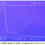 |
Figure 2: UV-Visible graph of AgNPs of Aqueous leaf extract of“Aerva lanata” Click here to View Figure |
Fourier Transform Infrared
In the AgNPs FTIR spectrum study (Figure 3) [25], the prominent peaks at 3849cm-1, 3624cm-1, 3645cm-1, and 3643cm-1 indicate the polyphenolic (Aromatic) O–H “stretching vibrations. Peaks seen at 2351cm-1 and 2331cm-1 represent -CH Aromatic stretching vibrations. Peaks at 1751cm-1 and 1539cm-1, respectively, represent C=O stretching vibrations and C=C aromatic stretching vibrations. Trans-CH out-of-plane bending vibrations are represented by 920cm-1, cis-CH in-plane bending vibrations by 743cm-1, -CN stretching vibrations by 1382 cm-1, and -CH stretching vibrations by 2831cm-1.These results suggested that the biological molecules are possibly responsible for the dual function of reduction and stabilization of AgNPs.25
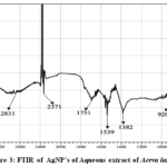 |
Figure 3: FTIR of AgNP’s of Aqueous extract of Aerva lanata Click here to View Figure |
Zeta Potential
One of the crucial characterization criteria that demonstrates the existence of charge on AgNPs in a given medium is the Zeta potential analysis. Because the nanoparticles (NPs) generally have a charge on their surface, a repulsive force can be generated among them, preventing agglomeration and ensuring the NPs’ stability. The biosynthesized AgNPs’ zeta potential analysis results, which were -22.0 ±0.01 mV (Figure 4), showed that they had a negative charge on their surface, which prevented agglomeration in the medium moreover produced moderate long-term stability. According to previous reports, NPs with charges between ±15 and ±25 mV were moderately stable, while those with charges between ±30 mV were very stable.26-28
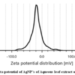 |
Figure 4: Zeta potential of AgNP’s of Aqueous leaf extract of Aerva lanata Click here to View Figure |
X-ray Diffraction Analysis
The Advanced characterization spectroscopic techniquethat was employed to assess the crystalline state of biosynthesized AgNPs was XRD examination. The elemental absorption peak was recorded at 3KeV in the EDAX analysis. The XRD pattern (Fig. 5) shows 4 distinctive peaks in the XRD spectra corresponding to their respective planes at 38.29 (111), 46.38 (200), 64.61 (220), and 77.43 (311), which suggests the crystalline nature of the NPs is the FCC structure, which identified that elemental silver is present in the sample. The XRD patterns matched the previous results.29,30
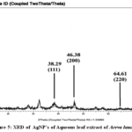 |
Figure 5: XRD of AgNP’s of Aqueous leaf extract of Aerva lanata Click here to View Figure |
Energy-Dispersive X-ray Spectroscopy
The “results of the EDAX study, which revealed a very strong signal in the silver region, validated the formation of AgNPs that might have been caused by the presence of biomolecules that are bound to the surface of” Aerva lanata AgNPs. The EDAX analysis demonstrated the presence of several elements, which are depicted in Table 1. Because of surface plasmon resonance (SPR), metallic AgNPs frequently displayed an absorption peak at around 20 keV.31
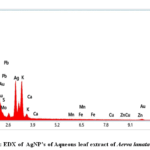 |
Figure 6: EDX of AgNP’s of Aqueous leaf extract of Aerva lanata Click here to View Figure |
Table 1: EDAX Charaterization of Silver Nanoparticles
|
S. No. |
Element | Weight % | S. No. | Element | Weight % |
| 1 | C | 20.2 | 7 | Mn |
0.3 |
|
2 |
O | 28.2 | 8 | Fe | 0.2 |
| 3 | Mg | 2.1 | 9 | Cu |
0.4 |
|
4 |
Al | 0.2 | 10 | Zn | 0.2 |
| 5 | K | 9.8 | 11 | Ag |
21.7 |
|
6 |
Ca | 0.1 | 12 | Au |
6.9 |
Field Emission Scanning Electron Microscopy:
The shape of green-synthesized AgNPs had been observed in FESEM. As depicted As seen in Fig. 7, the FESEM image demonstrated that the AgNPs generated were spherically shaped and well distributed, with diameters that range from 66.70 ±0.15 to 98.40 ±0.05 nm.32
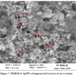 |
Figure 7: FESEM of AgNP’s of Aqueous leaf extract of Aerva lanata Click here to View Figure |
Conclusions
In this work, aqueous leaf extract from Aerva lanata was used for the green synthesis of AgNPs.This method for the synthesis of AgNPs offers an efficient, economic, and eco-friendly approach that does not need any special conditions such as vacuum, catalyst, hazardous chemicals,or sophisticated instruments.UV-Vis spectroscopy validated AgNPs synthesis and displayed characteristic optical absorption spectra in the UV-Vis region.The average particle size of biosynthesized crystalline, spherically shaped AgNPs was 83.06 ±0.05 nm. The FTIR analysis identified many active biocompounds that serve as capping and stabilizing agents during the AgNPs formation process. FESEM is used to evaluate the size and size distribution of AgNPs. AgNPs’ FCC crystal structure is confirmed by XRD. EDAX confirms that the various metals are present in the NPs. The zeta potential validates modest AgNPs stability.
Among Aerva lanata’s diverse ethnomedicinal applications, its use in bone fracture healing by indigenous communities is particularly notable.Aerva lanata leaf contains phytochemicals such as Quercetin and Kaempferol, which have the potential to enhancethe Bone Healing process. The Prepared AgNPs from the plant Aerva lanata can serve as Nanomedicine for the Bone Healing process. Afterin vivo studies, AgNPs may proceed to clinical trials.
Acknowledgement
The “authors are grateful to the Department of Physics and Astrophysics, Pt. Ravi Shankar University, Raipur, Chhattisgarh, “for providing facilities for FTIR and XRD analyses. We are also thankful to the Central Instrument facility, Indian Institute of Technology, Bhilai, Chhattisgarh,for providing facilities for FESEM and EDAX analysis. We are also obliged to the Department of Chemical Engineering, National Institute of Technology Raipur, Chhattisgarh, for providing facilities for Zeta potential analysis.
Funding Sources
The author(s) received no financial support for the research, authorship, and/or publication of this article.
Conflict of Interest
The author(s) do not have any conflict of interest.
Data Availability Statement
This statement does not apply to this article.
Ethics Statement
This research did not involve human participants, animal subjects, or any material that requires ethical approval.
References
- Mohammed, A. E., Al-Qahtani, A., Al-Mutairi, A., Al-Shamri, B., & Aabed, K. (2018). Antibacterial and Cytotoxic Potential of Biosynthesized Silver Nanoparticles by Some Plant Extracts. Nanomaterials, 8(6), 382. https://doi.org/10.3390/nano8060382
CrossRef - Ali, Z. A., Yahya, R., Sekaran, S. D., &Puteh, R. (2016). Green Synthesis of Silver Nanoparticles Using Apple Extract and Its Antibacterial Properties. Advances in Materials Science and Engineering, 4102196, 1-6.https://doi.org/10.1155/2016/4102196
CrossRef - Kummara, S., Patil, M. B., & Uriah, T. (2016). Synthesis, characterization, biocompatible and anticancer activity of green and chemically synthesized silver nanoparticles – A comparative study. Biomedicine & pharmacotherapy Biomedecine & pharmacotherapie, 84, 10-21. https://doi.org/10.1016/j.biopha.2016.09.003
CrossRef - Kumar, R., Ghoshal, G.,Jain, A., &Goyal, M. (2017). Rapid Green Synthesis of Silver Nanoparticles (AgNPs) Using (Prunus persica) Plants extract: Exploring its Antimicrobial and Catalytic Activities. Journal of Nanomedicine & Nanotechnology,8, 452. doi: 10.4172/2157-7439.1000452
CrossRef - Fatimah, I., & Aftrid, Z. H. V. I. (2019). Characteristics and antibacterial activity of green synthesized silver nanoparticles using red spinach (Amaranthus Tricolor) leaf extract. Green Chemistry Letters and Reviews, 12(1), 25–30. https://doi.org/10.1080/17518253.2019.1569729
CrossRef - Ijaz, M., Zafar, M., & Iqbal, T. (2020). Green synthesis of silver nanoparticles by using various extracts: a review. Inorganic and Nano-Metal Chemistry, 51(5), 744–755. https://doi.org/10.1080/24701556.2020.1808680
CrossRef - Sundar, N. S., Dhasarathan, P., Narayanan, K. R., & Thenmozhi, M. (2022). Screening of Anti Diuretic Activity Aerva Lanata Extracts against Furosemide Exposed Rodent Models. New Visions in Biological Science, 8, 160-164.
CrossRef - Tyagi, A., Chaudhary, P., & Garg, V. K. (2023). Pharmacological and Therapeutic Potential of Calotropis gigantea and Aerva lanata: A Review. Research Journal of Pharmacology and Pharmacodynamics, 15, 19-23.
CrossRef - Narayanan M., Krishnan, L., Natarajan, D., Kandasamy, S., El Askary, A., Elfasakhany, A., & Pugazhendhi, A. (2021). Evaluation of antibacterial, antioxidant, and nephroprotective proficiency of methanol extract of Aerva lanata. Process Biochemistry, 109, 98-103.https://doi.org/10.1016/j.procbio.2021.07.004.
CrossRef - Zuvairiya, U. S. M., Jayaraman, S., & Suresh, V. (2024). The Oncolytic Effect of Aerva lanata on Osteosarcoma Cell Lines via the Apoptotic Signaling Pathway. Cureus, 16(4), e58091. https://doi.org/10.7759/cureus.58091
CrossRef - Shanmuganathan, R., Devanesan, S., Oza, G., & Sharma, A. (2024). Assessment of antimicrobial, antidiabetic, and anti-inflammatory properties of acetone extract of Aerva lanata (L.) by in-vitro approach and bioactive compounds characterization. Environmental research, 248, 118348.https://doi.org/10.1016/j.envres.2024.118348
CrossRef - Ramachandra, Y. L., Shilali, K., Ahmed, M. Hegde, S., Kavitha, B. T., Gurumurthy, H., & Rai, P. (2011). Hepatoprotective properties of boerhaavia diffusa and Aerva lanata against carbon tetra chloride induced hepatic damage in rats. Pharmacologyonline, 3, 435-441.
- Nivrutti, G., Chandekar, A., & Tripathi, A. (2025). Phytochemical and pharmacological evaluation of medicinal plants with diuretic activity: A systematic review of experimental evidence. Journal of Medicinal Plants Studies, 13, 359-370. 10.22271/plants.2025.v13.i3e.1878.
CrossRef - Ghosh, R., Kumar, J., Roy, S., Kadahalli, A., & Thangammal, A. (2024). Anti-urolithiasis potential of Aerva lanata metabolites investigated in synthetic urine and cell-free in vitro assays. Asia-Pacific Journal of Science and Technology, 29(02), APST–29. https://doi.org/10.14456/apst.2024.32
- Ganesan, T., Muthukrishnan, S., Albeshr, M. F., Selvankumar, T., Pradeepkumar, S., Inumathi, K. P., & Anto, B. (2024).Aerva lanata flower extract mediated green synthesis of silver nanoparticles: Their characterization, in vitro antioxidants and antimicrobial investigations. Polymers for Advanced Technology, 35(3), e6354. doi:1002/pat.6354
CrossRef - Ajlouni, A. W., Hamdan, E. H., Alshalawi, R. A. E., Shaik, M. R., Khan, M., Kuniyil, M., Alwarthan, A., Ansari, M. A., Khan, M., Alkhathlan, H. Z., Shaik, J. P., & Adil, S. F. (2023). Green Synthesis of Silver Nanoparticles Using Aerial Part Extract of the Anthemis pseudocotula Plant and Their Biological Activity. Molecules, 28(1), 246. https://doi.org/10.3390/molecules28010246
CrossRef - Saiganesh, S., Krishnan, T., Narasimha, G., Almoallim, H. S., Alhari, S. A., Reddy, L. V., Mallikarjuna, K., Mohammed, A., & Prabhakar, V. S. V. (2021). Phytosynthetic Fabrication of Lanthanum Ion-Doped Nickel Oxide Nanoparticles Using Sesbania grandifloraLeaf Extract and Their Anti-Microbial Properties. Crystals, 11(2), 124. https://doi.org/10.3390/cryst11020124
CrossRef - Naidu, K. S. B. (2020). Engineered nanoparticles: Hazards and risk assessment upon exposure- A review. Current Trends in Biotechnology & Pharmacy, 14(1), 111–22.
CrossRef - Athira, P., & Nair, S. N. (2019). Pharmacognostic review of medicinal plant Aerva lanata. Journal of Pharmaceutical Sciences and Research, 9(9), 1420.
- Jyoti, K., & Singh, A. (2016). Green synthesis of nanostructured silver particles and their catalytic application in dye degradation. Journal of genetic engineering and biotechnology, Elsevier, 14, 311-317. https://doi.org/10.1016/j.jgeb.2016.09.005
CrossRef - Seku, K., Gangapuram, B.R., Pejjai, B., Kadimpati, K. K., & Golla, N. (2018). Microwave-assisted synthesis of silver nanoparticles and their application in catalytic, antibacterial and antioxidant activities. Journal of Nanostructure in Chemistry, 8, 179–188. https://doi.org/10.1007/s40097-018-0264-7
CrossRef - Vaishnavi, V., Sadasivuni, K. K., Ponnamma, D., & Golla, N. (2020). Green Synthesis of Silver Nanoparticles from Pterocarpus santalinusLeaf Broth and Their Antibacterial and Antioxidant Activities. Macromolecular Symposia, 392(1), 2000079. https://doi.org/10.1002/masy.202000079
CrossRef - Iqbal, T., Ara, G., Khalid, N. R., & Ijaz, M. (2019). Simple synthesis of Ag-doped CdS nanostructure material with excellent properties. Applied Nanoscience, 10, 23-28.https://doi.org/10.1007/s13204-019-01044-y.
CrossRef - Kirubha, R., & Alagumuthu, G. (2015). Investigation of Antibacterial Properties of Silver Nanoparticles Using Aerva LanataIndo American Journal of Pharmaceutical Sciences, 2(3), 668–675.
- Vidhya, R., & Udayakumar, R. (2015). Antibacterial Potential of Different Parts of Aerva lanata (L.) Against Some Selected Clinical Isolates from Urinary Tract Infections. British Microbiology Research Journal, 7(1), 35–47. doi:10.9734/BMRJ/2015/15738.
CrossRef - Riaz Ahmed, K. B., Nagy, A. M., Brown, R. P., Zhang, Q., Malghan, S. G., & Goering, P. L. (2017). Silver nanoparticles: Significance of physicochemical properties and assay interference on the interpretation of in vitro cytotoxicity studies. Toxicology in vitro : an international journal published in association with BIBRA, 38, 179–192. https://doi.org/10.1016/j.tiv.2016.10.012
CrossRef - Kotakadi, V. S., Gaddam, S. A., Venkata, S. K., Sarma, P. V., & Sai Gopal, D. V. (2016). Biofabrication and spectral characterization of silver nanoparticles and their cytotoxic studies on human CD34 +ve stem cells. 3 Biotech, 6(2), 216.https://doi.org/10.1007/s13205-016-0532-5
CrossRef - Kotakadi, V. S., Gaddam, S. A., Venkata, S. K., Sarma, P. V., & Sai Gopal, D. V. (2015).New generation of bactericidal silver nanoparticles against different antibiotic resistant Escherichia coli Applied Nanoscience, 5, 847–855. https://doi.org/10.1007/s13204-014-0381-7
CrossRef - Nagaratna, A., Prakash, Hegde, L., & Harini, A. (2015). A Pharmacological review on Gorakha ganja (Aerva lanata (Linn) Juss. Ex. Schult). Journal of Pharmacognosy and Phytochemistry, 3(5), 35-39.
- Khatami, M., Sharifi, I., Nobre, M. A. L., Zafarnia, N., & Aflatoonian, M. R. (2018). Waste-grass-mediated green synthesis of silver nanoparticles and evaluation of their anticancer, antifungal and antibacterial activity. Green Chemistry Letters and Reviews, 11(2), 125-134. https://doi.org/10.1080/17518253.2018.1444797
CrossRef - Gaddam, S. A., Kotakadi, V. S., Sai Gopal, D. V. R., Subba Rao, Y., & Varada Reddy, A. (2014). Efficient and robust biofabrication of silver nanoparticles by cassia alataleaf extract and their antimicrobial activity. Journal of Nanostructure Chemistry, 4, 82. https://doi.org/10.1007/s40097-014-0082-5
CrossRef - Ijaz, M., Aftab, M., Afsheen, S., & Iqbal, T. (2020). Novel Au nano-grating for detection of water in various electrolytes. Applied Nanoscience, 10(11), 4029-4036. doi:10.1007/s13204-020-01520-w.
CrossRef
Accepted on: 10 Apr 2026


![]()


