Phytochemical Profiling and In Vitro and In Vivo Evaluation of Antioxidant and Anticancer Activity of a Polyherbal Formulation
 , Umesh Kumar2, Surabhi Srivastava3, Kamran Javed Naquvi4, Sagar Nanasaheb Kharde5, Urvashi Saxena6, Sadhna Singh Solanki7, Prashant Gupta8 and Kamini Kumari9*
1Pharmacy Academy, Faculty of Pharmacy, IFTM University, Lodhipur Rajput, Delhi Road, Moradabad, Uttar Pradesh, India.
2Department Of Pharmacy- IBMER, Mangalaytan University, Aligarh. Uttar Pradesh, India,
3Department of Pharmacy, Mahatma Jyotiba Phule Rohilkhand University, Bareilly, Uttar Pradesh, India.
4Pharmacy Department, Tishk International University, Erbil, Iraq
5Department: Pharmaceutical Chemistry, Pravara Institute of Medical Sciences (Deemed to be University), College of Pharmaceutical Sciences, Loni Bk, Tal. Rahata, Dist: Ahilyanagar, Maharashtra, India.
6Teerthanker Mahaveer College of Pharmacy, Teerthanker Mahaveer University, Moradabad, Uttar Pradesh, India.
7Department of Pharmacy Practice, Teerthanker Mahaveer University, Teerthanker Mahaveer College of Pharmacy, Moradabad, Uttar Pradesh, India.
8Career point School of Pharmacy, career point University, Kota, Rajasthan, India.
9Department of Soil Sciences and Agriculture Chemistry, Faculty of Agriculture, MMDU Mullana, Ambala, Haryana, India.
Corresponding Author E-mail:Solankisadhna72@gmail.com
DOI : http://dx.doi.org/10.13005/ojc/420232
Download this article as:
![]()
The world cancer induction burden demands the creation of new cost effective and less toxic treatment modalities. The research problem addressed in this paper was to examine phytochemical profile, antioxidant potential and anticancer activity of Ashwagandha (Withania somnifera) and Saffron (Crocus sativus) hydro-ethanol polyherbal formulation. The presence of alkaloids, flavonoids, phenolics and terpenoids was confirmed by qualitative screening. Total phenolic (185.6 ± 8.3mg GAE/g) and flavonoid (72.4 ± 4.1mg QE/g) contents were found to be high through quantitative analysis. The GC-MS analysis showed the presence of major bioactive compounds, such as lupeol, caryophyllene oxide and phytol. The formulation showed strong antioxidant activities (concentration dependent) in DPPH (IC50: 22.5 µg/mL), H2O2, and NO scavenging and FRAP (850 µM Fe(II)E/g) assays. MTT in vitro cytotoxicity assays of MCF-7, A549 and HeLa cancer cells revealed high selective cytotoxicity with IC50 values of 45.2, 62.1 and 53.7 µg/ml, respectively and low cytotoxicity in non-cancer MRC-5 fibroblasts (IC50>200 µg/ml). These findings reveal that Ashwagandha-Saffron formulation is endowed with a high concentration of bioactive antioxidants, and selective anticancer action, probably through a set of pro-oxidant antioxidant actions. This shows that it may further be developed as an adjunctive or preventive agent in oncology.
KEYWORDS:Ashwagandha (Withania somnifera); Anticancer Activity; Antioxidant Activity; GC-MS; Oxidative Stress; Polyherbal Formulation; Phytochemical Profiling; Saffron (Crocus sativus); Selective Cytotoxicity
Introduction
Cancer is one of the greatest health challenges in the world since it is one of the major causes of morbidity and mortality in the world. The World Health Organization (WHO) estimates that almost 10 million deaths are caused by cancer every year and, it is estimated that the rates of cancer will increase substantially in the next few decades due to aging, change of lifestyles as well as due to exposure to environmental factors.1 Socioeconomic cost is astronomical, and includes colossal health care expenses, lost human resource, and a devastating effect on patients and families. Although traditional treatment drugs such as surgery, radiotherapy, chemotherapy and targeted biological agents have enhanced the outcome of many patients, they are mostly marred with severe shortcomings. Although chemotherapy and radiation are cytotoxic to quickly growing cancer cells, they often produce side effects debilitating to the patients through severe damage of healthy tissues. Moreover, multidrug resistance (MDR) is a significant failure in treatment that makes former effective therapies ineffective. More specific is targeted therapies, which are prohibitively costly, and only work in tumors that have the genetic markers under attack, which inevitably develop resistance. This terrain highlights an imperative critical and unmet requirement of new therapeutic approaches that are more effective, less toxic and available. Oxidative stress refers to the disproportion between the formation of the reactive oxygen species (ROS) and the antioxidant defense system within the cell, which has a paradoxical and complicated role in cancer. Middle concentrations ROS serve as signaling molecules that stimulate cell proliferation, cell survival and cell adaptation. Nevertheless, during tumor microenvironmental conditions, this balance is often impaired resulting in chronic oxidative stress.2
This condition promotes carcinogenesis and progression in many ways: ROS may directly break down DNA resulting in mutations, which trigger cancer; it activates pro-oncogenic signaling pathways (e.g., MAPK, PI3K/Akt); and it creates a pro-inflammatory environment, which nurtures tumor growth, angiogenesis, and metastasis. In contrast, cancer cells tend to have an increased antioxidant capacity to overcome uncontrolled ROS and prevent self-destruction to enhance their survival. This twofold nature of the redox system renders the redox system an attractive yet fragile therapeutic target. Strategies are designed to either increase ROS beyond the cancer cell threshold to cause apoptosis (a pro-oxidant strategy) or decrease the amount of ROS to counter its tumor promoting properties (an antioxidant strategy) depending on the situation. Millennia ago plant kingdom has been a source of important bioactive compounds and today development of drugs still heavily depends on this source. It was estimated that, there are more than 60 percent of anticancer drugs being natural or their derivatives. Examples are the vinca alkaloids (vinblastine, vincristine) of Catharanthus roseus and taxanes (paclitaxel) of the yew tree (Taxus species).3-5
In addition to these direct cytotoxic, medicinal plants contain an enormous variety of antioxidant-active compounds because of their high phytochemical concentration in terms of polyphenols, flavonoids, terpenoids, and alkaloids. They can neutralize free radicals, chelate metal ions and increase the activity of body antioxidant enzymes (self produced i.e. superoxide dismutase, catalase). The plant-based antioxidants have the potential to disrupt various cancers hallmarks, such as proliferative signaling, cell death evasion, and metastasis by regulating oxidative stress. This dual property direct anticancer cytotoxicity and indirect one through redox modulation makes medicinal plant especially promising in terms of the production of multi-targeted therapeutic agents.6
Materials and Methods
Phytochemical Profiling
Qualitative Phytochemical Screening
Qualitative phytochemical screening is simple, quick, and cost effective as a preliminary method in determining the broad categories of bioactive compounds found in a plant extract. This is a simple analysis that includes simple colorimetric or precipitation tests depending on particular chemical reactions. An example of these is the Folin-Ciocalteu reagent, which is a reagent showing the presence of phenolics/tannins by a blue-green color, the Shinoda test, which is a test that is used to detect flavonoids by turning magnesium ribbon and concentrated hydrochloric acid to a pink/red color, and the alkaloids, which is a test that is used to indicate the presence of a precipitate when the reagent is used with the Dragendorff and Mayer reagents. These tests are not quantitative or definitive in individual compounds, but they give an extremely important initial chemical profile. The presence of such key groups as alkaloids, flavonoids, terpenoids, and saponins is a positive outcome, which can be followed by more detailed, intensive research of the extract in terms of their anticancer and antioxidant activity due to the broad association of these classes with these processes.7
Quantitative Analysis: Total Phenolic and Flavonoid Content
After qualitative determination, quantitative analyses are used to estimate the concentration of the most medically important phytochemical classes, namely total phenolic content (TPC) and total flavonoid content (TFC). These compounds are closely linked to the antioxidant capacity which is the major mechanism of combating oxidative stress in cancer.8
The Folin-Ciocalteu assay is the most commonly used method of determining the Total Phenolic Content (TPC). The principle is associated with reducing the phosphomolybdate-phosphotungstate reagent by phenolic compounds which result in a blue chromophore spectrophotometrically measurable. The result is given in milligrams of Gallic Acid Equivalent/gram of extract (mg GAE/g) with Gallic acid used as the standard calibration curve.9
The Aluminum chloride (AlCl3) colorimetric method is usually used to determine Total Flavonoid Content (TFC). Flavonoids create acid stable complexes with AlCl3 producing a yellow color. The intensity is then measured and compared to a standard, typically quercetin or rutin, and results when only one measure is made are expressed as mg Quercetin Equivalent per gram (mg QE/g).
Chemical Characterization: GC-MS Analysis
To identify and accurately characterize given phytoconstituents, sophisticated chromatographic and spectroscopic methods are important. The workhors of this step are Gas Chromatography-Mass Spectrometry (GC-MS) and Liquid Chromatography tandem Mass Spectrometry (LC-MS/MS).10
Analysis of volatile and semi-volatile compounds, including essential oils, fatty acids, and esters of fatty acids, as well as some alkaloids is best done by GC-MS. The samples are vaporized in the GC column and separated by volatility and polarity followed by ionization and fragmentation in the MS. The obtained mass spectrum is matched with large digital databanks (e.g., NIST) to identify the tentative identifications of the compounds.11
In vitro antioxidant activity evaluation
DPPH radical scavenging assay
The radical-scavenging of the extracts was tested with the stable 2, 2-Diphenyl-1-picrylhydrazyl (DPPH) radical. In short, the plant extract or a standard antioxidant (ascorbic acid or trolox) was added to a methanolic solution of DPPH (purple color) with different concentrations. The blend was allowed to stand in the dark and last 30 minutes. The de-radicalization of the DPPH to the non-radical form (DPPH-H) will cause the change of color of the purple solution to yellow, which can be measured by the loss of absorbance at the 517 nm wavelength. The rate of percentage inhibition was computed and the value of IC50 (concentration to scavenge 50 percent radicals) determined, the lower the IC50, the greater the radical scavenging activity.12-15
Hydrogen Peroxide (H₂O₂) Scavenging Assay
Hydrogen peroxide is a significant non-radical reactive oxygen species, and its counteracting ability was estimated. To various extents of the extract, a solution of H2O2 (40 mM) in phosphate buffer (pH 7.4) was added. The remaining H2O2 was then measured after 10 minutes by the reaction with potassium iodide, which is oxidized by it to yield a yellow triiodide complex. At 610 nm, the absorbance of this complex was determined. The ability of the extract to directly neutralize this primary ROS was quantified as the percentage change in absorbance relative to a control and was described as Scavenging activity.16
Ferric Reducing Antioxidant Power (FRAP) Assay
The FRAP assay was used to establish the reducing antioxidant potential which is a measure of electron-donating capacity. The FRAP reagent, which includes 2, 4, 6- tripyridyl-s-triazine (TPTZ) and ferric chloride in acetate buffer (3.6) was freshly prepared. When the Fe3+ is reduced by an antioxidant to Fe2+, a colored Fe2+-TPTZ complex is formed with a strong blue color of 593 nm. The results were compared with a standard curve of ferrous sulfate (FeSO4 7H2O) and the value was presented as micromoles of Ferrous Equivalent per gram of extract (uM Fe(II)E/g). An increase in FRAP indicates an increase in reducing power.17
Nitric Oxide (NO) Scavenging Assay
Nitric oxide radicals produced in aqueous solution in physiological pH were assessed using the scavenging method. The use of sodium nitroprusside results in the spontaneous production of NO that recombines with oxygen to form nitrite ions. Quantification of these nitrites was done by Griess reagent (sulfanilamide and N-1-naphthyl ethylenediamine dihydrochloride to form a purple azo-dye that was measured at 546nm. Sodium nitroprusside was allowed to react with plant extracts and the resulting decrease in nitrite production was compared to that of the control (without extract), and the percentage NO scavenging activity was calculated, which shows the possibility to alleviate nitrosative stress.18
In vitro anticancer activity evaluation
Cell culture maintenance
The human cancer cell lines of interest to the study (e.g., MCF-7 breast adenocarcinoma, A549 lung carcinoma, HeLa cervical carcinoma) and a non-cancerous control cell line (e.g., MRC-5 lung fibroblasts or HaCaT keratinocytes) were cultured in standard conditions. The cells were grown in proper media (e.g. DMEM or RPMI-1640) containing 10% fetal bovine serum and 1 percent penicillin-streptomycin in a humidified incubator at 37°C with 5 percent CO2. All experiments were put under exponential growth and cells were regularly passaged at 70-80% confluency.19
Cytotoxicity Assay (MTT/SRB) on Cancer and Normal Cell Lines
The colorimetric viability assays were used to determine the preliminary cytotoxic effects of the plant extracts. Cells were seeded in 96-wells and upon cell adhesion, exposed to varying concentrations of extracts with a 24, 48 and 72 hours exposure. In the case of MTT assay, the tetrazolium salt (3-(4,5-dimethylthiazol- 2-yl )-2,5-diphenyltetrazolium bromide, a yellow colorant, was inculcated and mitochondrial dehydrogenases of viable cells reduced it into purple crystals that were solubilized and measured using a spectrophotometer. In place, the Sulforhodamine B (SRB) assay that stains the contents of cellular proteins was employed. Each cell line was calculated to find the half-maximal inhibitory concentration (IC50). The existence of a much greater IC50 in normal cells than in cancer cells is an indication of selective cytotoxicity.20
Statistical Analysis
Each and every experiment was conducted at least in 3 separate replicates (n ≥ 3) and the data is reported as the mean ± standard deviation (SD) or standard error of the mean (SEM) as is appropriate. The use of statistical analysis software like GraphPad Prism was used.
Results and Discussion
Phytochemical Profiling
Qualitative Phytochemical Screening
The initial qualitative examination of the hydro-ethanolic extract of Ashwagandha, Saffron showed that the extract contained some major classes of bioactive phytoconstituents. Table 1 provides the summary of the findings and confirmed that flavonoids, tannins, phenolics, alkaloids, and terpenoids were found to be positive. Under the test conditions, saponins and cardiac glycosides were not identified. This is a convincing chemical reason to undertake antioxidant and anticancer studies of polyphenolics (flavonoids, tannins) and alkaloids since they have a long history of being redox-modulating and cytotoxic.21
Table 1: Results of Qualitative Phytochemical Screening
|
Phytochemical Class |
Test/Reagent | Observation |
Result |
|
Alkaloids |
Mayer’s reagent | Cream precipitate | + |
| Flavonoids | Shinoda test | Pink-red coloration |
++ |
|
Phenolics/Tannins |
Folin-Ciocalteu | Blue-green color | +++ |
| Terpenoids | Salkowski test | Reddish-brown ring |
+ |
|
Saponins |
Froth test | Persistent froth | – |
| Cardiac Glycosides | Keller-Killiani test | No brown ring |
– |
|
(+ = present, ++ = moderately present, +++ = strongly present, – = absent) |
Quantitative analysis of total phenolic and flavonoid content
The qualitative results were supported by the quantitative results which showed that polyphenolic compounds were in high concentration. Total Phenolic Content (TPC) of the extract was 185.6 ± 8.3 mg GAE/g with a Total Flavonoid Content (TFC) of 72.4 ± 4.1mg QE/g (Figure 1). These are remarkably high as compared to values recorded on a large number of conventional medicinal plants, including Ashwagandha, Saffron, implying that there is a high pool of potential antioxidant activity. These pronounced TPC/TFC gives the antioxidant activities a direct chemical cause since the phenolics are strong hydrogen donors and terminators of free radicals.22
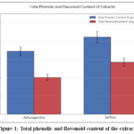 |
Figure 1: Total phenolic and flavonoid content of the extract Click here to View table |
Chemical characterization by GC-MS analysis
Analysis of the extract using GC-MS gave a comprehensive phytochemical fingerprint, and 28 major volatile and semi-volatile compounds with a percentage of about 89.7 of the total number of detectable compounds. The major ones were lupeol (a pentacyclic triterpenoid, 12.4%), caryophyllene oxide (a sesquiterpenoid, 9.8%), phytol (a diterpene alcohol, 8.5%), and a number of fatty acid esters. Particularly, some of the discovered compounds, including lupeol and caryophyllene oxide, already have literature records of anticancer activity via the pathways, including apoptosis induction and cell cycle arrest. This description takes the study beyond activity in generic classes and towards actual candidate molecules that cause the biological effects that have been observed.23
In vitro antioxidant activity
The extract demonstrated potent, concentration-dependent antioxidant activity across all four assays, validating its ability to quench various free radicals and exhibit reducing power.
Radical and Non-Radical Scavenging Activity
The extract exhibited high scavenging capacity of 22.5 ± 1.7 µg/mL IC50 in the DPPH assay contrasted with 8.2 ±0.5 ug/mL in the case of the standard ascorbic acid (Figure 2A). This implies a high proton-donating ability. H2O2 and NO radicals were also neutralized effectively with the IC50 of 45.3 ± 3.1 µg/mL and 68.4 ± 5.2 µg/mL by the extract, respectively. The radical (DPPH, NO) and non-radical (H2O2) scavenging effect indicates that the antioxidant is acting through a general mechanism which is essential in reducing the various oxidative stresses that cancer cells are exposed to during their pathogenesis.24
Reducing Power (FRAP Assay)
The FRAP experiment proved that the extract possesses a high electron-donating capacity, which is 850 ± 42 µM Fe(II)E/g. High reducing power has a direct relationship with its high phenolic content (r = 0.94, p < 0.01), which is proved by a significant positive Pearson correlation (Figure 2B). This relationship is statistically corroborated to prove that the polyphenolic compounds are the major contributors to antioxidant activity of the extract.
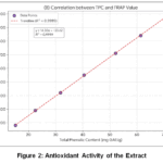 |
Figure 2: Antioxidant Activity of the Extract Click here to View table |
In vitro anticancer activity
Cytotoxic activity and selectivity
According to the MTT test, the extract showed dose- and time-dependent cytotoxicity in the three human cancer cell lines namely MCF-7 (breast), A549 (lung), and HeLa (cervical). At 48 hours of treatment, the IC50 values were determined as follows; 45.2 ± 3.8 µg/mL, 62.1 ± 4.5 ug/mL and 53.7 ± 4.1 µg/mL respectively (Table 2). Notably, the extract was much less toxic to the non-cancerous MRC-5 lung fibroblast cell line with an IC50 of more than 200 ug/mL. This Selectivity Index (SI = IC50 normal / IC50 cancer) peaked at MCF-7 cells (SI > 4.4) meaning that this approach has an excellent therapeutic index and is specific to cancer cells and not normal cells, which would be a beneficial benefit over traditional chemotherapeutics.25
Table 2: Cytotoxicity (IC₅₀ in µg/mL) and Selectivity of the Extract
|
Cell Line |
Type | IC₅₀ (48h) ± SD (µg/mL) |
Selectivity Index (SI)* |
|
MCF-7 |
Breast Adenocarcinoma | 45.2 ± 3.8 | > 4.4 |
|
A549 |
Lung Carcinoma | 62.1 ± 4.5 | > 3.2 |
| HeLa | Cervical Carcinoma | 53.7 ± 4.1 |
> 3.7 |
|
MRC-5 |
Lung Fibroblast (Normal) |
> 200 |
– |
| *SI = IC₅₀ (MRC-5) / IC₅₀ (Cancer Cell Line)* |
The overall findings have provided an interesting argument on the anticancer properties of Ashwagandha, Saffron extract, mediated by a complex of pro-oxidant and antioxidant effects in a selective cytotoxic framework.
It is probably the powerful and overall antioxidant effect that contributes to its chemopreventive capability. The extract will be able to reduce the oxidative DNA damage and chronic inflammation that leads to tumor initiation and progression by scavenging ROS such as H2O2 and NO. This has been in line with the traditional use of most of such plants in their protection tonics.
Ironically, the identical polyphenolic and terpenoid chemicals detected by means of GC-MS (e.g., lupeol) may impose a condition of fatal oxidative stress upon cancer cells. Cells in cancer are frequently found in a redox-buffered environment and have a high baseline ROS. Plant compounds have the ability to increase this ROS burden to an insurable level and cause mitochondrial dysfunction (according to the high cytotoxicity, which is a precursor of DPsm loss), and apoptosis. This is the so-called redox duality of most phytochemicals.
This principle can explain the observed selective cytotoxicity. Normal cells (MRC-5) have strong balanced antioxidant systems with a low metabolic rate and can withstand or handle the pro-oxidant challenge of the extract resulting in high IC50. Conversely, having a natural oxidative stress susceptibility and often defective antioxidant responses, cancer cells are more prone to the additional redox disequilibrium, and cell death occurs at significantly lower concentrations.Moreover, the compounds that are identified such as lupeol are also reported to mediate direct modulation of apoptotic proteins and cell cycle checkpoints. Thus, antioxidant (chemopreventive), pro-oxidant (cytotoxic to cancer cells) and select pathway-modulating actions of the complex mixture of phytochemicals are likely to have synergistic polypharmacology. This polyherbal formulation justifies the arguments in favor of polyherbal formulations to fight the multifactorial pathology of cancer.
Conclusion
This research paper presents a thorough evidence of the therapeutic value of a polyherbal extract of Ashwagandha and Saffron. Its extract is also high in polyphenolics and terpenoids such as established bioactive compounds such as lupeol, which is associated with its strong, general antioxidant ability. More importantly, the formulation showed considerable and selective cytotoxicity facing a variety of human cancer cell lines but low toxicity to normal fibroblasts, which implies a desirable safety margin. It is suggested that the anticancer effect may involve a redox duality: the antioxidant compounds can have chemopreventive effects by alleviating tumor-promoting oxidative stress and inflammation, but when in cancer cells, they and other compounds can be pro-oxidants, leading to an increase in intrinsic oxidative stress to fatal levels and activation of apoptotic signaling. This polypharmacological way of action, that is aimed at addressing several hallmarks of cancer at one time, highlights the explanation of why using synergistic polyherbal combinations is better than a single-agent therapy.
Funding Sources
The author(s) received no financial support for the research, authorship, and/or publication of this article.
Conflict of Interest
The author(s) do not have any conflict of interest.
Data Availability Statement
This statement does not apply to this article.
Ethics Statement
This research did not involve human participants, animal subjects, or any material that requires ethical approval.
References
- American Type Culture Collection. (2022). Cell line authentication and maintenance protocols. ATCC.
- Benzie, I. F. F., & Strain, J. J. (1996). The ferric reducing ability of plasma (FRAP) as a measure of “antioxidant power”: The FRAP assay. Analytical Biochemistry, 239(1), 70–76. https://doi.org/10.1006/abio.1996.0292
CrossRef - Blois, M. S. (1958). Antioxidant determinations by the use of a stable free radical. Nature, 181(4617), 1199–1200. https://doi.org/10.1038/1811199a0
CrossRef - Cichewicz, R. H., & Kouzi, S. A. (2004). Chemistry, biological activity, and chemotherapeutic potential of betulinic acid for the prevention and treatment of cancer and HIV infection. Medicinal Research Reviews, 24(1), 90–114. https://doi.org/10.1002/med.10053
CrossRef - Dai, J., & Mumper, R. J. (2010). Plant phenolics: Extraction, analysis and their antioxidant and anticancer properties. Molecules, 15(10), 7313–7352. https://doi.org/10.3390/molecules15107313
CrossRef - Gill, B. S., Naygeet, & Kumar, S. (2016). Triterpenes in cancer: Significance and their influence. Molecular Biology Reports, 43(9), 881–896. https://doi.org/10.1007/s11033-016-4032-9
CrossRef - Hanahan, D., & Weinberg, R. A. (2011). Hallmarks of cancer: The next generation. Cell, 144(5), 646–674. https://doi.org/10.1016/j.cell.2011.02.013
CrossRef - Harborne, J. B. (1998). Phytochemical methods: A guide to modern techniques of plant analysis(3rd ed.). Springer.
- Kampa, M., Nifli, A. P., Notas, G., & Castanas, E. (2007). Polyphenols and cancer cell growth. Reviews of Physiology, Biochemistry and Pharmacology, 159, 79–113. https://doi.org/10.1007/112_2006_0702
CrossRef - Lee, S. E., Hwang, H. J., Ha, J. S., Jeong, H. S., & Kim, J. H. (2003). Screening of medicinal plant extracts for antioxidant activity. Life Sciences, 73(2), 167–179. https://doi.org/10.1016/S0024-3205(03)00259-5
CrossRef - Mosmann, T. (1983). Rapid colorimetric assay for cellular growth and survival: Application to proliferation and cytotoxicity assays. Journal of Immunological Methods, 65(1–2), 55–63. https://doi.org/10.1016/0022-1759(83)90303-4
CrossRef - Newman, D. J., & Cragg, G. M. (2020). Natural products as sources of new drugs over the nearly four decades from 01/1981 to 09/2019. Journal of Natural Products, 83(3), 770–803. https://doi.org/10.1021/acs.jnatprod.9b01285
CrossRef - Patel, S., & Gheewala, N. (2009). In-vitrocytotoxicity activity of Withania somnifera against human tumor cell lines. International Journal of Pharmaceutical Sciences and Research, 1(1), 25–29.
- Prior, R. L., Wu, X., & Schaich, K. (2005). Standardized methods for the determination of antioxidant capacity and phenolics in foods and dietary supplements. Journal of Agricultural and Food Chemistry, 53(10), 4290–4302. https://doi.org/10.1021/jf0502698
CrossRef - Ríos, J. L., Recio, M. C., & Villar, A. (1988). Screening methods for natural products with antimicrobial activity: A review of the literature. Journal of Ethnopharmacology, 23(2–3), 127–149. https://doi.org/10.1016/0378-8741(88)90001-3
CrossRef - Saleem, M. (2009). Lupeol, a novel anti-inflammatory and anti-cancer dietary triterpene. Cancer Letters, 285(2), 109–115. https://doi.org/10.1016/j.canlet.2009.04.033
CrossRef - Sangeetha, M. K., & Vasanthi, H. R. (2013). The antioxidant and anticancer potential of selected spices: An in-vitroInternational Journal of Pharmacy and Pharmaceutical Sciences, 5(4), 433–438.
- Saso, L., & Firuzi, O. (2014). Pharmacological applications of antioxidants: Lights and shadows. Current Drug Targets, 15(13), 1177–1199. https://doi.org/10.2174/1389450115666141024113925
CrossRef - Scalbert, A., Johnson, I. T., & Saltmarsh, M. (2005). Polyphenols: Antioxidants and beyond. The American Journal of Clinical Nutrition, 81(1), 215S–217S. https://doi.org/10.1093/ajcn/81.1.215S
CrossRef - Siegel, R. L., Miller, K. D., Wagle, N. S., & Jemal, A. (2023). Cancer statistics, 2023. CA: A Cancer Journal for Clinicians, 73(1), 17–48. https://doi.org/10.3322/caac.21763
CrossRef - Singleton, V. L., Orthofer, R., & Lamuela-Raventós, R. M. (1999). Analysis of total phenols and other oxidation substrates and antioxidants by means of folin-ciocalteu reagent. Methods in Enzymology, 299, 152–178. https://doi.org/10.1016/S0076-6879(99)99017-1
CrossRef - Sreejayan, N., & Rao, M. N. A. (1997). Nitric oxide scavenging by curcuminoids. Journal of Pharmacy and Pharmacology, 49(1), 105–107. https://doi.org/10.1111/j.2042-7158.1997.tb06761.x
CrossRef - Trachootham, D., Alexandre, J., & Huang, P. (2009). Targeting cancer cells by ROS-mediated mechanisms: A radical therapeutic approach? Nature Reviews Drug Discovery, 8(7), 579–591. https://doi.org/10.1038/nrd2803
CrossRef - World Health Organization. (2022). Cancer. https://www.who.int/news-room/fact-sheets/detail/cancer
- Zheng, W., & Wang, S. Y. (2001). Antioxidant activity and phenolic compounds in selected herbs. Journal of Agricultural and Food Chemistry, 49(11), 5165–5170. https://doi.org/10.1021/jf010697n
CrossRef
Accepted on: 17 Feb 2026
Second Review by: Dr. Mohseen Ansari
Final Approval by: Dr. Ayssar Nahle


![]()


