Photocatalytic Degradation of Emerging Contaminants Using Sargassum-derived silver nanoparticle and Alpha-cyclodextrin inclusion complex
 and Prema Kumari. J*
Department of Chemistry, Scott Christian College (Autonomous), Nagercoil. Affiliated to Manonmaniam Sundaranar University, Tirunelveli. Tamil Nadu.
Corresponding Author E-mail: premaisaac67@gmail.com
DOI : http://dx.doi.org/10.13005/ojc/420225
Download this article as:
![]()
This paper outlines an eco-friendly biosynthesis of silver nanoparticle (SAA) from the marine brown algae Sargassum, sourced from the Koodankulam shoreline, as verified by XRD analysis. The substance is subsequently treated with an organic compound known as alpha cyclodextrin to yield a firm inclusion complex (S1). UV and IR are employed to characterize the synthesized nanoparticle. The significant peak of absorbance at 415 nm confirms the synthesis of silver nanoparticle produced by the algae Sargassum. The functional components in SAA have been characterized using IR spectroscopy. IR technology, ultraviolet (UV) spectroscopy, and 1H NMR spectroscopy techniques are employed to analyze the synthesized alpha-cyclodextrin inclusion complex (S1). Fluorescence analyses conducted under daylight demonstrated that extracts from various solvents derived from granular Sargassum samples exhibited a spectrum of colours. The synthesized silver nanoparticle and it’s alpha-cyclodextrin inclusion complex are principally examined for photocatalytic applications against the contaminants (Dyes).
KEYWORDS:Alpha cyclodextrin; Inclusion complex; Photocatalytic; Sargassum, Silver nanoparticle
Introduction
Green nano-biology involves the synthesis of silver nanoparticles from diverse biological sources1-4, exhibiting varying morphologies and dimensions, with prospective industrial uses5. With the advent of green nanoparticle production, there has been an increasing desire for environmentally acceptable methods of metal-nanoparticle synthesis that do not utilize harmful chemicals6-10. Nanoparticles have been regarded as significant throughout the past decades and are being employed in many applications, including water purification, catalytic activity, biological and chemical sensors, memory systems and wireless electronic logic5,11–12. The predominant method for synthesizing green nanoparticles involves utilizing aqueous extracts from a variety of organisms, including fungi13, an alga14, and the plants15-18. Sargassum species, a marine macroalga classified within the category Phaeophyceae, is extensively found in tropical and arid oceans. It is part of the aquatic family Sargassaceae and the order Fucales. A broad spectrum of bioactive characteristics of Sargassum has been documented19. Furthermore, the Sargassum species, including, Sargassum tenerrimum20, Sargassum polycystum, and Sargassum glaucescens21 Sargassum latifolium22 have been investigated and demonstrated notable antibacterial and antifungal properties. Alpha-cyclodextrins are cyclical oligosaccharides composed of six 1,4-linked d-glucose units. The glucose chair conformation results in a hollow truncated cone structure, imparting a hydrophobic nature to the inner cavity of the cyclodextrin, in contrast to its hydrophilic external surface. Alpha cyclodextrin exhibit functional features including moderate water solubility, tastelessness, Odor lessness, thermal stability (up to 200 °C), and stability in both alkaline and acidic solutions23. Cyclodextrin molecules possess a distinctive structure, allowing for the entrapment of a guest molecule. Cyclodextrin can create inclusion complexes with a diverse array of liquid, solid and gaseous substances. The complex formation is multidimensional, geometrically constrained interaction between the cavity of cyclodextrin and a foreign molecule24. Cyclodextrins are appropriate for use in food preservation and flavouring, medication, the cosmetics industry, agricultural, and chemical sectors.
This study synthesizes silver nanoparticle using the algae Sargassum. An appealing alpha cyclodextrin inclusion complex is formed by combining the synthesized silver nanoparticle with the compound alpha cyclodextrin. Subsequently, their photocatalytic breakdown of the pollutants is analysed and quantified.
Materials and methods
Compilation of specimen
The algae known as Sargassum have been taken from the shoreline area of Koodankulam. It is further purified with seawater to eliminate contaminants such as grains of sand and organisms known as epiphytes. To eliminate contaminants, particularly pathogenic creatures, it is thoroughly purified using double-distilled water. The specimen was permitted to desiccate prior to being pulverized into a smooth powder.
Fabrication of Silver Nanoparticles from Sargassum
A sustainable production of silver nanoparticle was conducted in a glass container contains 70 mL of a 1 mM aqueous AgNO3 solution and 30 mL of a micro-alga extract. The mixture was positioned in the darkness and agitated for an entire day. Consistent ocular assessments of silver nanoparticle synthesis indicated that a light-yellow solution transformed into a deep brown after 20 minutes, signifying the formation of silver nanoparticle. Sequential centrifugation and lyophilization were employed to enhance the developed nanoparticle.
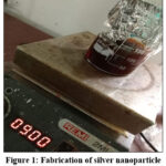 |
Figure 1: Fabrication of silver nanoparticle from the algae Sargassum Click here to View Figure |
Fluorescence Examination
A range of chemical solvents are employed to investigate the behaviour of a pulverised Dictyota dichotoma algae sample. The hue variations in the pulverised substance are examined under both ultraviolet and visible light.
Reagents Required
Synthesized silver nanoparticle (SAA) from the algae Sargassum, alpha cyclodextrin (Sigma-Aldrich) and double distilled water are used for the preparation of Alpha cyclodextrin inclusion complex.
Instruments Required
Systronics Double Beam Spectrophotometer-2203 (Ultraviolet Visible Spectroscopy)
JASCO Spectrofluorometer FP-8200 (Fluorescence Spectroscopy)
Preparation of solid and liquid inclusion complex
For the preparation of solid inclusion complex about 0.301g of alpha cyclodextrin in water is mixed with 0.121g of synthesized silver nanoparticle (SAA) in water and kept on stirring for 96 hrs. The prepared alpha cyclodextrin inclusion complex with SAA (Silver nanoparticle) is obtained as a precipitate, which is then oven dried and utilized for characterization and application studies.
Similarly, for liquid inclusion complex about 0.063g of alpha cyclodextrin in water is mixed with 0.008g of SAA in 10 ml of water in a clean beaker. The concentration of the alpha cyclodextrin taken can also be vary for the production of liquid inclusion complex. This is then characterized by IR and proton NMR spectroscopy.
Catalytic activity
The catalytic activity of the synthesized silver nanoparticle (SAA) and the alpha cyclodextrin inclusion complex (S1) is studied using the contaminants Congo red and methylene blue. The decomposition study involving light from the sun irradiation is conducted outdoors in direct sunlight during midday (summer time) from 12.30 to 2.30 p.m. on clear, hot days. The investigations utilised synthesised quantities of silver nanoparticle (SAA) and alpha cyclodextrin inclusion complex (S1). In the general experiment, six distinct beakers are filled with 20 ml of Congo red dye solution and 0.002 g of SAA. Similarly, another six beakers are filled with 20 ml of methylene blue dye solution and 0.002 g of SAA, which are then placed in a sonicator under direct sunlight to investigate photocatalytic degradation. The degradation of environmental contaminants in relation to the synthesised nanoparticle SAA is observed at 30-minute intervals (0, 30, 60, 90, 150, 180). The identical protocol should be adhered to conduct the photocatalytic degradation investigation of the alpha cyclodextrin inclusion complex S1.
Result and Discussion
XRD of SAA (synthesized silver nanoparticle)
Figure 2 illustrates the X-ray diffraction patterns of SAA. The crystalline quality of the synthesised nano SAA is indicated by a strong peak. The crystalline dimension of the biosynthesised Sargassum nanoparticle was determined using the Debye-Scherrer equation. 2.7×10⁻⁹m was the mean crystal size of the Sargassum silver metal nanoparticle.
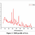 |
Figure 2: XRD profile of SAA Click here to View Figure |
Ultraviolet visible spectroscopy of SAA
The initial indication of silver nanoparticle (SAA) generation was observed through visual inspection of the reaction media. The introduction of algae extracts of Sargassum to the AgNO3 solution resulted in a gradual colour change from pale yellow to brown, indicating the synthesis of silver nanoparticle SAA. The characteristic brown coloration resulted from the stimulation of surface plasmon resonance (SPR) in silver nanoparticles25.
The synthesis of silver nanoparticle SAA was corroborated by the conspicuous absorption peak at 415 nm in the ultraviolet visible spectrum as shown in (Figure. 3), indicative of the surface plasmon resonance of silver nanoparticle.
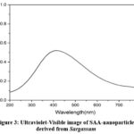 |
Figure 3: Ultraviolet-Visible image of SAA-nanoparticle derived from Sargassum Click here to View Figure |
Ultraviolet visible absorption study of SAA: Alpha cyclodextrin inclusion complex
The spectrum of absorption for SAA at various doses of Alpha cyclodextrin are portrayed in Figure 4 and Table 1. When a blue shift (hypochromic) or red shift (bathochromic) occurs, it is considered that the SAA and Alpha cyclodextrin connect. A wavelength shift from λmax⁓415 nm to λmax⁓445 nm occurs in this case as a consequence of a Bathochromic (red) shift that is noticeable as absorbance increases. The results shown above prove that an inclusion complex is formed when the generated silver nanoparticle (SAA) gets wrapped up in the Alpha cyclodextrin cavity.
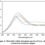 |
Figure 4: Ultraviolet visible absorption spectra of SAA: Alpha cyclodextrin inclusion complex Click here to View Figure |
Table.1 Ultraviolet Visible absorption spectra of SAA: Alpha cyclodextrin inclusion complex
|
Conc. of Alpha cyclodextrin |
Λmax | Absorbance (A) |
| 0 | 415 |
0.317 |
|
0.002 |
423 | 0.319 |
| 0.004 | 430 |
0.332 |
|
0.006 |
435 | 0.327 |
| 0.008 | 440 |
0.344 |
|
0.01 |
445 |
0.362 |
Fluorescent emission study of SAA: Alpha cyclodextrin inclusion complex
The inclusion complex of SAA and Alpha cyclodextrin is discernible in the emission spectra (Figure 5 and Table 2). From λmax⁓510 nm to λmax⁓516 nm, the intensity in this case improves as the wavelength increases, which is the red (Bathochromic) shift. The foregoing finding confirms that the silver nanoparticle that is formed gets entrained in the Alpha cyclodextrin cavity and forms an inclusion complex.
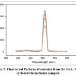 |
Figure 5: Fluorescent Patterns of emission from the SAA: Alpha cyclodextrin inclusion complex Click here to View Figure |
Table 2: Fluorescent Emission spectra of SAA: Alpha cyclodextrin inclusion complex
|
Concentration of α-CD |
Λmax | Intensity (I) |
| 0 | 510 |
2112.5 |
|
0.002 |
511.7 | 2200 |
| 0.004 | 513.3 |
2873 |
|
0.006 |
514.1 | 3075 |
| 0.008 | 515.5 |
3250 |
|
0.01 |
516 |
3500 |
IR Spectroscopy of SAA and S1
The IR spectra of SAA and S1 is depicted in figure 6(a) and 6(b), revealing the O-H stretching of hydroxyl bonds at 3419.56 cm-1 in SAA, which shifts to 3403.16 cm-1 in S1. The signal at 2945.10 cm⁻¹ in nano SAA is displaced to 2927.74 cm⁻¹ in sample S1, indicating the existence of C-H bond stretching. The C=C stretching vibration of SAA at 1607.56 cm⁻¹ is displaced to 1647.27 cm⁻¹. The C-O stretching at 1083.97 cm⁻¹ is displaced to 1154.32 cm⁻¹. The aforementioned alterations in the FT-IR spectra of the synthesized silver nanoparticle (SAA) and its inclusion complex (S1) demonstrate that the host Alpha cyclodextrin cavity accommodates the guest molecule SAA.
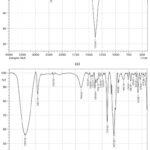 |
Figure 6: IR spectra of the produced silver nanoparticle and its Alpha cyclodextrin inclusion complex (a) SAA and (b) S1. Click here to View Figure |
Proton NMR spectroscopy of S1
The picture (Figure 7a & 7b) and Table 3 illustrate the proton NMR of the synthesized alpha-cyclodextrin inclusion complex (S1) and the corresponding proton shifts, respectively. The up-field shift of H1 from 4.978 to 4.970 indicates a modification in its local electronic environment, presumably resulting from the incorporation of a guest molecule into the α-cyclodextrin cavity. The downfield change of H3 from 3.906 to 3.918 ppm clearly indicates the creation of an inclusion complex, as it signifies direct interactions between H3 and the guest molecule within the α-cyclodextrin cavity. The upfield change of H5 from 3.753 to 3.749 ppm corroborates that the guest molecule is situated within the cyclodextrin cavity, modifying the shielding environment of H5. The downfield shift of H6 from 3.462 to 3.475 signifies that the H6 proton is located at the principal hydroxyl side of Alpha cyclodextrin, resulting in a broader cavity opening.
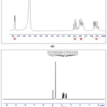 |
Figure 7: Proton NMR of the produced Alpha cyclodextrin inclusion complex with SAA and Alpha cyclodextrin (a) S1 (b) Alpha cyclodextrin Click here to View Figure |
Table 3: Proton Shifts for the Alpha-Cyclodextrin Inclusion Complex with SAA and Alpha-Cyclodextrin
|
|
H1 | H2 | H3 | H4 | H5 | H6 |
| Alpha cyclodextrin | 4.978 | – | 3.906 | – | 3.753 |
3.462 |
|
Inclusion complex of SAA with Alpha cyclodextrin |
4.970 | – | 3.918 | – | 3.749 | 3.475 |
| δ | 0.008 | – | 0.012 | – | 0.004 |
0.013 |
Fluorescence Examination
Table 4 shows the Fluorescence characteristics of Sargassum. The C2H5OH and CHCl3 extracts of the sample appear green when exposed to visible light, whereas H2O and C6H14 extracts are brown and dark brown, respectively. Longer wavelengths of ultraviolet light render C2H5OH and C6H14 extracts dark orange and orange, respectively. The H2O extract of algae has a bright green hue, while CHCl3 displays a yellow colouration. CHCl3 and C2H5OH extracts of algae yield a pale green hue at shorter wavelengths, while H2O and C6H14 display dark green and green hues, respectively, at shorter wavelengths.
Table 4: Fluorescence Characteristics of Sargassum
|
Solvents |
Visible light | Ultraviolet light | |
| Longer wavelength |
Shorter wavelength |
||
|
C2H5OH |
Green | Dark orange | Pale Green |
| H2O | Dark brown | Fluorescent green |
Dark Green |
|
C6H14 |
Brown | Orange | Green |
| CHCl3 | Green | Yellow |
Pale Green |
Catalytic activity of SAA and S1
The synthesised nano-scale SAA and its inclusion complex S1 extract, serving as catalysing agents, were employed to decompose the prevalent organic pollutants methylene blue (MB) and Congo red (CR). Figure 8 depicts the catalytic efficacy of SAA and S1 with both pollutants. The catalytic data for SAA indicates that after a full 180 minutes of exposure to sun, the reduction of cationic pollutant (MB) and anionic pollutant (CR) was recorded at 91% and 87%, respectively, while S1 achieved reductions of 97% for cationic pollutant (MB) and 91% for anionic pollutant (CR). A thorough analysis of the data suggests that a cationic pollutant (MB), degrades more efficiently than an anionic pollutant (CR). This disparity may be attributed to the molecular structure of the cationic contaminant, which features an oxygen group and a positive charge.
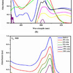 |
Figure 8: Catalytic activity of Anionic pollutants (CR) and Cationic pollutants (MB) by SAA and S1 (a) SAA MB (b) SAA CR (c) S1 MB (d) S1 CR Click here to View Figure |
Conclusion
The Sargassum, an alga, was retrieved from the Koodankulam coast for the purpose of this study. An eco-friendly method of preparing silver nanoparticle from the seaweed extract Sargassum is the focus of this investigation. Alpha cyclodextrin and a synthetic silver nanoparticle, SAA, were used to create an inclusion complex. Infrared, ultraviolet, 1H nuclear magnetic resonance, XRD, absorption and fluorescence emission spectroscopy, and other analytical tools verified that silver nanoparticle were successfully produced from the seaweed and its inclusion complex. The results also showed that the nanocatalyst SAA and its inclusion complex S1 had excellent catalytic activity for breaking down cation and anion pollutants when exposed to sunlight. Cationic pollutants (MB) photodegrade at a faster rate than anionic pollutants (CR) in SAA and S1, according to a more thorough analysis of the data. The chosen contaminants are easily broken down by the synthetic nano and its inclusion complex. Researchers found that SAA, a nanomaterial made from algae, and its inclusion complex S1 have environmental applications that could benefit the planet.
Acknowledgement
The authors sincerely thank the respective institutions for providing essential instrumental facilities.
Funding Sources
The author(s) received no financial support for the research, authorship, and/or publication of this article.
Conflict of Interest
The author(s) do not have any conflict of interest.
Data Availability Statement
This statement does not apply to this article.
Ethics Statement
This research did not involve human participants, animal subjects, or any material that requires ethical approval.
Reference
- Govarthanan, M.; Selvankumar, T.; Manoharan, K.; Rathika, R.; Shanthi, K.; Lee, K. J.; Cho, M.; Kamala-Kannan, S.; Oh, B. T. J. Nanomed, 2014, 9, 1593-1599.
CrossRef - Govarthanan, M.; Seo, Y. S.; Lee, K. J.; Jung, I. B.; Ju, H. J.; Kim, J. S.; Cho, M.; Kamala-Kannan, S.; Oh, B. T. Cells Nanomed. Biotechnol, 2016, 44, 1878–1882.
CrossRef - Govarthanan, M.; Selvankumar, T.; Mythili, R.; Sudhakar, C.; Selvam, K. Sci. Pollut Res, 2017, 24, 19459–19464.
CrossRef - Mythili, R.; Selvankumar, T.; Kamala-Kannan, S.; Sudhakar, C.; Ameen, F.; Sabri, K. Selvam, A. A.; Govarthanan, M.; Kim, H. Lett, 2018, 225, 101–104.
CrossRef - Sun, Y.; Xia, Y. Science, 2002, 298, 2176–2179.
CrossRef - Chen, J.; Wang, J.; Zhang, X.; Jin, Y. Chem. Phys, 2008, 108, 421–424.
CrossRef - MubarakAli, D.; Thajuddin, N.; Jeganathan, K.; Gunasekaran, M. Colloids Surf. B Biointerfaces, 2011, 85, 360–365.
CrossRef - Padalia, H.; Moteriya, P.; Chanda, S. J. Chem, 2015, 8, 732–741.
CrossRef - Thakkar, K.N.; Mhatre, S.S.; Parikh, R.Y. Nanotechnol. Biol Med, 2010, 6, 257–262.
CrossRef - Thovhogi, N.; Diallo, A.; Gurib-Fakim, A.; Maaza, M. Alloys Compd, 2015, 647, 392–396.
CrossRef - Vijayan, S. R.; Santhiyagu, P.; Ramasamy, R.; Arivalagan, P.; Kumar, G.; Ethiraj, K.; Ramaswamy, B. R. Enzyme Microb. Technol, 2016, 95, 45–57.
CrossRef - Pugazhendhi, S.; Kirubha, E.; Palanisamy, P. K.; Gopalakrishnan, R. Surf. Sci., 2015, 357, 1801–1808.
CrossRef - Balaji, D. S.; Basavaraja, S.; Bedre, M. D.; Prabhakar, B. K.; Venkataraman, A. Insciences Journal, 2011, 1, 65–79
- Sahayaa, K.; Rajesh, S.; Rahi, J. M. Journal of Nanomaterials and Biostructures, 2012, 7, 1557–1567
- Amkamwar, B.; Damle, C.; Ahmad, A.; Sastry, M. Journal of Nanoscience and Nanotechnology, 2005, 5,1665–1671
CrossRef - Chandran, S. P.; Chaudhary, M.; Pasricha, R.; Ahmad, A.; Sastry, M. Biotechnology Progress, 2006, 22, 577–583
CrossRef - Li, S.; Shen, Y.; Xie, A.; Yu, X.; Qiu, L.; Zhang, L. Green Chemistry, 2007, 9, 852–858
CrossRef - Shankar, S. S.; Ahmad, A.; Sastry, M. Biotechnology Progress, 2003, 19, 1627–1631
CrossRef - Devi, J. A. I.; Balan, G. S.; Periyanayagam, K. of Coastal Life Med., 2013, 1(3), 199-204.
- Kausalya, M.; Narasimha Rao, G. M. Algal Biomass Utln., 2015, 6(1), 78-87.
- Mahianeh, A.; Ghaednia, B.; Mirbakhsh, M.; Velayatzadeh, M.; Mohammadi, E.; Jafari, M.; Ghaedifard B. J of Biosciences, 2014, 5(12), 399-405.
CrossRef - Dashtiannasab, A.; Kakoolaki, S.; Sharif Rohani, M.; Yeganeh, V. Iranian J of Fisheries Sci., 2012, 11(4), 765-775.
- SarahFoods, Nutrients and food ingredients with authorised EU health claims, 2017, 3, 219-228.
CrossRef - Marques, H. M. C. Flavour Frag J, 2010, 25, 313–326.
CrossRef - Mulvaney, P. Langmuir, 1996, 12, 788–800.
CrossRef
Accepted on: 12 Feb 2026


ISSN Online: 2231-5039

![]()


