Nano-structure of the cristobalite and tridymite stacking sequences in the common purple opal from the Gevrekseydi deposit, Seyitömer-Kütahya, Turkey
Murat Hatipoğlu1,2*, Yaşar Kibici3, Gürsel Yanık3, Cafer Özkul3, Yasemin Yardımcı1
1Dokuz Eylül University, İMYO, İzmir Multidisciplinary Vocational School, Gemmology and Jewellery Programme TR-35380 Buca-İzmir, Turkey 2Dokuz Eylül University, The Graduate School of Natural and Applied Sciences, Natural Building Stone and Gemstone Programme TR 35370 Buca-İzmir, Turkey 3Dumlupınar University, Faculty of Engineering, Department of Geological Engineering TR-43100 Kampüs, Kütahya, Turkey
DOI : http://dx.doi.org/10.13005/ojc/310104
Article Received on :
Article Accepted on :
Article Published : 23 Mar 2015
The strata that include the fine purple opal formations examined in this paper from the magmagenetic hydrothermal dis solutions. The opals are locally known as Gevrekseydi purple opals. These opal-bearing strata are deposited in volcanic lavas and tuffs in the Seyitömer-Kütahya region of western Turkey. The purple opals are common-type and attractive gem-quality, and they are sold on the worldwide gem markets. We conducted mineralogical investigations to clarify their silica building components, measure their nano-size, and determine their origins. The opals are an opal-CT (opal-cristobalite/tridymite)- type silica polymorph with the variable number of cristobalite and tridymite layers. The cristobalite and tridymite stacking sequences were evaluated in terms of maturation, crystallite size, and genesis of the opaline silica material. These sequences are modelled using X-ray diffraction patterns. The relationship between the crystallite size and full width at half maximum values of the Gevrekseydi purple opals were also examined using X-ray diffraction patterns. The crystallite sizes were found to be L=17 nm for the main opal-CT peak (4.09 Å), L=23 nm for the shoulder opal-CT peak (4.29 Å), and L=27 nm for the opal-CT/C peak (2.51 Å). Given the relationship between the crystallite size and maturation of opal-CT, the Gevrekseydi volcanic common purple opals are most likely in a stage of early to mid maturation
KEYWORDS:Gevrekseydi common purple opals; XRD; Opal-CT; Kütahya-Turkey
Download this article as:| Copy the following to cite this article: Hatipoğlu M, Kibici Y, Yanık G, Özkul C, Yardımcı Y. Nano-structure of the cristobalite and tridymite stacking sequences in the common purple opal from the Gevrekseydi deposit, Seyitömer-Kütahya, Turkey. Orient J Chem 2015;31(1). |
| Copy the following to cite this URL: Hatipoğlu M, Kibici Y, Yanık G, Özkul C, Yardımcı Y. Nano-structure of the cristobalite and tridymite stacking sequences in the common purple opal from the Gevrekseydi deposit, Seyitömer-Kütahya, Turkey. Orient J Chem 2015;31(1). Available from: http://www.orientjchem.org/?p=8015 |
Introduction
The gemmological classification of opal as precious, common, or hyalite (semi-precious) is based on whether the opal shows a play-of-color phenomenon caused by visible light diffraction on the stacking and whether it is transparent and vividly colored. An opal without play-of-color or vivid colors is called a common (semiprecious) opal. Precious opals are widely known as either sedimentary opaque-translucent black and white opals or volcanic transparent-translucent fire opals (JONES & SEGNIT 1971, 1972; NAGASE & AKIZUKI 1997). However, the common or hyalite opals have the more varied origins. They occur as sedimentary or volcanic rocks as well as volcaniclastic sediments, and are almost entirely opaque. Their appearance and colors are caused by their absorbance and by the inclusion of some metal transition trace elements (LEECHMAN 1975, FLÖRKE et al. 1991, ELZEA & RICE 1996, FRITSCH et al. 2002). In this study, we are interested in a sample of gem-quality, volcanic common opals with an attractive purple coloration (Fig. 1). However, some of the materials of biogenic origin or the concretions (such as diatomites and geyserites) are described as “opaline silica”, instead of as “opal” (JONES & SEGNIT 1971).
Many of the common opals, of course, have significant commercial value (GAILLOU et al. 2008). Common colored opals have been mined worldwide. These include purple opals from Mexican and the United States, blue-green opals from Peru, green opals from Tanzania and Brazil, yellow opals from Saint-Nectaire, France, pink opals from France, Peru and Mexico (SANDERS 1981, KOIVULA & FRYER 1984, FRITSCH 1991, KOIVULA & KAMMERLING 1991, KAMMERLING & FRYER 1994, FRITSCH et al. 2002, FRITSCH ET AL., 2004, RONDEAU et al. 2004).
There is a significant body of research on common opals, that have attractive colors, because of their geological significance. Some common purple opals are mined in some parts of Mexico, and they are sold under the trade name “Morado” opal on the worldwide gem market, because they can be cut into the beautiful cabochons (ELSASS & DUBROEUCQ 2000, FRITSCH et al. 2004).
Accordingly, the present study is the first investigation of the gemmological and geological properties of an opaque-translucent common opal with attractive purple coloration. The opals described here originate a gem-quality common opal mine deposit in the Seyitömer (Kütahya) region in Turkey. The distinctive color of these opals is a vivid purple with swirls of white (Fig. 1). In addition, many samples are multicoloured with a range of colors from lilac to orange, some with whitish features, in the same deposit. Their structure is compact, without macroscopic inclusions. Therefore, the bulk specimens can be fashioned into cabochons, carvings, or slabs. Because the compact is hard enough to take a polish, the structure makes it a superb lapidary material. The gem’s beauty is also notably more natural than the product of heating or irradiation, which adds to its appeal.
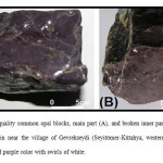 |
Figure1: Gem-quality common opal blocks, main part (A), and broken inner part (B). They are newly found in near the village of Gevrekseydi (Seyitömer-Kütahya, western Turkey), and display a vivid purple color with swirls of white. Click here to View figure |
Therefore, this paper aims (1) to study the geological settling and petrological features of this new opal mine-field, (2) reveal the chemical, spectroscopic and microscopic mineralogical fingerprint of the purple opal, and (3) survey the often under-appreciated “common” opal and its versatility as a lapidary gem to identify their characteristics and compare them to other common purple opals.
Material and Methods.
Gem-quality purple common opals were obtained near the village of Gevrekseydi in the Seyitömer-Kütahya region in the western Turkey.
To verify that the rough opal samples were indeed a pseudo-crystalline (or para-crystalline) silica, gemmological (non-destructive) characterization tests were performed on representative samples. The tests were performed in the DGL-Gemmological Testing Laboratory at Dokuz Eylül University.
Mineralogical (destructive) tests were also performed. Polarizing microscope images of thin sections of the purple opals were obtained using an Olympus BX41 binocular polarizing microscope with a high-intensity 6 V, 30 W halogen light source combined with U-CPA and U-OPA optical systems, after thin sections of the samples had been mounted on glass lamellae. For the digital images, the microscopic magnification (MM) was 5X (a combined magnification of 0.5 X objective and 10 X ocular) under crossed nicols (+N) (active polarizer and analyzer) and parallel nicols. Polarizing microscope investigations were performed in the Optical Mineralogy Laboratory of the Department of Geology at Dumlupınar University.
Chemical analyses of the Gevrekseydi purple opals used X-ray fluorescence (XRF) for major oxides, inductively coupled plasma-atomic emission spectroscopy (ICP-AES) for trace elements, and WST-SIM (loss on igneous for whole rock package-XRF) to determine ignition losses. These analyses were performed, and certified with the code number ‘‘IZ 11037803’’, under contract with the accredited ALS Chemex Laboratory in Canada.
The base silica-building components of the purple opals were detected using X-ray powder diffraction patterns of a Cubi-XRD device with a Cu tube and a graphic monochromator. The samples were analyzed with Cu radiation and a 0.3 mm collimator at atmospheric pressure for 10 minutes each, in the range between 5 and 70 o2-theta. The d-spacing [Å] diffraction matching using the comparative matching technique is based on the positions of peaks with relative intensities of (I/Io) % ≥1, 2-theta values below 70 degree, and a tolerance range of ±0.01. The FWHM values were calculated using the Broker AXS diffract plus software. X-ray diffraction XRD patterns were taken in the Material Research Laboratory of the Batı Anadolu Cement Factory in İzmir.
The dispersive confocal micro-Raman spectroscopy of the purple opals was performed on a dark background at room temperature using a HORIBA Jobin Yvan Scientific XPLORA dispersive confocal micro-Raman spectrometer (DCµRS) with a high-throughput integrated spectrograph. The spectrometer uses one laser excitation of approximately 532 nm. The operating temperature is between 15 and 28 oC. The operation features are as follows: 799.75 of spectro, 1.17066 of exposition, 1200 T of grating, 5 of accumulation, 500 of hole, 100 of slit, 50% filter, 11.4315 of spectrum width, 50 X objective, and 1 binning. Spectral manipulation as baseline adjustment was carried out using the device’s software. The Raman record was carried out in the DGL-Gemmological Testing Laboratory at Dokuz Eylül University.
Results and Discussion
Opaline Material Features
To verify that the investigated rough opal samples were indeed a pseudocrystalline (or paracrystalline) silica variety, non-destructive gemmological characterization tests were performed on representative samples. First, the average specific gravity (SG) values of the eight representative samples were measured using an electronic balance scale (measurement sensitivity of 0.001) using an SG kit, based on the formula (SG = Wair / Wair –Wwater). The specific gravity values ranged from 2.14 and 2.17 gr/cm3. Second, because they are opaque, the optical character, optical sign, and refractive index values of the eight representative opal samples were determined via the spot method, using an Eickhorst SR/XS standard refractometer device with an optical contact liquid of 1.79 RI, and a quartz lamp with a wavelength of 589 nm. The refractive index value of the purple opals was between N=1.428 and 1.436. Third, ultraviolet (UV) luminescence excitation of the purple samples was observed using a System Eickhorst UV 240 shortwave (255 nm) and long wave (366 nm) 4W UV lamp. The samples are inert under UV beams. Thus, when the average specific gravity value 2.15 is considered along with the other verifying results, the investigated opal samples appear to have a typical pseudo- or para-crystalline structure (AREM 1987, ROSSMAN 1994, BACK & MANDARINO 2008).
Provenance, Geology, Petrology, and Genesis of the Opals
The region surrounding the gem-quality common opal mine-deposit near the Gevrekseydi village was examined for its geological and petrological features. Western Turkey is dominated by large E-W grabens and a number of approximately N-S-trending grabens. The Tavşanlı–Kütahya zone has been the subject of several previous by geologists (LEBKUCHNER 1957, 1959, NEBERT 1961, KALAFATÇIOĞLU 1962, OKAY 1981, VICIL 1982, BAŞ 1986, SARIYILDIZ 1987, YANIK 1997, ÖZGENÇ & İLBEYLİ 2008, TÜRKMENOĞLU & YAVUZ-IŞIK 2008, ÖZBURAN & GÜRER 2011, AKKIRAZ et al. 2012). Both Paleozoic metamorphic rocks and upper Cretaceous ophiolitic melange are found on the floor of the Seyitömer–Kütahya basin. Neogene volcanic and sedimentary rocks also occur in the region. However, the Upper Miocene to Lower Pleistocene volcanic rocks where the purple opal-bearing in volcanics occurred. These volcanics are predominantly intermediate-felsic lavas and pyroclastics, as a W-E outcropping in the Tavşanlı Zone.
The volcanics occur as flowing units with thicknesses of 5 m to 25 m. Petrographically, the lavas mainly consist of plagioclase, quartz, biotite, pyroxene, amphibole, and glassy matrix. They have aphanitic texture with grey color in fresh samples and with light-grey color in weathering (altered/decomposed) samples. The volcanic tuffs are made of a combination of rock and mineral fragments (e.g. feldspar, quartz) in volcanic ash matrix. The rock fragments in the tuffs are predominantly volcanic and rarely sedimentary in origin. The lavas and tuffs show locally silica and clay minerals alteration zones. Thin sections of the purple opal show mostly negative elongation that are sometimes undulatory-damped, indicating the presence of cristobalite and tridymite like layers (opal-CT and opal-C) (PRETOLA 2001). They are referred to as pseudo-crystalline (or para-) textures in this study (Fig. 2.A and 2.B).
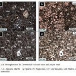 |
Figure2A: Microphotos of the Gevrekseydi volcanic units and purple opals. 6.A -7.A: Andesite /Dacite, (Q: Quartz, Pl: Plagioclase, Cly: Clay minerals, Mat; Matrix, Lit: Litic materials). Click here to View figure |
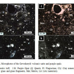 |
Figure2B: Microphotos of the Gevrekseydi volcanic units and purple opals. 10. Andesitic tuff, 2.B: Purple Opal (Q: Quartz, Pl: Plagioclase, Cly: Clay minerals, Vg: Volcanic glass and glass fragments, Mat; Matrix, Lit: Litic materials). Click here to View figure |
The purple opal-bearing zone and its surrounding area were geologically re-mapped, which revealed that the fine purple opal strata become intensely clear on the field at the northeast of the village of Gevrekseydi in the region: such stones are therefore called Gevrekseydi opals. These opals are an attractive gem-quality common opal. The opals are deposited in the large strata of the volcanic tuffs. However, the main silica source of the opals is derived from the underlain andesitic and dacitic rocks, and all of them are called the Gevrekseydi volcanics.
A fault zone that developed in the Gevrekseydi volcanics allowed the magmagenetic hydrothermal dissolutions to rise. After the silicate minerals were dissolved by a hydrothermal alteration in the andesite and dacite, a silicic acid (H4SiO4)-rich fluid was released. Thus, the dissolved silicon ions were transported through the crack zone in the volcanics. These fluids then combined with ground water. However, because the Gevrekseydi volcanics do not have large pores and cavities, the silicic acid-rich hydrothermal fluids continued into the overlain tuffs, forming a colloidal silica gel and ultimately opal in a relatively low-pressure and low-temperature environment with the requisite pH conditions. The silicic acid-rich solutions coagulated as pseudo-crystalline-structured opals of cryptocrystalline or crystalline structured silica varieties in the relatively cooled cavities and pores. It is well known that the opals occur at temperatures below 100 oC and lower pressure conditions.
A range of opal colors have been observed in an investigated area. These levels were filled with the major purple and minor lilac, violet, and yellow colored opals. In addition, light, dark brown, white and pink opals are also exposed in the same area.
Chemistry and Mineralogical Properties of Purple Opal
Chemical Analyses and Coloration
The average composition (Table 1) of the Gevrekseydi common purple opals are as follows; SiO2 (86.86%), Al2O3 (0.66%), Fe2O3 (2.31%), CaO (0.11%), MgO (0.16%), Na2O (0.05%), and K2O (0.08%). In addition, some trace element concentrations (Table 1) are also very distinctive, such as Cu (2500 ppm), Ti (800 ppm), Ba (150 ppm), Zn (317 ppm), Mn (206 ppm), and Pb (127 ppm). The transition metal elements above are characteristic of the Gevrekseydi opals. The distribution of the elements is dictated especially by cristallo-chemical substitution in the pseudo crystalline structure. The trace elements are most likely derived from hydrothermal alteration of the rock-forming minerals in the primary source rocks, such as andesite, dacite and tuffs of the Gevrekseydi volcanics.
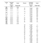 |
Table1: Average bulk and trace element analyses of the Gevrekseydi common purple opals. Click here to View table |
The ICP-AES analysis suggests that purple coloration of the Gevrekseydi opals is due to the transition metals, i.e., the higher abundance of Fe, Cu, Zn, Mn, and Pb. However, the main determination of color in the opal is not only the iron ion content but also the copper and titanium ion contents. Similar elemental contents for the purplish coloration have been reported for some common purple opals from Mexico, which are sold under the trade name “Morado” opal on the worldwide gem market (GÜBELIN 1986; ELSASS et al. 2000; FRITSCH et al. 2004).
X-Ray Diffraction
The XRD patterns of the opals do not show sharp diffraction peaks but rather a broad and diffuse reflection at approximately 21.5° 2-theta with d-values between 4.0 and 3.8 Å. This broad peak is called the “glass-peak”, because of its resemblance with the diffuse peak produced by glass and because it merges with the background without interruptions (DODONY & TAKACS 1980, GHISOLI et al. 2010).
The XRD patterns belonging to the Gevrekseydi common purple opals are signed opal-CT (Fig. 3). The main peak (4.09 Å) on the representative pattern is relatively intense and has a broad diffraction band with 4.29 Å peak on its shoulders. The main peak at 4.09 Å is evidence of opal-CT. In particular, the higher d-value of the main peak (>4.05 Å) and its shape comply with the definition of opal-CT, as do the peaks at 2.51 Å, 2.03 Å, and 1.62 Å, and the lack of peaks at 3.13 Å and 2.84 Å which are characteristic of opal-C (MONROE et al. 1969, FLÖRKE et al. 1975, FLÖRKE et al. 1976, FLÖRKE et al. 1991, ELZEA & RICE 1996, ESENLİ et al. 2003, ESENLİ & EKİNCİ-ŞANS 2013,).
The XRD patterns of the common opals formed from magmagenetic hydrothermal dissolutions show the presence of more than one pseudo-crystalline phase: opal-CT, containing disordered α-cristobalite, and a disordered stacking of sub-equal amounts of cristobalite and tridymite-like layers that were formed in relatively low temperature environments (SANDERS 1975, KASTNER et al. 1977, SANDERS & MURRAY 1978, NAGASE & AKIZUKI 1997, CAUCIA et al. 2013). However, in the literature, the constituents of opal-CT are often expressed as opal-T and opal-C, to separate them from the real crystalline phases (JONES & SEGNIT 1971, 1972). Therefore, the opals derived from the magmagenetic dissolutions are generally classified as pseudo-, quasi-, para- or micro-crystalline (KOIVULA 1984, GRAETSCH & FLÖRKE 1991, KAMMERLING & FRYER 1994, NAGASE & AKIZUKI 1997, POP et al. 2004, HATİPOĞLU 2009, 2010). Thus, while opal-CT and opal-C are mentioned as pseudo-crystalline, opal-A is non-crystalline. The XRD pattern of opal-CT generally results in peaks reflecting both the cristobalite and tridymite-like layers. In the current opal terminology, the term opal-C represents disordered α-cristobalite with 20-30% tridymite-like layers, the term opal-CT represents very disordered α-cristobalite with ~50% tridymite like layers. In the literature, opals made of tridymite-like layers have been called opal-T. However, no separate opal-T was observed in the XRD patterns of the Gevrekseydi purple opals. The broad peak at ~4.1 Å is present in both opal-C (4.05 Å) and opal-T (4.107 Å). The peaks observed on the shoulders of the~4.1 Å peak at 4.328 Å and 3.818 Å are attributed to opal-T (SANDERS 1975, FLÖRKE et al. 1976, KANO 1982, FLÖRKE et al. 1991, GRAETSCH 1994, GRAETSCH et al. 1994, GUTHRIE et al. 1995, DE PABLO-GALAN 1997, SMITH 1997, 1998, NAGASE & AKIZUKI 1997, FRITSCH et al. 2002, ESENLİ et al. 2003, LYNNE & CAMPBELL 2003, BERMANEC 2004, GAILOU & MOCQUET 2004, FRITSCH et al. 2004, RONDEAU et al. 2004,).
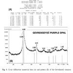 |
Figure3: X-ray diffraction numerical data (A) and pattern (B) of the Gevrekseydi common purple opal. Click here to View figure |
The XRD pattern evaluation, which gives largely qualitative estimates (DE PABLO-GALAN 1997), is the most commonly used method to evaluate crystalline degree, crystallite size, amount of tridymite, interstratifications of cristobalite and tridymite, present crystalline inclusions, relative proportions of cristobalite and tridymite layers, degree of ordered stacking, and site of the H2O molecules in the opals (GRAETSCH 1994, GAILOU & MOCQUET 2004). Two main interpretive techniques were used to evaluate the XRD patterns of the opals; the comparative matching technique, and the graphical modelling technique, the latter of which was modified from SANDERS & MURRAY 1978, GRAETSCH et al. 1994, GUTHRIE et al.1995, and ELZEA & RICE 1996.
Crystallinity determination using the comparative matching technique
The observed peaks of the XRD patterns of the purple opal samples were modelled and compared with the XRD patterns reported for pseudo-crystalline opals from different parts of the world using comparative matching (HATİPOĞLU 2009, 2010) to identify the pseudo-crystalline phases in the Gevrekseydi purple opals.
The phase assignments of the XRD peaks for the pseudo-crystalline silica phases of the Gevrekseydi purple opals are given in Table 2. The XRD patterns of the opals represent opal-CT. The d-spacing of the broad diffraction band of opal-CT and associated shoulder are a 4.09 Å (both opal-T and opal-C) peak and a 4.29 Å (opal-T) shoulder. The broad diffraction band between 19 and 24o2θ, which results from the overlap of the [101] from the cristobalite layers at 4.05 Å and the [404] from the interstratified tridymite-like layers at 4.09 Å, is called opal-CT. Therefore, a characteristic peak at ~4.0 Å (~21.7o2θ) must be observed to demonstrate the presence of opal-CT in pseudo-crystalline opals. Additionally, the main XRD peak at 2.51 Å (~35.8o2θ) is an indicator for the structural ordering and represents the existence of both cristobalite and tridymite layers.
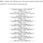 |
Table2: Summary of the XRD peak values of the pseudo-crystalline silica phases (Opal-CT and Opal-CT/C) of the Gevrekseydi common purple opals. Click here to View table |
Modelling the crystallite size of the opal matrix
Though the width (FWHM) of 2.51 Å peak (~35.9o2θ) of opal-CT/C is primarily sensitive to the size of the opal crystallites, the main peak widths (FWHM) of opal-CT (d-value= 4.09 Å, angle value= ~21.7o2θ) and shoulder opal-CT (d-value= 4.29Å, angle value= ~20.7o2θ) are also somewhat sensitive to the size of the opal crystallites (ELZEA & RICE 1996, DE PABLO-GALAN 1997, LYNNE & CAMPBELL 2003, BERMANEC et al. 2004). The relation between the crystallite size (L) and width values (FWHM) of the opal-CT/C and opal-CT peaks of Gevrekseydi purple opals is given in Fig. 4. Accordingly, the crystallite sizes were found to be L=17 nm for main opal-CT, L=23 nm for shoulder opal-CT, and L=27 nm for opal-CT/C. These sizes agree with the range of the crystallite sizes of opal-CT and opal-CT/C in other type of opals (e.g., fire opals) that have been previously reported in the literature (KOIVULA & FRYER 1984, LU et al. 1995, FRITSCH et al. 2004, GAILOU & MOCQUET 2004, GAILLOU et al. 2008, ESENLİ, F. & EKINCİ-ŞANS 2013, RONDEAU et al. 2013). The crystallite size for the opals between 11 and 23 nm, which was revealed by modelling XRD patterns, was reported by GUTHRIE et al. 1995. In addition, a crystallite size between 10 and 50 nm (average 20 nm) was obtained using SEM images and AFM by FRITSCH et al. 2004. Guthrie and his colleagues reported that the crystallite size increases as opal-CT’s mature. Given the relationship between the increasing crystallite size and maturation of opal-CT, volcano-sedimentary Gevrekseydi common purple opals are likely to reflect an early- to mid-level of maturation (GUTHRIE et al. 1995).
![Fig. 4. Relations between crystallite size (L) and width value of the peak at ~35.9o2θ, which represents the main peak of opal-C (d-value= ~2.5 Å) of the Gevrekseydi common purple opals [Full-Width Half-Maximum (FWHM) value of at ~35.9o2θ], (modified from GUTHRIE et al. 1995).](http://www.orientjchem.org/wp-content/uploads/2015/03/Vol31_No1_Nano_Mura_Fig4-150x150.jpg) |
Figure4: Relations between crystallite size (L) and width value of the peak at ~35.9o2θ, which represents the main peak of opal-C (d-value= ~2.5 Å) of the Gevrekseydi common purple opals [Full-Width Half-Maximum (FWHM) value of at ~35.9o2θ], (modified from GUTHRIE et al. 1995). Click here to View figure |
Proportions of cristobalite and tridymite stacking using the modelling technique
The opal-CT band (between 19 and 24o2θ) is sensitive to the relative proportions of the cristobalite and tridymite-like layers, whereas the amount of tridymite present is calculated by modelling the entire diffraction pattern (GUTHRIE et al. 1995). The amount of tridymite obtained by modelling the Gevrekseydi purple opals is given in Table 3. According to this model, a larger peak suggests the presence of more tridymite, 65-70%. Conversely, Ghisoli and his colleagues offered an alternative method for the estimate (GHISOLI et al. 2010). The ratio of tridymite and cristobalite in opaline polymorphs defined as “C/T= d(4.11)-d (measured) / d(4.11)-d(4.04)”. According to this formula, for Gevrekseydi purple opal the C/T= 4.11-4.09 / 4.11-4.04, and the ratio is 0.29 (C/T= 29/71). In this case, the Gevrekseydi opal includes approximately 30% cristobalite and 70% tridymite, and should be designed opal-CT with disordered structure. Moreover, the tridymite variety in the Gevrekseydi purple opals could form in lower-temperature monoclinic tridymite. If it was in high-temperature tridymite, the shoulder of 4.09 Å peak would be lower than 4.29 Å (FLÖRKE et al. 1975).
![Table 3. Modelling the probable amount of tridymite of the Gevrekseydi common purple opals, According to the peak width of 20.710 o2θ which represents the shoulder peak of Opal-CT (d-Value= 4.29 Å), and probable amount of tridymite (modified from Guthrie et al. [52] and Elzea and Rice [5]).](http://www.orientjchem.org/wp-content/uploads/2015/03/Vol31_No1_Nano_Mura_T3-150x150.jpg) |
Table3: Modelling the probable amount of tridymite of the Gevrekseydi common purple opals, According to the peak width of 20.710 o2θ which represents the shoulder peak of Opal-CT (d-Value= 4.29 Å), and probable amount of tridymite (modified from Guthrie et al. [52] and Elzea and Rice [5]). Click here to View table |
Sharpness and breadth of opal-CT peaks using the modelling technique
The main peak of opal-CT in the XRD patterns of Gevrekseydi purple opals is unusually sharp, giving small crystallite sizes (17 nm) and relating directly to long-range order of crystallinity (SANDERS & MURRAY 1978, KANO 1982, GRAETSCH et al 1994, ELZEA & RICE 1996). Some modelling graphics related to the sharpness of opal-CT peaks are given in Figs. 5-7. Gevrekseydi purple opals show the principle d-spacing values at 4.09 Å, and principle FWHM at 0.480o2θ (Fig. 5A). The peak breadth and sharpness relate to the opaline crystallite size (Fig. 5B). These findings are consistent with the interpretation of Elzea and Rice 1996. In the classification schemes recommended in previous studies (ELZEA & RICE 1996, RONDEAU et al. 2013), Gevrekseydi purple opals fall into the opal-CT category (Figs. 6 and 7).
![Fig. 5. Modelling the sharpness and broadening of opal-CT by plotting the peak width (o2θ) [Full-Width Half-Maximum (FWHM)] (A), and the opaline crystallite size (nm) (B) against the principle d-spacing values (Å) of the opal-CT peaks obtained from XRD patterns of the Gevrekseydi common purple opals (modified from ELZEA & RICE 1996).](http://www.orientjchem.org/wp-content/uploads/2015/03/Vol31_No1_Nano_Mura_Fig5-150x150.jpg) |
Figure5: Modelling the sharpness and broadening of opal-CT by plotting the peak width (o2θ) [Full-Width Half-Maximum (FWHM)] (A), and the opaline crystallite size (nm) (B) against the principle d-spacing values (Å) of the opal-CT peaks obtained from XRD patterns of the Gevrekseydi common purple opals (modified from ELZEA & RICE 1996). Click here to View figure |
 |
Figure6: Modelling the sharpness of opal-CT by plotting the ratio of the principle peak d-spacing of the shoulder peak d-spacing against the principle d-spacing values (Å) of the opal-CT peaks obtained from XRD patterns of the Gevrekseydi common purple opals. The 4.29 Å peak is the main shoulder of the opal-CT peak (4.09 Å) (modified from GRAETSCH et al. 1994 and JONES & SEGNIT 1971). Click here to View figure |
![Fig. 7. Modelling the sharpness of opal-CT by plotting the ratio of the principle peak d-spacing (Å) of the shoulder peak d-spacing (Å) against the principle peak width (o2θ) [Full-Width Half-Maximum (FWHM)] of the opal-CT peaks obtained from XRD patterns of the Gevrekseydi common purple opals. The ~4.3 Å peak is the main shoulder of the ~4.1 Å peak (modified from ELZEA & RICE 1996; GRAETSCH et al. 1994; JONES & SEGNIT 1971).](http://www.orientjchem.org/wp-content/uploads/2015/03/Vol31_No1_Nano_Mura_Fig7-150x150.jpg) |
Figure7: Modelling the sharpness of opal-CT by plotting the ratio of the principle peak d-spacing (Å) of the shoulder peak d-spacing (Å) against the principle peak width(o2θ) [Full-Width Half-Maximum (FWHM)] of the opal-CT peaks obtained from XRD patterns of the Gevrekseydi common purple opals. The ~4.3 Å peak is the main shoulder of the ~4.1 Å peak (modified from ELZEA & RICE 1996; GRAETSCH et al. 1994; JONES & SEGNIT 1971). Click here to View figure |
Confocal Micro-Raman
The confocal micro-Raman spectrum of Gevrekseydi purple common opal is given in Fig. 8. In addition, its band assignments and comparisons are listed in Table 4. The representative micro-Raman spectrum reveals the presence of opal-CT/C with the characteristic band at 292 cm-1 and the presence of opal-CT with characteristic bands at 363 and 404 cm-1 (KINGMA & HEMLEY 1994, GÖTZE, et al. 1998, OSTROOUMOV et al. 1999, MCCREERY 2004, POP et al. 2004, OSTROOUMOV 2007, DUMANSKA-SLOWIK et al. 2013). However, some vibration bands in the sample cannot be assigned to the specific molecules. We can state that these bands are due to complex heterogeneous inclusions. The two bands at 65 and 144 cm-1 can be assigned to low quartz found as inclusion in the purple opals. The micro-Raman bands below 1000 cm-1 (such as, at 783, 829, 911, and 961 cm-1) indicate the presence of nanocrystalline molecules of Fe, Cu, Ti, Zn, Mn, and Pb oxides, as detected in the purple opals. Finally, the presence of carbonaceous matter is marked by 1055 and 1729 cm bands (KINGMA & HEMLEY 1994, GÖTZE, et al. 1998, POP et al. 2004, DUMANSKA-SLOWIK et al. 2013).
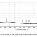 |
Figure8: The confocal micro-Raman spectrum of the Gevrekseydi common purple opal. Click here to View figure |
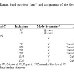 |
Table4: Micro-Raman band positions (cm-1) and assignments of the Gevrekseydi common purple opal.Click here to View table |
Conclusions
- Gevrekseydi opals are an attractive gem-quality common opal. The opals are deposited in the large strata of the volcanic tuffs. However, the main silica source of the opals is derived from the underlain andesitic and dacitic rocks, and all of them are called the Gevrekseydi volcanic because of their proximity to the village of Gevrekseydi. The purple opal strata in the Seyitömer (Kütahya) region are genetically associated with magmagenetic hydrothermal activity. This formation is a true dissolution process in which the spheres remain intact, but are dispersed into the gel.
- Some transition metal elements such as Cu (2500 ppm), Ti (800 ppm), Zn (317 ppm), Mn (206 ppm), and Pb (127 ppm) are characteristic of the Gevrekseydi purple opals.
- The variability in chemical composition, physical properties and micro-textures indicate that the purple opals formed from different solutions at variable growth rates.
- Individual modelling techniques using X-ray diffraction (XRD) patterns are suitable detection method for determining the crystallite size of the opaline building phases, as confirmed by the scanning electron microscope (SEM) images and atomic force microscope (AFM) graphics.
- The degree of crystallinite of the Gevrekseydi common purple opals was interpreted. The opals have a nano-structure at crystallite sizes between 10 and 50 nm comprising cristobalite and tridymite stacking sequences according to the type of opal (precious, common or hyalite) and its origin (sedimentary or volcanic rocks, or volcaniclastic sediments). The crystallite size of the main opaline building phase (opal-CT) in the Gevrekseydi opals is 17 nm. This value indicates a relatively small crystallite size and porosity that is filled with the molecular and silanol waters in opal texture. Accordingly, we can state that the pores in the “volcano-sedimentary” common opals are smaller and of more uniform size compared with those found in “sedimentary and volcanic” common opals.
- There is no evidence for the presence of any accessory minerals in the Gevrekseydi purple common opal.
- The band assignments and comparisons of the Opal-CT and opal-CT/C in the confocal micro-Raman spectrum of the Gevrekseydi purple common opal are presented. The Raman bands reveal many silica inclusions, transition metal oxides and carbonaceous matters in the Gevrekseydi purple opals.
- Further investigations on the Gevrekseydi opals with various body colors would be desirable FT-IR analyses would be particularly interesting, and could indicate the gneiss of this type of opal formations.
Acknowledgements
We would like to express our gratitude and special thanks to three anonymous reviewers and to Prof.Dr. Fahri ESENLİ-İstanbul Technical University- for the useful comments and suggestions on the interpretation of our draft manuscript. We appreciate their time and effort in helping to revise this manuscript and greatly value their assistance, valuable comments and language corrections.
- AKKİRAZ, M.S., AKGÜN, F., UTESCHER, T., WILDE, W., VOLKER-BRUCH, A.A., MOSBRUGGER, V. & ÜÇBAŞ, S.D. (2012): Palaeoflora and Climate of Lignite-bearing Lower−Middle Miocene Sediments in the Seyitömer and Tunçbilek Sub-basins, Kütahya Province, Northwest Turkey. Turkish Journal of Earth Sciences, 21, 213–235.
- AREM, J.E. (1987): Color Encyclopaedia of Gemstones, 2nd Ed. Van Nostrand Reinhold. Co., New York, 675.
- BACK, M. & MANDARİNO, J. (2008): Fleischer’s Glossary of Mineral Species; 10th ed. The Mineralogical Record, Inc., Tucson, 342.
- BAŞ, H. (1986): Domaniç-Tavşanlı-Kütahya-Gediz yöresinin Tersiyer Jeolojisi. Jeoloji Mühendisliği Dergisi, 27, 11-18.
- BERMANEC, V., TOMASIC, N., KARFUNKEL, J., SCHOLZ, R., KNIEWALD, G. & JAKSCH, H. (2004): Nanocrystals of tridymite in opal cavities, in: Proceedings 8th International Congress on Applied Mineralogy, Aguas De Lindoia-Brazil, 525-527.
- CAUCIA, F., MARINONI, L. & LEONE, A. (2013): Physical, geochemical and gemological properties of opals from Faroe Islands. Neues Jahrbuch fur Mineralogie-Abhandlungen, 191, 33-43.
- DE PABLO-GALAN, L., DE LAURDES CHAVEZ-GARCIA, M. & HUANOSTA TERA, A. (1997): Electrical properties of opal-CT. Physics and Chemistry of Minerals, 24, 90-101.
- DODONY, I. & TAKACS, J. (1980): Structures of precious opal from Cervenica. Ann. Univ. Sci. Budap. Rolando Eotvos Nominatae, Sect. Geol., 22, 37–50.
- DUMANSKA-SLOWIK, M., NATKANIEK-NOWAK, L., WASELUCHA-BIRCZYNSKA, A., GAWEL, A., LANKOSZ, M. & WROBEL, P. (2013): Agates from Sidi Rahal, in the Atlas Mountains of Morocco: Gemological characteristics and proposed origin. Gems & Gemology, 49, 148-159.
- ELZEA, J.M. & RICE, S.B. (1996): TEM and X-ray diffraction evidence for cristobalite and tridymite stacking sequences in opal. Clays Clay Minerals, 44, 492-500.
- ELSASS, F., DUBROEUCQ, D. & THIRY, M. (2000): Diagenesis of silica minerals from clay minerals in volcanic soils of Mexico. Clay Minerals, 35, 477-489.
- ELZEA, J.M. & RICE, S.B. (1996): TEM and X-ray diffraction evidence for cristobalite and tridymite stacking sequences in opal. Clays and Clay Minerals, 44, 492-500.
- ESENLİ, F., KUMBASAR, I., ESENLİ, V. & KIRIKOĞLU, S. (2003): A study on the characteristics of some opals from Turkey. Neues Jahrbuch für Mineralogie Mh., 4, 177-192.
- ESENLİ, F. & EKINCİ-ŞANS, B. (2013): XRD studies of opals (4 A peak) in bentonites from Turkey; Implications for the origin of bentonites. Neues Jahrbuch für Mineralogie-Abhandlungen, 191, 45-53.
- FLÖRKE, O.W., JONES, J.B. & SEGNIT, E.R. (1975): Opal-CT crystals. Neues Jahrbuch für Mineralogie-Abhandlungen, 8, 369-377.
- FLÖRKE, O.W., HOLLMANN, R., RAD, U. & RÖSCH, H. (1976): Intergrowth and twinning in opal-CT lepispheres. Contribution to Mineralogy and Petrology, 58, 235-242.
- FLÖRKE, O.W., GRAETSCH, H., MARTIN, B., RÖLLER, K., WIRTH, R. (1991): Nomenclature of micro- and non-crystalline silica minerals, based on structure and microstructure. Neues Jahrbuch für Mineralogie-Abhandlungen, 163, 19-42.
- FRITSCH, E. (1991): Gem News: Fine greenish-blue opal. Gems & Gemology, 27, 120-121.
- FRITSCH, E., OSTROOUMOV, M., RONDEAU, B., BARREAU, A., ALBERTINI, D., MARIE, A.M., LASNIER, B. & WERY, J. (2002): Mexican gem opal: nano- and micro-structure, origin of colour and comparison with other common opals of gemmological significance. The Australian Gemmologist, 21, 230- 233.
- FRITSCH, E., GAILLOU, E., OSTROUMOV, M., RONDEAU, B., DEVOUARD B. & BARREAU, A. (2004): Relationship between nanostructure and optical absorption in fibrous pink opals from Mexico and Peru. European Journal of Mineralogy, 16, 743-752.
- GAILOU, E. & MOCQUET, B. (2004): A new gem material from Madagascar: A mixture of cristobalite and opal. Gem & Gemology, 40, 339-340.
- GAILLOU, E., FRITSCH, E., AGUILAR-REYES, B., FONDEAU, B., POST, J. BARREAU, A. & OSTROUMOV, M. (2008): Common gem opal: An investigation of micro- to nano-structure. American Mineralogist, 93, 1865-1983.
- GHISOLI, C., CAUCIA, F. & MARINONI, L. (2010): XRPD patterns of opals: A brief review and new results from recent studies. Powder Diffraction, 25, 274-282.
- GÖTZE, J., NASDALA, L., KLEEBERG, R. & WENZEL, M. (1998): Occurrence and distribution of “moganite” in agate/chalcedony: a combined micro-Raman, Rietveld, and cathodoluminescence study. Contributions to Mineralogy and Petrology, 133, 96-105.
- GRAETSCH, H. & FLÖRKE, O.W. (1991): X-ray powder diffraction patterns and phase relationship of tridymite modifications. Zeitschrift fur Kristallographie, 195, 31-48.
- GRAETSCH, H. (1994): Structural characteristics of opaline and microcrystalline silica minerals. Reviews in Mineralogy, 29, 209-232.
- GRAETSCH, H., GIES, H. & TOPOLOVIC, I. (1994): NMR, XRD, and, IR study on microcrystalline opals. Physics and Chemistry of Minerals, 21, 166-175.
- GUTHRIE, G.D., BISH, D.L., REYNOLDS, R.C. (1995): Modelling the X-ray diffraction pattern of opal-CT. American Mineralogist, 80, 869-872.
- GÜBELIN, E.J. (1986): Opal from Mexico. The Australian Gemmologist, 16, 45-51.
- HATİPOĞLU, M. (2009): Moganite and quartz inclusions in the nano-structured Anatolian fire opals from Turkey. Journal of African Earth Sciences, 54, 1-21.
- HATİPOĞLU, M. (2010): The nano-grain sizes of the opalline matrix components in Anatolian fire opals. Journal of Non-Crystalline Solids, 356, 1408-1415.
- JONES, J.B. & SEGNIT, E.R. The nature of opal. Part I. Nomenclature and constituent phases. Journal of the Geological Society of Australia 1971, 18, 57-68.
- JONES, J.B. & SEGNIT, E.R. (1972): Genesis of cristobalit and trydimite at low temperatures. Journal of the Geological Society of Australia, 18, 419-422.
- KALAFATÇIOĞLU A. (1962): Tavşanlı-Dağardı arasındaki bölgenin jeoloji ve serpantin ile kalkerlerin yaşı hakkında not. Bulletin of M.T.A. Ankara, 58, 34.
- KAMMERLING, R.C. & FRYER, C.W. (1994): Gem news: Opal translucent greenish blue. Gems & Gemology, 30, 43-44.
- KANO, K. (1982): Taguchi, K. Experimental study on the ordering of opal-CT. Geochemical Journal, 16, 33-41.
- KASTNER, M., KEENE, J.B., GIESKES, J.M. (1977): Diagenesis of siliceous oozes-I. Chemical controls of the rate of opal-A to opal-CT transformation. An experimental study. Geochimica and Cosmochimica Acta, 41, 1041-1059.
- KINGMA, K.J. & HEMLEY, R.J. (1994): Raman spectroscopic study of microcrystalline silica. American Mineralogist, 79, 269-273.
- KOIVULA, J.I. & FRYER, C.W. (1984): Green opal from East Africa. Gems & Gemology, 20, 226-227.
- KOIVULA, J.I. (1991): Kammerling, R.C. Gem news: Fine greenish blue opal. Gems & Gemology, 27,120-121.
- LEECHMAN, F. (1975): The Opal Book; 5th Ed. Ure Smith, Lansdowne Press, Sidney, 264.
- LEBKÜCHNER, R.F. (1957): Linyit bulunması muhtemel Ayvalı (Kütahya) sahasında yapılan jeolojik ve madencilikle ilgili etüdlerin neticesi hakkında rapor. M.T.A. Report No: 2984, Ankara (unpublished).
- _________ (1959): Seyitömer-Kütahya Neojen sahasında jeoloji ve linyit yatakları ile ilgili olarak yapılan etüdler hakkında rapor: M.T.A. Report No: 2985, Ankara (unpublished).
- LU, T., ZHANG, X., SUNAGAWA, I. & GROVES, G.W. (1995): Anometre scale texture in agate and Beltane opal. Mineralogical Magazine, 59, 103-109.
- LYNNE, B.Y. & CAMPBELL, K.A. (2003): Diagenetic transformation (opal-A to quartz) of low- and mid-temperature microbial textures in siliceous hot-spring deposits, Taupo Volcanic Zone, New Zealand. Canadian Journal of Earth Sciences, 40, 1679-1696.
- MC CREERY, R.L. (2004): Raman spectroscopy for chemical analysis. In: Winefordner JD (ed), Chemical Analysis: A series of monographs on analytical chemistry and its applications, John Wiley & Sons, New York, 448 pp.
- MONROE, E.A. SASS, D.B., COLE, S.H. (1969): Stacking faults and polytypism in opal, SiO2nH2O. Acta Crystalografica, A25, 578-580.
- NAGASE, T. & AKIZUKI, M. (1997): Texture and structure of opal-C in volcanic rocks. Canadian Mineralogist, 35, 947-958.
- NEBERT, K. (1961): Tunçbilek havzasının (Kütahya) detay jeolojik lövelere dayanan montan jeolojik durum hakkında rapor. M.T.A. Report No: 3002, Ankara (unpublished).
- OKAY, A. (1981): The geology and blueschist metamorphism of the ophiolites in Northwest Turkey (Tavşanlı-Kutahya). Türkiye Jeoloji Kurumu Bülteni, (in Turkish with English Abstract), 24, 85–95.
- OSTROOUMOV, M., FRITSCH, E., LASNIER, B., LEFRANT, S. (1999): Raman spectra of opals: Diagnostic aspect and classification tool. European Journal of Mineralogy, 11, 899-908.
- OSTROOUMOV, M. (2007): A Raman, infrared and XRD analysis of the instability in volcanic opals from Mexico. Spectrochimica Acta Part A, 68, 1070–1076.
- ÖZBURAN, M. & GÜRER, Ö.F. (2011): Late Cenozoic polyphase deformation and basin development, Kütahya region, western Turkey. International Geology Review, 54.1401-1418.
- ÖZGENÇ, İ. & İLBEYLİ, N. (2008): Petrogenesis of the Late Cenozoic Egrigöz pluton in western Anatolia, Turkey: implications for magma genesis and crustal processes. International Geology Review, 50, 375-391.
- POP, D., CONSTANTINA, C., TATAR, D. & KIEFER, W. (2004): Raman spectroscopy on gem-quality microcrystalline and amorphous silica varieties from Romania. Studia Universitatis babe-Bolyai Geologia, 49, 41-52.
- PRETOLA, J.P. (2001): A feasibility study using silica polymorph ratios for sourcing chert and chalcedony lithic materials. Journal of Archaeological Science, 28, 721-739.
- RONDEAU, B., FRITSCH, E., GUIRAUD, M. & RENAC, C. (2004): Opals from Slovaks (“Hungarian” opals): a re-assessment of the conditions of formation. European Journal of Mineralogy, 16, 789-799.
- RONDEAU, B., GAUTHIER, J-P., MAZZERO, F., FRITSCH, E., BODEUR, Y. & CHAUVIRE, B. (2013): On the origin of Digit patterns in gem opal. Gems & Gemology, 49, 138-146.
- ROSSMAN, G.R. (1994): Colored varieties of the silica minerals. Silica: Physical Behavior. Geochemistry and Materials Applications, 29, 433–467.
- SANDERS, J.V. (1975): Microstructure and crystallinity of gem opals. The American Mineralogist, 60, 749-757.
- SANDERS, J.V. & MURRAY, M.J. (1978): Ordered arrangements of spheres of two different sizes in opal. Nature, 275, 201-203.
- SANDERS, J.V. (1981): Jelly opal from White Cliffs. The Australian Gemmologist, 14, 161-168.
- SARIYILDIZ, M. (1987): Seyitömer (Kütahya) KB’sindeki kömürlü Neojen kayaların jeolojisi [Geology of Neogene Rocks in NW of Seyitömer (Kütahya)]. MSc Thesis, Dokuz Eylül University, Geology Dept., İzmir, Turkey (Unpublished), 153 pp.
- SMITH, D.K. (1997): Evaluation of the detectability and quantification of respirable crystalline silica by X-ray powder diffraction. Powder Diffraction, 12, 200–227.
- SMITH, D. K. (1998): Opal, cristobalite, and tridymite; noncrystallinity versus crystallinity nomenclature of the silica minerals and bibliography. Powder Diffraction, 13, 2-19.
- TÜRKMENOĞLU, A.G. & YAVUZ-IŞIK, N. (2008): Mineralogy, chemistry and potential utilization of clays from coal deposits in the Kütahya province, Western Turkey. Applied Clay Science, 42, 63–73.
- VICIL, M. (1982): Gümüşköy (Kütahya) Aktepe Pb-Zn- Sb-Ag cevherleşmesi. PhD Thesis, Ege University, Geology Dept., İzmir, Turkey (Unpublished), 258 pp.
- YANIK, G. (1997): Seyitömer (Kütahya) kömür havzası killerinin jeolojisi, mineralojisi ve seramik endüstrisindeki kullanım olanakları. Msc Thesis, Dumlupınar University, Kütahya, Turkey (Unpublished), 89 pp.

This work is licensed under a Creative Commons Attribution 4.0 International License.
![]()
A New Edition of Web of Science
Journal Impact Factor
2022: 0.5
Five Year: 0.8
Journal is Indexed in

Cabells Whitelist
![]()


